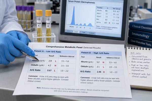
The albumin/globulin ratio compares the amount of albumin in blood with the calculated amount of globulins. It is usually reported with a total protein test or comprehensive metabolic panel. A low ratio can develop because albumin has fallen, globulins have risen, or both changes occurred together. A high ratio usually points to relatively low globulins or, less often, dehydration raising albumin.
The A/G ratio is useful as a pattern-recognition clue, not as a stand-alone diagnosis. Albumin changes with inflammation, liver production, kidney or intestinal protein loss, nutrition risk, and fluid balance. Globulins include antibodies, complement proteins, transport proteins, and acute-phase proteins, so their total can shift with infection, autoimmune disease, liver disease, immune deficiency, and plasma-cell disorders. The most informative approach is to inspect the albumin, calculated globulin, and total protein values separately, then decide whether repeat testing, urine studies, liver tests, immunoglobulins, or serum protein electrophoresis are needed.
- Many laboratories use an A/G ratio near 1.0–2.5 as a general adult reference interval, but the printed laboratory range is the correct one to use.
- A low ratio usually means low albumin, high globulins, or both, often with inflammation, liver disease, kidney protein loss, or increased antibody production.
- A high ratio most often reflects low globulins or dehydration, not excessive albumin production.
- The ratio is calculated rather than directly measured: albumin ÷ (total protein − albumin).
- Persistent abnormalities, especially with weight loss, recurrent infections, swelling, jaundice, bone pain, anemia, or kidney problems, deserve medical follow-up.
Table of Contents
- How the A/G ratio is calculated
- How to interpret the number
- What causes a low A/G ratio
- What causes a high A/G ratio
- Inflammation and immune-system meaning
- Follow-up tests that clarify an abnormal ratio
- Practical next steps and warning signs
How the A/G ratio is calculated
The A/G ratio is albumin divided by globulin, and the globulin value is commonly calculated by subtracting albumin from total protein. For example, if total protein is 7.2 g/dL and albumin is 4.2 g/dL, calculated globulin is 3.0 g/dL. The A/G ratio is 4.2 ÷ 3.0, or 1.4.
Albumin is one specific protein made by the liver. It helps keep fluid inside blood vessels and transports hormones, fatty acids, bilirubin, calcium, and many medicines. Globulin is not one protein. It is a collective label for hundreds of proteins, including:
- immunoglobulins, or antibodies, made by plasma cells;
- complement proteins involved in immune defense;
- acute-phase proteins that change during inflammation;
- carrier proteins and enzymes; and
- clotting-related and other liver-produced proteins.
Because the two sides of the ratio represent different biological systems, the same numerical result can arise through different pathways. A ratio of 0.8 might come from albumin of 3.2 g/dL and globulin of 4.0 g/dL, from albumin of 2.4 g/dL and globulin of 3.0 g/dL, or from another combination. Those patterns do not carry the same meaning.
The ratio may appear on a comprehensive metabolic panel, liver panel, or total protein report. Some laboratories display “globulin” and the ratio automatically; others report only total protein and albumin. The albumin result and globulin result should always be reviewed alongside the ratio.
A small technical point matters: calculated globulin inherits any measurement error in both total protein and albumin. Different albumin assay methods can also shift the result slightly. This is one reason not to compare values from different laboratories too rigidly.
How to interpret the number
A/G ratio interpretation starts with the laboratory’s own reference range and then asks which component is abnormal. Many adult reports show a range roughly between 1.0 and 2.5, but lower and upper limits differ. Children, pregnancy, hydration, acute illness, and assay method can affect the result.
| Pattern | Possible explanation | Useful next question |
|---|---|---|
| Low ratio, low albumin, normal globulin | Inflammation, liver dysfunction, protein loss, dilution, or nutrition risk | Why is albumin low? |
| Low ratio, normal albumin, high globulin | Chronic infection, autoimmune activity, liver disease, or monoclonal protein | Are globulins broadly or narrowly increased? |
| Low ratio, low albumin, high globulin | Combined inflammation and immune activation; chronic liver disease is one example | What do liver tests and protein electrophoresis show? |
| High ratio, normal albumin, low globulin | Low immunoglobulins, protein loss, medication effect, or less common inherited cause | Are quantitative immunoglobulins low? |
| High ratio, high albumin, normal globulin | Usually dehydration or hemoconcentration | Does the value normalize after hydration? |
A result just outside the range is often nonspecific. For example, 0.9 may be a minor change caused by a temporary infection, a slightly low albumin level, or normal biological variation. A ratio does not become dangerous at a single universal cutoff. Severity depends on the component values, trend, symptoms, and cause.
The total protein level adds context. A low ratio with high total protein suggests that excess globulin is contributing. A low ratio with low total protein points more toward reduced albumin, loss of several proteins, dilution, or impaired production. A normal total protein value can hide an important imbalance because low albumin and high globulin may offset each other.
Timing can change the picture. Albumin often falls during hospitalization because of inflammation and IV-fluid dilution, while antibody-related globulins may change more slowly. A value drawn during an acute infection should not automatically be treated as a person’s stable baseline. Conversely, a persistent abnormal ratio across months is more likely to represent an ongoing process. Laboratories also calculate globulin rather than measuring every component directly, so a repeat panel from the same laboratory is often the cleanest way to confirm a small change.
What causes a low A/G ratio
A low A/G ratio is more common than a high one and usually reflects falling albumin, rising globulins, or both. The following mechanisms are the most important.
Inflammation and infection
Inflammation lowers albumin through capillary leakage, altered liver priorities, increased breakdown, and fluid shifts. At the same time, several globulin proteins may rise. Acute infections can produce a temporary change, while tuberculosis, chronic viral infections, endocarditis, and other persistent infections can cause a longer-lasting polyclonal immunoglobulin increase.
The ratio may therefore act as a broad inflammation-related signal, but it cannot tell whether the source is bacterial, viral, autoimmune, malignant, or another process. More direct markers such as C-reactive protein and ESR help describe inflammatory activity but still do not identify the cause by themselves.
Liver disease
Advanced liver disease may reduce albumin production. Cirrhosis can also produce a polyclonal increase in immunoglobulins, creating the classic combination of low albumin and high globulin. The ratio can become low even when total protein remains within range.
The A/G ratio is not a sensitive screening test for early liver disease. Albumin often stays normal until disease is advanced or complicated, and many liver conditions alter AST, ALT, alkaline phosphatase, bilirubin, platelets, or INR before the ratio changes. Alcohol-related liver disease, chronic hepatitis, autoimmune hepatitis, and cirrhosis can each produce different protein patterns.
Kidney or intestinal protein loss
Kidney filters normally retain albumin. Glomerular damage can cause albumin to leak into urine, reducing the ratio. Nephrotic syndrome can also alter specific globulin fractions: some proteins are lost while larger proteins are retained or rise. Urinalysis and quantitative urine protein testing are essential when swelling, foamy urine, diabetes, high blood pressure, or kidney disease is present.
Protein-losing enteropathy can remove albumin and other proteins through the gastrointestinal tract. It may occur with severe intestinal inflammation, celiac disease, lymphatic disorders, or certain heart conditions. Diarrhea, abdominal symptoms, edema, and weight loss may guide testing.
Autoimmune and chronic inflammatory disease
Lupus, rheumatoid arthritis, Sjögren syndrome, inflammatory bowel disease, and other immune-mediated conditions can raise multiple antibody populations while lowering albumin through inflammation. This produces a polyclonal pattern—many immunoglobulin clones are active. The ratio may track broadly with disease burden in some populations, but it is not specific enough to diagnose or monitor an autoimmune disease alone.
Monoclonal gammopathy and blood-cell disorders
A single plasma-cell or B-cell clone may produce a large amount of one immunoglobulin or light chain. This can raise globulin and lower the ratio in monoclonal gammopathy of undetermined significance, multiple myeloma, Waldenström macroglobulinemia, and some lymphomas. The ratio cannot distinguish a harmless monoclonal gammopathy from cancer, and a normal ratio does not exclude one.
Clues that increase concern include unexplained anemia, kidney dysfunction, high calcium, bone pain, fractures, recurrent infections, neuropathy, weight loss, or a large protein gap. Serum protein electrophoresis, immunofixation, and the serum free light chain test are more specific.
What causes a high A/G ratio
A high A/G ratio usually means globulins are relatively low; true excess albumin production is rare. The first step is to inspect whether albumin is high, globulin is low, or both.
Dehydration can concentrate albumin and other blood components. The ratio may rise if albumin increases proportionally more than the calculated globulin value. Clues include thirst, dry mouth, reduced urine, vomiting, diarrhea, heat exposure, or other concentrated laboratory values such as high sodium, blood urea nitrogen, hematocrit, or total protein. A repeat test after recovery may normalize.
Low globulins can occur when antibody production is reduced. Causes include primary immunodeficiency, acquired immune suppression, some blood cancers, medications such as B-cell-depleting therapy, and protein loss. Recurrent sinus infections, pneumonia, ear infections, chronic diarrhea, unusual infections, or poor vaccine responses may prompt an immunoglobulin panel.
Kidney disease can lower selected globulins as well as albumin, although albumin often falls enough to keep the ratio low rather than high. The exact pattern depends on which proteins are lost and how the liver responds. Severe burns, intestinal loss, and malnutrition can also reduce several proteins.
Some laboratory reports or educational sources associate high ratios with leukemia or inherited disorders, but the ratio itself is too nonspecific to support those diagnoses. A mildly high value in a healthy person with normal albumin, total protein, blood counts, and no infection history is often less concerning than a high ratio caused by a clearly low globulin level.
Inflammation and immune-system meaning
The A/G ratio compresses two opposite inflammatory tendencies into one number: albumin often falls while immune-related globulins often rise. This makes a low ratio appealing as an “inflammation-nutrition” index in research, but its clinical role is limited.
Studies have linked lower ratios with worse outcomes in inflammatory bowel disease, infection, cancer, stroke-associated pneumonia, and many other conditions. These associations do not mean the ratio causes disease or that one cutoff works across populations. Most studies are observational and use disease-specific thresholds chosen for prediction, not standard laboratory diagnosis.
The globulin portion also includes proteins unrelated to antibodies. During acute inflammation, alpha-1 and alpha-2 globulins may rise because proteins such as alpha-1 antitrypsin, haptoglobin, and ceruloplasmin are acute-phase reactants. During chronic immune activation, gamma globulins may increase because antibody-producing plasma cells are active. A single calculated globulin value cannot separate those fractions.
Serum protein electrophoresis solves part of this problem by dividing proteins into albumin, alpha-1, alpha-2, beta, and gamma regions. A broad gamma-region increase suggests polyclonal immune stimulation. A narrow spike suggests a monoclonal protein and requires confirmation. The SPEP test is therefore far more informative than the A/G ratio when abnormal antibody production is suspected.
The ratio should also not be treated as a direct measure of nutrition. Albumin can fall rapidly during inflammation, while globulins rise because of immune activity. Weight history, food intake, muscle and fat loss, functional status, and a nutrition-focused examination are needed to diagnose malnutrition.
Follow-up tests that clarify an abnormal ratio
Follow-up should target the component that changed and the clinical clues present. A clinician may repeat the panel first if the abnormality is mild, unexpected, or drawn during an acute illness.
Common next steps include:
- Repeat total protein, albumin, and calculated globulin: confirms persistence and shows the trend.
- Liver testing: AST, ALT, alkaline phosphatase, bilirubin, INR, platelets, hepatitis testing, and imaging when indicated.
- Kidney testing: creatinine, estimated glomerular filtration rate, urinalysis, urine albumin-to-creatinine ratio, or urine protein-to-creatinine ratio.
- Inflammation testing: CRP, ESR, CBC, ferritin, and other tests chosen for symptoms.
- Quantitative immunoglobulins: measures IgG, IgA, and IgM separately when globulins are low or immune activation is suspected.
- SPEP and immunofixation: evaluates broad versus monoclonal protein increases.
- Serum free light chains: helps detect or characterize plasma-cell disorders, especially when kidney function and symptoms raise concern.
- Targeted autoimmune or infection tests: selected from the history and examination rather than ordered as a broad fishing expedition.
A useful derived value is the protein gap, calculated as total protein minus albumin—the same number often reported as globulin. A markedly increased gap can prompt evaluation for immunoglobulin excess, but there is no single gap threshold that diagnoses infection or monoclonal gammopathy.
Medication review matters. Immunosuppressive drugs, corticosteroids, monoclonal antibodies, chemotherapy, and some anticonvulsants can affect immunoglobulins or liver proteins. Recent IV immunoglobulin therapy can temporarily raise measured globulins and complicate electrophoresis interpretation.
Practical next steps and warning signs
For an isolated abnormal ratio, look at the three underlying numbers before drawing conclusions. Write down albumin, total protein, and globulin, compare them with prior results, and note whether the blood was drawn during infection, dehydration, hospitalization, pregnancy, or treatment with IV fluids.
A practical sequence is:
- Confirm whether the ratio is truly outside that laboratory’s range.
- Identify whether albumin, globulin, or both drive the change.
- Check the trend rather than focusing on one decimal point.
- Review symptoms, medications, hydration, recent illness, and known liver or kidney conditions.
- Discuss whether repeat testing or specific follow-up is warranted.
Routine fasting is not required for the ratio itself. However, a comprehensive metabolic panel may be drawn with glucose or lipid testing that requires fasting under local instructions. Do not stop medications or supplements unless the ordering clinician says to do so.
Arrange timely medical review if an abnormal ratio accompanies unexplained weight loss, persistent fever, swollen lymph nodes, recurrent infections, jaundice, edema, foamy urine, chronic diarrhea, anemia, bone pain, numbness, or declining kidney function. Seek urgent care for confusion, severe shortness of breath, chest pain, fainting, very low urine output, signs of sepsis, or rapidly worsening swelling.
The key is to avoid two common errors: assuming every low ratio means autoimmune disease or cancer, and dismissing a persistent abnormality because the total protein is normal. The ratio is a starting point. The separate protein values and the clinical story determine what it means.
One final safeguard is to avoid self-diagnosis from online cutoffs. The same ratio may be clinically trivial in one setting and important in another. Age, pregnancy, hydration, assay method, medicines, and recent illness all affect interpretation, so follow-up should be proportionate rather than alarm-driven.
References
- Total Protein and Albumin/Globulin (A/G) Ratio 2024
- Accuracy of Albumin, Globulin, and Albumin–Globulin Ratio for Diagnosing Periprosthetic Joint Infection: A Systematic Review and Meta-Analysis 2023 (Systematic Review)
- Serum Albumin to Globulin Ratio is Associated with the Presence and Severity of Inflammatory Bowel Disease 2022
- Clinical significance of albumin to globulin ratio among patients with stroke-associated pneumonia 2022
- A study investigating how the albumin-globulin ratio relates to depression risk within U.S. adults: a cross-sectional analysis 2024
- Interpretations of the Role of Plasma Albumin in Prognostic Indices: A Literature Review 2023 (Review)
Disclaimer
This article is for general education and cannot determine the cause of an individual A/G ratio. Laboratory ranges, symptoms, and related test results must be interpreted together by a qualified healthcare professional.