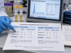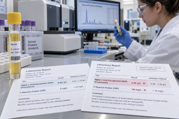
Alpha-1 acid glycoprotein is a blood protein that often rises when the body is responding to inflammation, infection, injury, or major physical stress. It is also called AGP, orosomucoid, or ORM. Unlike a test aimed at one disease, the alpha-1 acid glycoprotein test measures a broad acute-phase response. A high result can confirm that inflammatory signaling is active, but it cannot identify the cause by itself. The number must be interpreted with symptoms, medical history, other laboratory findings, and changes over time. AGP also has an unusual second role: it binds several medications in the bloodstream. Large changes in its concentration may therefore affect how much unbound, active drug is available. This article explains what the test measures, why levels increase, what low values may mean, how AGP compares with familiar inflammatory markers, and what questions are useful when reviewing an abnormal result.
- Acute-phase marker: Alpha-1 acid glycoprotein is a positive acute-phase protein made mainly by the liver.
- Nonspecific signal: A high level supports inflammation or physiological stress but does not diagnose one condition.
- Range differences: Reference intervals vary by laboratory, age, sex, pregnancy status, and testing method.
- Drug binding: AGP can bind basic and lipophilic medicines, affecting drug distribution and total levels.
- Trends matter: Companion tests and changes over time are usually more informative than one isolated measurement.
Table of Contents
- What the Alpha-1 Acid Glycoprotein Test Measures
- Normal Range and Result Interpretation
- Why Alpha-1 Acid Glycoprotein Rises
- High Levels and Possible Causes
- Low Levels and Special Situations
- Drug Binding and Clinical Importance
- Testing, Follow-Up, and Next Steps
What the Alpha-1 Acid Glycoprotein Test Measures
Alpha-1 acid glycoprotein is a heavily glycosylated plasma protein. “Glycosylated” means that carbohydrate chains are attached to the protein. Those chains account for a large part of its mass and help shape its interactions with immune cells, blood vessels, and circulating compounds. Two closely related genes, ORM1 and ORM2, encode the main forms found in human blood.
The liver is the primary source of circulating AGP. When inflammatory messengers such as interleukin-6 signal that tissue has been injured or an infection is present, liver cells shift protein production. They make more positive acute-phase proteins, including AGP, C-reactive protein, fibrinogen, haptoglobin, and ceruloplasmin. At the same time, concentrations of some negative acute-phase proteins, especially albumin and transferrin, may fall.
AGP is not simply an inflammation counter. It has transport and regulatory functions. Its negatively charged, highly branched carbohydrate surface allows it to bind a range of basic or lipophilic molecules. It may also help limit excessive immune activation, affect neutrophil behavior, and influence interactions between blood cells and the vessel lining. Researchers are studying whether changes in AGP’s carbohydrate pattern carry additional information beyond its concentration, but glycoform testing remains largely a research tool rather than routine clinical practice.
A standard AGP blood test reports the total concentration in serum or plasma, commonly in grams per liter or milligrams per deciliter. It does not usually separate ORM1 from ORM2, identify individual glycoforms, or reveal the source of inflammation. In practice, clinicians may order it to assess an acute-phase response, add context to another protein measurement, investigate unusual medication binding, or monitor selected inflammatory conditions.
AGP is less commonly ordered than the C-reactive protein test. This does not make it unimportant; it means its best use is more specialized. Its slower behavior, drug-binding properties, and relationship to other serum proteins can provide information that CRP alone does not provide.
Normal Range and Result Interpretation
There is no single universal AGP reference interval. Laboratories establish ranges based on their assay, equipment, calibration, and local population. Age and sex can matter, and pregnancy and infancy can lower expected concentrations. Always compare the result with the interval printed on the same laboratory report.
One NHS laboratory, for example, lists adult intervals of 0.6–1.2 g/L for males and 0.4–1.0 g/L for females from ages 5 to 50, and 0.8–2.0 g/L for people older than 50. These figures illustrate how much ranges can vary across demographic groups; they should not replace the interval supplied by the testing laboratory.
| Result pattern | General interpretation | What is usually needed next |
|---|---|---|
| Within the laboratory range | No clear AGP elevation at the time of sampling | Interpret with symptoms and other tests; normal AGP does not exclude localized or early disease |
| Mildly high | Possible low-grade, recovering, or chronic acute-phase response | Review recent illness, medications, chronic conditions, and companion markers |
| Clearly high | Stronger evidence of systemic inflammatory signaling or major physiological stress | Look for the clinical cause rather than treating the number itself |
| Low | May reflect age, pregnancy, reduced production, protein loss, or biological variation | Consider liver function, kidney or intestinal protein loss, nutritional context, and repeat testing when appropriate |
A value just beyond the upper limit is not automatically evidence of serious disease. Reference intervals usually contain about 95% of a healthy comparison population, so a small proportion of healthy people will fall outside them. Hydration, temporary illness, age-related changes, and analytical variation can also shift the result.
The degree of elevation is only one part of interpretation. Timing matters. AGP generally rises more gradually than CRP and may remain elevated during recovery. A person whose fever and pain are improving may still have a raised AGP because liver production and clearance have not yet returned to baseline. Conversely, testing very early in an inflammatory event may show a less dramatic increase than a later sample.
A trend can therefore be more useful than one number. A falling value may support recovery when it agrees with symptoms and other findings. A persistent or rising level may justify a search for ongoing inflammation, but it still does not distinguish infection from autoimmune disease, tissue damage, cancer, or another trigger. The same principle applies to an erythrocyte sedimentation rate: nonspecific markers describe a biological response, not a final diagnosis.
Why Alpha-1 Acid Glycoprotein Rises
The acute-phase response is a coordinated defense program. Damaged tissues and activated immune cells release cytokines. These chemical messengers act on the brain, bone marrow, blood vessels, and liver. They can produce fever, increase white blood cell production, change iron handling, promote clotting readiness, and alter the mix of proteins circulating in plasma.
AGP is called a positive acute-phase protein because its concentration increases during this response. Its rise is thought to serve several purposes. By binding endogenous molecules and medicines, it changes how compounds move through blood and tissues. Its carbohydrate chains can interact with immune receptors. Experimental and clinical studies also suggest that AGP may temper excessive neutrophil activation and help protect tissues from uncontrolled inflammation. These effects are complex: AGP is a marker of inflammation, but it may simultaneously participate in limiting inflammatory injury.
AGP does not move in perfect lockstep with every other marker. Different proteins have different production rates and half-lives. CRP can rise rapidly and dramatically, making it useful for many acute infections and inflammatory flares. ESR changes indirectly because inflammation alters red-cell settling, especially through fibrinogen and immunoglobulin effects. AGP may provide a steadier, somewhat slower signal.
| Marker | What it reflects | Typical interpretive strength | Important limitation |
|---|---|---|---|
| Alpha-1 acid glycoprotein | Liver acute-phase production plus transport and immune-regulatory functions | Specialized assessment of systemic inflammatory response and drug binding | Not widely standardized for general diagnostic use |
| CRP | Rapid liver response to inflammatory cytokines | Useful for detecting and following many acute inflammatory states | Still nonspecific |
| ESR | How quickly red cells settle under the influence of plasma proteins and red-cell factors | Helpful in selected chronic inflammatory disorders | Affected by age, anemia, pregnancy, and blood-cell characteristics |
| Fibrinogen | Acute-phase production and clotting capacity | Connects inflammation with coagulation | Can be altered by clotting, liver disease, pregnancy, and smoking |
Because these markers respond differently, a clinician may compare them rather than expecting exact agreement. High CRP with a modest AGP increase can fit an early acute event. Persistently raised AGP after CRP falls can fit a lagging recovery pattern. A high ESR with normal CRP and AGP may point toward factors that influence sedimentation rather than a strong current acute-phase response. These are patterns, not diagnostic rules.
An inflammatory marker panel can be helpful only when each result is interpreted in context. Ordering many markers without a focused clinical question can create confusing minor abnormalities and unnecessary repeat testing.
High Levels and Possible Causes
A high alpha-1 acid glycoprotein level means the body is producing more of the protein or clearing it more slowly. In most clinical settings, increased production during an acute-phase response is the leading explanation. The test cannot determine which condition is responsible, so the likely causes are narrowed by symptoms, examination, risk factors, imaging, cultures, and more specific laboratory tests.
Common categories include:
- Infection: Bacterial, viral, fungal, and other infections can raise AGP, particularly when they cause a systemic response.
- Autoimmune or inflammatory disease: Active rheumatoid arthritis, vasculitis, inflammatory bowel disease, connective-tissue disease, and other immune-mediated disorders may increase acute-phase protein production.
- Tissue injury: Surgery, trauma, burns, heart injury, and extensive tissue damage can trigger a rise even when no infection is present.
- Cancer: Some malignancies are associated with elevated AGP, especially when they produce inflammation, tissue injury, infection, or a substantial disease burden. AGP is not a stand-alone cancer screening test.
- Metabolic or organ disease: Obesity, insulin resistance, chronic kidney disease, and inflammatory liver conditions may be associated with persistent low-grade elevations.
- Major physiological stress: Severe illness can activate the acute-phase response through several pathways at once.
The clinical pattern matters more than the category list. Fever, cough, painful urination, or a wound may direct attention toward infection. Morning stiffness, swollen joints, rash, mouth ulcers, or unexplained vascular symptoms may support autoimmune evaluation. Weight loss, persistent night sweats, unexplained bleeding, or a new mass require timely assessment but do not prove cancer.
Liver disease deserves careful interpretation. Because the liver makes AGP, inflammatory liver injury can stimulate production, while advanced loss of liver synthetic capacity can limit it. A person with active steatohepatitis may have a high level, whereas someone with severe liver failure may not mount the expected rise. Albumin, bilirubin, clotting tests, and liver enzymes help clarify the pattern. The albumin blood test often moves in the opposite direction during inflammation because albumin is a negative acute-phase protein.
A high result should not be used to decide automatically that antibiotics, steroids, or another anti-inflammatory treatment is needed. Treatment must target the underlying condition. Lowering AGP itself is not normally a clinical goal. When the cause improves, AGP generally moves toward baseline over time.
Low Levels and Special Situations
Low AGP receives less attention than high AGP because the test is mainly used as a positive acute-phase marker. A low value can still be meaningful, especially when it appears with other abnormal proteins or symptoms suggesting reduced production or excessive loss.
Possible explanations include:
- Pregnancy and infancy: Lower concentrations may be physiological, and adult reference intervals should not be applied without adjustment.
- Reduced liver synthesis: Advanced liver dysfunction can reduce production of AGP along with other liver-made proteins.
- Protein loss: Significant urinary protein loss, protein-losing intestinal disease, extensive burns, or other conditions may reduce circulating proteins.
- Malnutrition or severe systemic illness: These can alter liver protein synthesis, although AGP may rise if strong inflammation is present, creating a mixed pattern.
- Genetic or individual variation: Rare inherited factors and ordinary biological differences may contribute.
- Laboratory variation: A borderline low result may not persist when repeated.
Low AGP by itself does not diagnose liver failure, kidney disease, or malnutrition. Clinicians usually review the complete protein picture. Total protein, albumin, globulins, urine protein, kidney function, liver enzymes, bilirubin, and clotting measurements may be more directly informative. When protein loss is suspected, the location of loss matters: urine testing evaluates renal leakage, while gastrointestinal investigations may be needed for intestinal loss.
Mixed results are common. For example, inflammation pushes AGP upward while advanced liver dysfunction pulls production downward. The final concentration may be normal even though both processes are active. This is one reason a “normal” result should not be interpreted in isolation.
Other acute-phase proteins can provide clues. Haptoglobin may rise with inflammation but fall when red blood cells are being destroyed faster than the liver can replace it. Comparing AGP with a haptoglobin blood test can therefore reveal different biological forces acting at the same time. Ceruloplasmin is another liver-made acute-phase protein, but it also carries copper and has its own disease-specific interpretation.
Drug Binding and Clinical Importance
AGP’s drug-binding role is one of the most distinctive reasons the test may matter. Many medicines circulate in two forms: bound to plasma proteins and unbound. The unbound fraction can generally cross membranes, reach receptors, be metabolized, and be eliminated more readily. Laboratory drug assays often report total concentration, which combines bound and unbound drug.
Albumin is the main binding protein for many acidic drugs. AGP tends to bind basic and some neutral lipophilic drugs. Examples discussed in clinical laboratory resources include propranolol, imipramine, and quinidine. Other drugs with clinically relevant AGP binding may include lidocaine and certain antipsychotic, antidepressant, antiarrhythmic, anesthetic, or anticancer agents, although the practical effect differs by drug.
When AGP rises sharply during critical illness, more of a highly AGP-bound medication may become protein-bound. The total drug concentration can remain normal or rise while the active unbound fraction changes. When inflammation resolves and AGP falls, binding can decrease again. Dose changes made during the inflammatory phase may then have different effects during recovery.
This does not mean that every elevated AGP requires medication adjustment. Several factors determine clinical significance:
- How strongly the drug binds to AGP
- Whether it has a narrow therapeutic range
- Whether dosing is guided by symptoms, total levels, or free levels
- Liver and kidney clearance
- Interactions with other medicines
- The speed and magnitude of the AGP change
Patients should not change a prescription based on an AGP result without medical guidance. The clinician or pharmacist may review adverse effects, treatment response, timing of doses, organ function, and therapeutic drug monitoring. In selected cases, measuring the free drug concentration is more informative than measuring total concentration.
The same principle helps explain why severe illness can complicate pharmacology. Inflammation can alter AGP, albumin, liver enzymes, kidney filtration, blood flow, and drug transporters simultaneously. A mathematically “therapeutic” total drug level may not perfectly represent exposure at the target tissue. AGP is one part of that larger picture.
Testing, Follow-Up, and Next Steps
The AGP test uses a blood sample, usually serum. Most laboratories do not require fasting, although the ordering clinician may request fasting when other tests are collected at the same time. Tell the healthcare team about prescription medicines, over-the-counter products, supplements, recent surgery, infection, pregnancy, and major changes in health because these details can alter interpretation.
After an abnormal result, follow-up should answer a specific question rather than simply repeat every inflammatory marker. Useful questions include:
- Does the result fit the clinical picture? A high value is more meaningful when symptoms or examination findings also suggest inflammation.
- Is the abnormality new or persistent? Previous results can distinguish a temporary response from a chronic baseline.
- Do companion tests agree? CRP, ESR, blood counts, liver tests, kidney tests, cultures, and disease-specific antibodies may identify the direction of investigation.
- Could protein production or loss be affecting the value? This is especially important for low or unexpectedly normal results.
- Could medication binding matter? Review drugs with narrow therapeutic windows or unexplained changes in effectiveness or toxicity.
The interval for repeat testing depends on the reason for ordering it. In an acute illness, a clinician may repeat it after treatment or clinical change. In chronic inflammatory disease, measurements may be spaced to follow activity over weeks or months. Repeating the test too soon may not add useful information because AGP can change more slowly than faster markers.
Seek prompt medical attention when an abnormal result accompanies warning signs such as trouble breathing, confusion, severe weakness, persistent high fever, chest pain, rapidly worsening swelling, reduced urine output, yellowing of the skin, uncontrolled bleeding, or signs of a serious medication reaction. The urgency comes from the symptoms, not from the AGP number alone.
For most people, the central message is straightforward: alpha-1 acid glycoprotein is evidence about the body’s response, not a diagnosis. A high level supports active or recent systemic stress. A low level may raise questions about production, loss, or physiological state. Its greatest value comes from combining the result with timing, symptoms, other proteins, organ function, and medication context.
References
– Alpha-1 Acid Glycoprotein (AGP) 2026 – Glycosylation Variability of Serum α1-Acid Glycoprotein in the Context of Physiological and Pathological Conditions—A Review 2025 – Acute Phase Protein Orosomucoid (Alpha-1-Acid Glycoprotein) Predicts Delayed Cerebral Ischemia and 3-Month Unfavorable Outcome after Aneurysmal Subarachnoid Hemorrhage 2023 – The Clinical Utility of Serum Alpha-1-Acid Glycoprotein in Reflecting the Cross-Sectional Activity of Antineutrophil Cytoplasmic Antibody-Associated Vasculitis 2024 – Alpha 1-Acid Glycoprotein Is Upregulated in Severe COVID-19 Patients and Decreases Neutrophil NETs in SARS-CoV-2 Infection 2024 – α1-Acid Glycoprotein with Highly Fucosylated Glycans as a Potential Biomarker for Hepatobiliary and Pancreatic Cancers 2024
Disclaimer
This article is for general educational purposes and does not diagnose the cause of a high or low alpha-1 acid glycoprotein result. Reference ranges, testing methods, and clinical uses differ among laboratories, and medication-binding effects require individualized assessment. Discuss your result and any treatment decisions with a qualified healthcare professional.