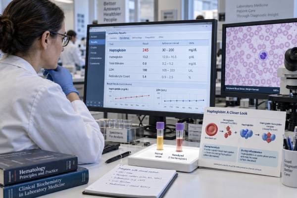
The haptoglobin blood test is best known for helping evaluate hemolysis—the premature destruction of red blood cells. Haptoglobin is made by the liver and binds free hemoglobin released into plasma. The haptoglobin-hemoglobin complex is then cleared, limiting iron loss and oxidative injury. When substantial intravascular hemolysis occurs, haptoglobin is consumed and the blood level usually falls. Yet haptoglobin is also a positive acute-phase protein, so inflammation, infection, or tissue injury can raise it. This creates an important interpretive tension: a high result usually reflects an inflammatory response and does not mean red blood cells are being destroyed, while a normal result may sometimes mask mild hemolysis if inflammation is simultaneously pushing production upward. Clinicians therefore read haptoglobin with hemoglobin, reticulocytes, lactate dehydrogenase, indirect bilirubin, blood smear, liver function, and symptoms. This article explains high and low patterns, the limitations of the test, and how a hemolysis workup is assembled.
- Primary role: Haptoglobin captures free hemoglobin released from red blood cells and carries it for clearance.
- Classic hemolysis pattern: Low haptoglobin with high LDH, high indirect bilirubin, and an increased reticulocyte count.
- Meaning of a high value: Most often an acute-phase response to inflammation, infection, tissue injury, or another inflammatory condition.
- Key limitation: Inflammation can keep haptoglobin normal or high despite concurrent hemolysis.
- Clinical rule: No haptoglobin value should be interpreted without the rest of the blood count and hemolysis markers.
Table of Contents
- What haptoglobin does and how the test works
- Reference ranges and result patterns
- Why haptoglobin levels become high
- Low haptoglobin and hemolysis
- When inflammation masks hemolysis
- How clinicians investigate an abnormal result
- Treatment, follow-up, and urgent warning signs
What Haptoglobin Does and How the Test Works
Red blood cells normally circulate for about 120 days before macrophages remove them, mainly in the spleen and liver. Most of their hemoglobin is recycled inside those cells. If red cells rupture within blood vessels, hemoglobin escapes directly into plasma. Free hemoglobin can filter through the kidneys, consume nitric oxide, promote oxidative injury, and release iron-containing heme.
Haptoglobin binds free hemoglobin with high affinity. The complex is recognized by receptors on macrophages and cleared, helping conserve iron and protect tissues. This protective system has limited capacity. With brisk or sustained intravascular hemolysis, haptoglobin is used faster than the liver can replace it, so the measured concentration falls, sometimes below the assay’s detection limit.
The liver also increases haptoglobin synthesis during the acute-phase response. Cytokines released in infection, inflammation, trauma, burns, and tissue necrosis can drive a large increase. This dual behavior—consumption by hemolysis and production during inflammation—is the central feature of interpretation.
The test uses serum or plasma from a routine venous blood draw. No special preparation is usually needed. The clinician should know about recent transfusion, infection, surgery, vigorous exercise, liver disease, pregnancy, medications, and any personal or family history of anemia. Results may be reported in milligrams per deciliter, grams per liter, or another laboratory-specific unit.
Haptoglobin is generally ordered when hemolytic anemia is suspected, including after a possible transfusion reaction. Symptoms that may lead to testing include fatigue, pallor, jaundice, dark urine, back or abdominal pain, shortness of breath, rapid heartbeat, or an unexplained fall in hemoglobin. It is rarely useful as a stand-alone screen for inflammation because more familiar markers are available and haptoglobin has strong hemolysis and liver-related confounders.
The protein exists in genetically determined forms that can influence measured concentration and biological behavior. Laboratories account for normal population variation through their reference intervals, but an individual’s baseline may differ. Some people have inherited very low or absent haptoglobin without active hemolysis, and prevalence varies by ancestry. This possibility becomes relevant when the value remains low despite no other evidence of red-cell destruction.
Reference Ranges and Result Patterns
Haptoglobin intervals differ substantially by laboratory, assay, age, and population. Many adult laboratories use a range around 30–200 mg/dL, but broader or narrower intervals are common. Infants naturally have lower concentrations, making the test unreliable for hemolysis in early life. The reference range on the report is the appropriate starting point.
A result is interpreted as a pattern rather than a binary high or low flag.
| Haptoglobin pattern | Possible interpretation | Companion findings that matter |
|---|---|---|
| Low or undetectable | Intravascular hemolysis, severe liver dysfunction, inherited low level, or less commonly another cause | LDH, indirect bilirubin, reticulocytes, hemoglobin, smear, urine hemoglobin |
| Normal with no inflammatory evidence | Substantial intravascular hemolysis is less likely, but not completely excluded | Timing, severity, extravascular hemolysis, other markers |
| Normal with active inflammation | Hemolysis may be masked by increased hepatic production | CRP, trend, LDH, bilirubin, reticulocytes |
| High | Inflammation, infection, tissue injury, or another acute-phase stimulus | Symptoms, CRP/ESR, liver and metabolic context |
| Falling over time | New or worsening consumption may be developing | Concurrent hemoglobin fall and hemolysis markers |
Timing can change the answer. Haptoglobin may fall after hemolysis begins, while reticulocytes take several days to rise because the marrow needs time to respond. After hemolysis stops, haptoglobin does not immediately normalize. A transfusion can introduce donor red cells and change several markers. Serial testing may therefore be more informative than one sample.
Intravascular and extravascular hemolysis also differ. Intravascular hemolysis releases hemoglobin directly into plasma and strongly consumes haptoglobin. Extravascular hemolysis occurs mainly when macrophages remove abnormal or antibody-coated red cells in the spleen or liver. Less free hemoglobin reaches plasma, so haptoglobin may be only mildly low or occasionally normal, especially if inflammation is present.
The laboratory flag should never be used to grade the severity of anemia by itself. A person can have severe anemia from bleeding or low production with normal haptoglobin. Another person can have low haptoglobin without anemia because the marrow compensates or because the low value is inherited.
Why Haptoglobin Levels Become High
A high haptoglobin result usually means the liver has increased production as part of an acute-phase response. It does not mean there is excess hemoglobin in circulation; binding to free hemoglobin tends to consume haptoglobin rather than raise the measured level.
Acute Infection and Tissue Injury
Severe bacterial infection, some viral illnesses, surgery, trauma, burns, and tissue necrosis can increase haptoglobin. The rise may occur alongside fever, leukocytosis, high CRP, and other acute-phase changes. Because different proteins have different timing, haptoglobin may remain elevated after the fastest inflammatory markers begin to fall.
Chronic Inflammatory and Autoimmune Disease
Rheumatoid arthritis, inflammatory bowel disease, vasculitis, and other chronic inflammatory conditions may keep haptoglobin above the usual range. The value is nonspecific and cannot identify which disease is active. It may contribute to a broader inflammatory pattern but is not commonly used as a treatment target.
A CRP test is generally more direct for tracking rapid acute-phase changes. The ESR may reflect longer-lasting protein and red-cell effects. Haptoglobin adds a different question: is its concentration consistent with or against substantial hemoglobin release into plasma?
Metabolic and Other Associations
Higher concentrations have been reported with diabetes, obesity, smoking, corticosteroid exposure, and some cancers. These associations often reflect low-grade inflammation, tissue stress, or altered liver protein production. A high result alone is not a cancer marker and should not trigger a broad malignancy search without other clinical clues.
Pregnancy and estrogen-related states may influence acute-phase and binding proteins, although haptoglobin behavior is not as uniform as some other markers. Kidney disease can complicate anemia interpretation through reduced erythropoietin, blood loss, inflammation, and dialysis-related factors. A high haptoglobin in kidney disease does not explain the anemia by itself.
What High Haptoglobin Does Not Mean
A high level does not prove that the blood is “too thick,” that a clot is present, or that iron stores are excessive. It is not the same as hemoglobin, ferritin, or hematocrit. It also does not diagnose inflammation in the absence of symptoms and supporting findings. The cause is usually addressed by evaluating the underlying illness rather than lowering haptoglobin.
If high haptoglobin appears with low hemoglobin, clinicians do not assume the two are contradictory. The anemia may come from iron deficiency, inflammation-related restricted production, kidney disease, bleeding, vitamin deficiency, marrow disease, or hemolysis partially hidden by an acute-phase response. The rest of the pattern separates these possibilities.
Low Haptoglobin and Hemolysis
Low haptoglobin is most useful when it fits a coherent hemolysis pattern. Hemolysis means red cells are destroyed faster than they can be replaced. The marrow usually responds by releasing more reticulocytes, while red-cell contents and breakdown products change other laboratory values.
A typical set of findings includes:
- Falling hemoglobin or anemia
- Increased reticulocyte count, unless marrow response is impaired
- Elevated lactate dehydrogenase (LDH)
- Elevated unconjugated, or indirect, bilirubin
- Low haptoglobin
- Characteristic blood-smear findings in some causes
This constellation confirms that destruction is likely but does not identify why it is happening. Causes include autoimmune hemolytic anemia, transfusion reactions, mechanical destruction from heart valves or circulatory devices, microangiopathic processes, infections, burns, inherited membrane or enzyme disorders, sickle-cell disease, thalassemia, and certain medications.
The peripheral blood smear can show spherocytes, schistocytes, bite cells, sickled cells, parasites, agglutination, or other clues. The direct antiglobulin test checks whether antibodies or complement are attached to red cells and helps distinguish immune from nonimmune causes. It is not the same as haptoglobin and may be negative in many forms of hemolysis.
Low haptoglobin can occur without hemolysis. Severe liver disease may reduce production. Large hematomas can cause red-cell breakdown outside the circulation and alter markers. Regular strenuous exercise may cause low-grade mechanical hemolysis, particularly in endurance athletes. Congenital low or absent haptoglobin is another explanation. Therefore, isolated low haptoglobin with normal hemoglobin, LDH, bilirubin, reticulocytes, and smear should be interpreted cautiously.
Not every hemolytic episode produces reticulocytosis. Bone-marrow suppression, nutrient deficiency, kidney disease, infection, or a very recent onset can blunt or delay the response. A “normal” reticulocyte percentage may also be inadequate for the degree of anemia; clinicians may use an absolute count or corrected index.
When Inflammation Masks Hemolysis
The most important limitation of haptoglobin is the possibility of opposing forces. Hemolysis consumes the protein, while inflammation increases production. If both occur, the measured concentration may land within the reference interval or even above it. A normal haptoglobin cannot safely exclude hemolysis when there is a strong inflammatory response.
Consider a person with bacterial sepsis and a mechanical heart valve. Infection may markedly stimulate haptoglobin synthesis, while valve-related red-cell damage consumes it. The result may be “normal,” even though LDH is high, indirect bilirubin is rising, and the smear shows fragmented cells. The correct interpretation comes from the combined trajectory.
Conversely, a very low haptoglobin during inflammation strengthens evidence for significant intravascular hemolysis because production is expected to be increased. It still needs confirmation, particularly if severe liver disease limits synthesis.
Several strategies reduce misinterpretation:
- Compare haptoglobin with CRP or other evidence of an acute-phase response.
- Use serial values rather than relying on a single result.
- Review absolute reticulocytes, LDH, bilirubin fractions, and hemoglobin together.
- Inspect the blood smear when hemolysis is suspected.
- Consider liver synthetic function and baseline haptoglobin variation.
- Match laboratory timing to transfusion, surgery, medication exposure, and symptom onset.
LDH is also nonspecific: it rises with liver, muscle, tissue, and malignant injury. Bilirubin can rise from liver or biliary disease. Reticulocytes can increase after bleeding. Each marker has limitations, but their agreement creates a much stronger conclusion than any one result.
An inflammatory marker panel may document systemic inflammation, yet it cannot replace the specific hemolysis workup. The clinical problem is not simply whether inflammation exists, but whether red cells are being destroyed and why.
How Clinicians Investigate an Abnormal Result
The investigation begins with the complete blood count. Hemoglobin and hematocrit establish whether anemia is present. Mean cell volume and red-cell distribution width offer clues but do not classify hemolysis reliably. Platelets and white cells can point toward a broader marrow, infection, or microangiopathic process.
When hemolysis is suspected, common tests include:
- Absolute reticulocyte count
- LDH
- Total and direct bilirubin, allowing indirect bilirubin calculation
- Haptoglobin
- Peripheral blood smear
- Urinalysis for hemoglobin and urobilinogen
After hemolysis is supported, testing is directed toward cause. The direct antiglobulin test assesses immune coating. Coagulation studies and organ-function tests may be needed for suspected disseminated intravascular coagulation. Flow cytometry is used for paroxysmal nocturnal hemoglobinuria. Enzyme testing, hemoglobin analysis, membrane studies, infection testing, or imaging may be appropriate in selected cases.
A recent transfusion changes priorities. Fever, chills, back pain, chest tightness, dark urine, low blood pressure, or breathing difficulty during or soon after transfusion requires immediate evaluation. The transfusion is stopped, blood-bank protocols are activated, and samples are checked for incompatibility and hemolysis. Delayed reactions can appear days to weeks later with falling hemoglobin and jaundice.
For isolated high haptoglobin, clinicians usually review infection and inflammatory symptoms, CRP, liver tests, diabetes, medications, and recent tissue injury. If the reason is obvious and the person is recovering, no haptoglobin-specific workup may be needed. Persistent unexplained elevation is interpreted as one clue among many, not as a disease category.
Liver assessment deserves care because the organ makes haptoglobin. Severe synthetic failure can produce a low value even without red-cell destruction. Mild liver inflammation, however, may raise it as an acute-phase reactant. Albumin, INR, bilirubin, enzymes, imaging, and clinical findings clarify which process predominates.
Treatment, Follow-Up, and Urgent Warning Signs
There is no routine treatment for high haptoglobin itself. The value generally falls as infection, inflammation, or tissue injury resolves. Treatment targets the underlying condition. Repeating the test is useful only when it answers a clinical question, such as whether a suspected hemolytic process is improving.
Hemolytic anemia treatment depends on cause and severity. Immune hemolysis may require corticosteroids, other immune therapy, or treatment of a trigger. A transfusion reaction requires immediate supportive and blood-bank management. Mechanical hemolysis may require evaluation of a valve or circulatory device. G6PD-related episodes are managed by stopping triggers and treating infection. Microangiopathic hemolysis can signal a life-threatening condition requiring urgent specialist therapy.
Supportive care may include transfusion when anemia is severe or symptomatic, folate replacement in chronic high-turnover states, kidney protection, and monitoring for thrombosis or organ injury. Iron should not be started solely because anemia is present; hemolysis recycles iron and some chronic disorders can accumulate it. The cause and iron studies should guide supplementation.
Seek emergency care for sudden dark or red urine, jaundice with severe weakness, chest or back pain after transfusion, breathing difficulty, fainting, confusion, fever with rapid deterioration, or rapidly worsening anemia symptoms. New neurologic changes, very low platelets, kidney injury, or fragmented cells on a smear may indicate a thrombotic microangiopathy or DIC and need urgent action.
Contact a clinician promptly for persistent fatigue, pallor, yellowing of the eyes, unexplained tachycardia, recurrent dark urine, or a falling hemoglobin. A high haptoglobin result by itself is rarely an emergency. Its value lies in context: it shows the balance between hepatic production and consumption by free hemoglobin, while the rest of the hemolysis panel reveals which force is likely dominant.
References
- Haptoglobin (HP) Test 2024
- HAPT – Overview: Haptoglobin, Serum 2026
- Hemolytic Anemias 2026
- Reticulocyte Count 2024
- Blood Smear 2024
- Clinical Benefits and Risks of Haptoglobin Therapy in Patients With Hemolytic Disorders: A Systematic Review 2025
Disclaimer
This article provides general education and cannot diagnose hemolysis, anemia, liver disease, or inflammation. Haptoglobin reference ranges and reliability vary with age, assay, liver function, and acute illness, so personal results require interpretation with the complete hemolysis panel. Seek urgent care for severe anemia symptoms, a possible transfusion reaction, dark urine with weakness, or rapid clinical deterioration.