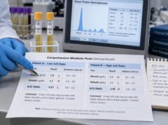
A chronic inflammation blood test panel does not diagnose one specific disease. Instead, it looks for patterns that suggest ongoing immune activity, infection, tissue injury, anemia, iron disruption, or another condition that may be driving inflammation. The most common starting tests are C-reactive protein (CRP), erythrocyte sedimentation rate (ESR), a complete blood count (CBC) with differential, and ferritin. Depending on symptoms, a clinician may add iron studies, liver and kidney tests, autoimmune antibodies, complement proteins, immunoglobulins, or other targeted markers.
The most useful result is rarely a single high number. Timing, symptoms, medications, age, pregnancy, recent illness, body weight, and chronic medical conditions can all affect the panel. Trends are often more informative than one isolated test. A mildly abnormal marker may need repeat testing, while a strongly abnormal result combined with fever, weight loss, joint swelling, breathing problems, or other warning signs may require prompt evaluation.
- CRP usually changes faster than ESR, making it useful for tracking active inflammation over days.
- ESR changes more slowly and is affected by age, anemia, pregnancy, and blood-protein levels.
- CBC results can show infection patterns, anemia, platelet changes, or unusual white blood cell counts.
- Ferritin reflects iron storage but can rise during inflammation, so a normal or high value does not always rule out iron deficiency.
- No single blood test proves chronic inflammation or identifies its cause; interpretation depends on the full clinical picture.
Table of Contents
- What a chronic inflammation panel measures
- Core tests: CRP, ESR, CBC, and ferritin
- How to interpret common result patterns
- When immune and autoimmune markers are added
- Preparation, timing, and repeat testing
- What abnormal results can and cannot tell you
- Next steps and when to seek care
What a chronic inflammation panel measures
There is no single universally defined “chronic inflammation panel.” The term usually describes a group of tests selected to answer three practical questions:
- Is there evidence of inflammation or immune activation?
- Is the process affecting blood cells, iron balance, or organ function?
- Does the pattern point toward infection, autoimmune disease, metabolic disease, tissue injury, or another cause?
CRP and ESR are broad inflammatory markers. They can show that inflammation may be present, but they cannot identify where it is occurring. A CBC adds information about white blood cells, red blood cells, hemoglobin, and platelets. Ferritin adds context about stored iron and the body’s acute-phase response.
The word “chronic” usually refers to inflammation that persists or repeatedly returns over weeks, months, or years. A blood draw cannot determine duration by itself. Chronicity is established through symptoms, medical history, prior results, imaging, examination findings, and repeated testing.
Clinicians may order this panel for persistent fatigue, unexplained fever, night sweats, weight loss, joint pain, prolonged stiffness, recurring infections, swollen lymph nodes, abdominal symptoms, skin rashes, or anemia. It may also be used to monitor a known inflammatory condition or treatment response.
A useful panel is targeted rather than oversized. Ordering dozens of markers without a clear clinical question can produce false positives and confusing borderline results. A focused first round often gives more actionable information and guides the next step.
Core tests: CRP, ESR, CBC, and ferritin
C-reactive protein
CRP is a protein made mainly by the liver in response to inflammatory signals. It can rise within hours of infection, tissue injury, surgery, or an autoimmune flare, then fall relatively quickly as the stimulus improves. This makes the CRP blood test useful for following change over time.
Many laboratories report standard CRP in milligrams per liter (mg/L). A common upper reference limit is about 8–10 mg/L, although the laboratory’s own range should be used. Some reports use mg/dL; 1 mg/dL equals 10 mg/L.
A mildly elevated CRP can occur with obesity, smoking, poor sleep, periodontal disease, minor infection, or other low-grade inflammatory states. Moderate or marked elevations are more concerning for active infection, major tissue injury, or a stronger inflammatory process, but the value alone does not establish the cause.
Standard CRP should not be confused with high-sensitivity CRP, or hs-CRP. The high-sensitivity method measures very low concentrations and is often used for cardiovascular risk assessment in otherwise stable people. It is not a better general test for diagnosing an acute infection.
Erythrocyte sedimentation rate
ESR measures how quickly red blood cells settle in a vertical tube over one hour. Inflammatory proteins can make red cells form stacks or clumps that settle faster. The ESR test therefore reflects both inflammation and several non-inflammatory features of the blood.
ESR tends to rise more slowly than CRP and may remain elevated after symptoms improve. Reference ranges vary by laboratory, age, and sex. Many laboratories use approximate adult upper limits near 15–20 mm/hour for younger adults, with higher expected values in older adults. The report’s range is more reliable than a universal cutoff.
Anemia can increase ESR even when inflammation is not severe. Pregnancy, kidney disease, obesity, aging, and changes in immunoglobulins can also raise it. Polycythemia, abnormal red-cell shapes, and very high white blood cell counts may lower or distort it.
CRP and ESR sometimes disagree. A high CRP with a normal ESR may occur early in inflammation. A high ESR with a normal CRP may reflect anemia, age-related change, certain autoimmune patterns, kidney disease, or abnormal blood proteins.
Complete blood count with differential
A CBC measures red cells, white cells, hemoglobin, hematocrit, platelets, and red-cell indices. A differential separates white cells into neutrophils, lymphocytes, monocytes, eosinophils, and basophils.
Typical adult reference ranges often place total white blood cells around 4.0–11.0 × 10⁹/L and platelets around 150–450 × 10⁹/L, but ranges differ by laboratory and population. Results should always be compared with the printed range.
Inflammation may appear as:
- High neutrophils, often seen with bacterial infection, corticosteroid use, smoking, physical stress, or tissue injury
- High lymphocytes, sometimes seen with viral infections or certain blood disorders
- High platelets, which may occur as a reactive response to inflammation or iron deficiency
- Low hemoglobin, which may suggest iron deficiency, blood loss, kidney disease, or anemia of inflammation
- Low white cells or platelets, which can occur with autoimmune disease, medication effects, viral illness, bone marrow disorders, or severe infection
The white blood cell count is helpful but nonspecific. The neutrophil-to-lymphocyte ratio can be calculated from the differential, but it is not standardized enough to diagnose chronic inflammation by itself.
Ferritin and iron studies
Ferritin is the main blood marker used to estimate stored iron, but it is also an acute-phase protein. Inflammation can raise ferritin even when the body has limited usable iron. This is why a ferritin blood test should often be interpreted with serum iron, total iron-binding capacity or transferrin, transferrin saturation, hemoglobin, and red-cell indices.
Ferritin reference ranges vary substantially by sex, age, laboratory, pregnancy status, and method. A low ferritin strongly supports iron deficiency. A normal ferritin does not always exclude it when CRP or ESR is elevated. In inflammatory states, transferrin saturation below about 20% can add evidence that iron availability is reduced.
High ferritin has many possible causes, including infection, liver disease, alcohol-related injury, metabolic dysfunction, malignancy, autoimmune inflammation, iron overload, and rare hyperinflammatory syndromes. It should not automatically be interpreted as excess iron. Iron overload is more likely when ferritin is elevated together with a high transferrin saturation, often above about 45%, though diagnostic thresholds depend on the clinical setting.
How to interpret common result patterns
Patterns are more useful than isolated numbers. The following examples are common starting points, not diagnoses.
| Result pattern | What it may suggest | Common follow-up |
|---|---|---|
| High CRP and high ESR | Active infection, autoimmune inflammation, inflammatory disease, or tissue injury | Repeat markers, examination, cultures, imaging, or disease-specific tests |
| High CRP with normal ESR | Early or rapidly changing inflammation, acute infection, recent injury, or a short-lived response | Recheck based on symptoms and clinical risk |
| Normal CRP with high ESR | Anemia, older age, kidney disease, increased immunoglobulins, or selected autoimmune patterns | CBC, kidney tests, protein studies, and autoimmune evaluation if symptoms fit |
| High platelets with low hemoglobin | Iron deficiency, inflammation, blood loss, or a mixed pattern | Ferritin, transferrin saturation, reticulocyte count, and bleeding evaluation |
| High ferritin with high CRP | Ferritin acting as an inflammatory marker; infection, liver disease, autoimmune disease, or metabolic inflammation | Iron studies, liver tests, and symptom-directed evaluation |
| High ferritin with high transferrin saturation | Possible iron overload, especially if persistent | Repeat fasting iron studies, liver evaluation, and genetic testing when appropriate |
| Low hemoglobin with normal or high ferritin | Anemia of inflammation, kidney disease, mixed iron deficiency, or another anemia | Iron studies, kidney function, reticulocyte count, and blood smear |
| High neutrophils and low lymphocytes | Infection, corticosteroid effect, smoking, acute stress, or systemic inflammation | Review medicines, symptoms, and trends; investigate infection when indicated |
| Low complement with positive autoimmune markers | Possible immune-complex activity, especially in lupus-related patterns | Disease-specific antibodies, urine testing, kidney function, and specialist review |
| High immunoglobulins with high ESR | Chronic infection, autoimmune activity, liver disease, or abnormal antibody production | Quantitative immunoglobulins, protein electrophoresis, and targeted evaluation |
The degree of abnormality matters, but so does the direction of change. A CRP falling from 120 mg/L to 30 mg/L may show improvement even though it remains high. An ESR may lag behind and stay elevated longer. Conversely, a gradual rise across several tests can be more meaningful than a single borderline result.
Laboratory ranges are designed to include most healthy people, not to create a perfect boundary between health and disease. A result just outside the range may be unimportant, while a result technically inside the range may still deserve attention if it has changed sharply from a person’s usual baseline.
When immune and autoimmune markers are added
Broad inflammatory markers are often followed by more specific tests only when symptoms or examination findings support them.
ANA and disease-specific antibodies
An antinuclear antibody test looks for antibodies that react with components of the cell nucleus. A positive ANA test can occur in lupus, Sjögren syndrome, systemic sclerosis, autoimmune liver disease, thyroid disease, infections, medication reactions, and some healthy people. It is not a general wellness screen and does not diagnose an autoimmune disease on its own.
When ANA is positive and symptoms fit, follow-up may include anti-dsDNA, anti-Sm, SSA/Ro, SSB/La, RNP, Scl-70, centromere antibodies, or an ENA panel. Joint swelling and prolonged morning stiffness may prompt rheumatoid factor and anti-CCP testing. Muscle weakness may lead to creatine kinase and a myositis antibody panel. Blood or protein in the urine may require kidney-focused testing.
A structured autoimmune disease blood test panel is most useful when selected according to the symptom pattern rather than ordered as an indiscriminate screen.
Complement proteins
Complement proteins help antibodies clear microbes and damaged material. C3 and C4 are commonly measured, while CH50 assesses overall activity of the classical complement pathway. Low levels can occur when complement is being consumed in immune-complex disease, but they may also reflect inherited deficiency, liver disease, protein loss, or other conditions.
Complement results are especially useful when monitoring certain forms of lupus, kidney inflammation, vasculitis, or recurrent unusual infections. They are not universal markers of chronic inflammation. Some inflammatory states raise complement proteins rather than lower them.
Immunoglobulins and protein studies
Quantitative immunoglobulins measure IgG, IgA, and IgM. High levels may occur with chronic infection, autoimmune disease, liver disease, or abnormal antibody production. Low levels may suggest an immune deficiency, protein loss, medication effect, or a blood disorder.
Serum protein electrophoresis and immunofixation may be added when ESR is very high, globulin is elevated, anemia is unexplained, kidney function is abnormal, calcium is high, or bone pain is present. These tests look for broad or monoclonal patterns of antibody production.
A complement and immunoglobulin panel can be valuable in selected cases, but it is not part of every inflammation workup.
Other targeted markers
Additional tests depend on the suspected source:
- Procalcitonin may help assess serious bacterial infection in acute-care settings.
- Fecal calprotectin can help detect intestinal inflammation when chronic diarrhea, blood in stool, or abdominal pain is present.
- Creatine kinase can identify muscle injury.
- Liver enzymes and albumin can show liver or systemic effects.
- Creatinine and urinalysis can reveal kidney involvement.
- Thyroid tests may explain fatigue, weight change, or temperature intolerance.
- Infection testing may include blood cultures, hepatitis tests, HIV testing, tuberculosis testing, or other targeted studies.
- Cytokine panels are rarely useful for routine chronic inflammation evaluation and are generally reserved for specialized clinical questions.
Preparation, timing, and repeat testing
Most CRP, ESR, and CBC tests require no fasting. Ferritin alone may not require fasting, but some clinicians prefer a morning fasting sample when a full iron panel is ordered because serum iron varies during the day and after meals. Follow the laboratory’s instructions.
Before testing, tell the clinician about prescription medicines, over-the-counter drugs, supplements, recent vaccinations, infections, surgery, strenuous exercise, pregnancy, menstrual bleeding, smoking, and alcohol use. Corticosteroids can change white-cell patterns. Nonsteroidal anti-inflammatory drugs may affect symptoms and, in some situations, inflammatory markers. Iron supplements can influence iron studies.
Timing matters. Testing during a cold, dental infection, injury, or immediately after intense exercise may capture temporary inflammation rather than a chronic process. When symptoms are mild and the result is only slightly abnormal, repeat testing after recovery may be more informative than expanding immediately to a large panel.
Repeat intervals depend on the concern:
- Several days to two weeks: acute infection, rapidly changing symptoms, or treatment response
- Four to twelve weeks: mild unexplained abnormalities after a temporary trigger has resolved
- Every few months: monitoring a known inflammatory or autoimmune condition
- Sooner than planned: new fever, worsening pain, weight loss, shortness of breath, neurologic symptoms, or organ-specific warning signs
Use the same laboratory when possible, because methods and reference ranges can differ. Compare actual values, not only “high” or “normal” flags. A simple timeline of symptoms, medications, infections, and test results can reveal patterns that a single report misses.
What abnormal results can and cannot tell you
An abnormal panel can support the presence of inflammation, but it cannot tell you the exact cause, location, or duration without other evidence. Infection, autoimmune disease, cancer, obesity, smoking, diabetes, liver disease, kidney disease, tissue injury, pregnancy, anemia, and medication effects can overlap.
Normal results also have limits. Some inflammatory diseases remain localized and do not produce a large CRP or ESR response. Markers can be normal between flares. Early disease may not yet change blood tests. Certain autoimmune conditions may show active symptoms with a relatively modest CRP.
Common interpretation mistakes include:
- Treating a single elevated CRP as proof of autoimmune disease
- Assuming a normal ESR rules out inflammation
- Interpreting high ferritin as iron overload without transferrin saturation
- Using ANA as a screening test for vague fatigue alone
- Comparing results from different laboratories without checking units
- Ignoring anemia, kidney disease, pregnancy, or age when interpreting ESR
- Chasing every borderline value without considering symptoms or repeat testing
- Taking iron or anti-inflammatory supplements before the cause is clear
The goal is not to “lower the marker” at any cost. The goal is to identify and treat the underlying process. A marker that improves after treatment can support that the plan is working, but symptom improvement, examination findings, organ function, and imaging may matter more.
Chronic low-grade inflammation associated with obesity, smoking, poor sleep, periodontal disease, or metabolic dysfunction may produce only mild abnormalities. In these settings, clinicians may also assess blood pressure, glucose or hemoglobin A1c, cholesterol, liver enzymes, sleep apnea risk, and lifestyle factors. These are not substitutes for investigating red-flag symptoms.
Next steps and when to seek care
The next step depends on the pattern and the person’s symptoms. A practical approach is:
- Confirm the units and laboratory reference ranges.
- Review recent infection, injury, exercise, medication changes, and other temporary influences.
- Compare the results with previous tests.
- Identify whether the pattern points toward infection, anemia, iron disruption, autoimmune disease, organ involvement, or a blood disorder.
- Repeat or expand testing only when it answers a clear clinical question.
- Arrange imaging or specialist review when symptoms and laboratory findings point to a specific organ system.
A primary care clinician can manage many initial evaluations. Rheumatology may be appropriate for persistent inflammatory joint symptoms, autoimmune features, or disease-specific antibodies. Hematology may be needed for unexplained severe anemia, very abnormal blood counts, abnormal protein studies, or possible iron overload. Gastroenterology, infectious disease, nephrology, or other specialties may be appropriate when the pattern is organ-specific.
Seek urgent medical care for chest pain, severe shortness of breath, confusion, fainting, new one-sided weakness, a stiff neck with fever, severe abdominal pain, uncontrolled bleeding, rapidly worsening illness, or signs of sepsis such as fever with fast breathing, fast heart rate, low blood pressure, or altered alertness. Prompt assessment is also important for persistent fever in an immunocompromised person, a new severe headache with vision symptoms, or marked weakness with dark urine.
For non-urgent abnormalities, bring the full report rather than one highlighted value. Ask what the leading possibilities are, which findings support them, what needs to be repeated, and what symptoms should trigger faster follow-up. That turns a broad inflammation panel into a focused diagnostic plan.
References
- C-Reactive Protein: Clinical Relevance and Interpretation 2025 (Review)
- Erythrocyte Sedimentation Rate 2025 (Review)
- Complete Blood Count (CBC): MedlinePlus Medical Test 2024 (Official Page)
- Ferritin Blood Test: MedlinePlus Medical Test 2025 (Official Page)
- WHO guideline on use of ferritin concentrations to assess iron status in individuals and populations 2020 (Guideline)
- C-Reactive Protein (CRP) Test: MedlinePlus Medical Test 2025 (Official Page)
Disclaimer
This information is for general education and cannot diagnose the cause of abnormal inflammatory markers. Test ranges and next steps vary by laboratory, age, pregnancy status, symptoms, medicines, and medical history. Discuss persistent, marked, or worsening abnormalities with a qualified healthcare professional.