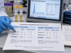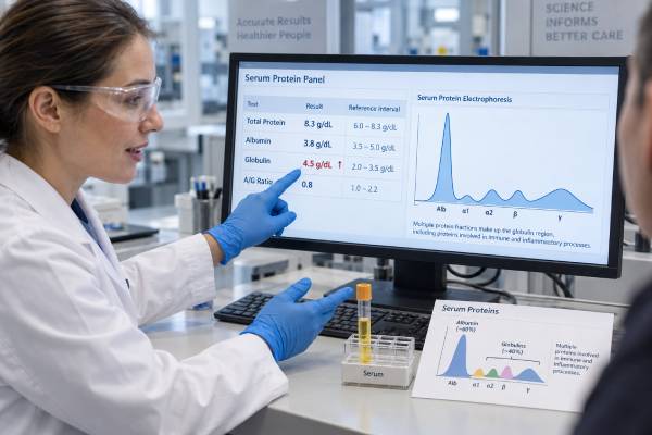
A globulin blood test result usually appears on a comprehensive metabolic panel or liver panel, but “globulin” is not one single protein. It is a calculated or measured group that includes antibodies, transport proteins, complement proteins, and several acute-phase proteins. A high value may result from immune activation, chronic infection, autoimmune disease, liver disease, dehydration, or an abnormal clone of antibody-producing cells. The most important distinction is whether many immunoglobulin types are increased together, called a polyclonal pattern, or one cell clone is producing a monoclonal protein. A routine globulin value cannot make that distinction. Clinicians interpret it with total protein, albumin, the albumin-to-globulin ratio, symptoms, liver and kidney tests, and—when indicated—serum protein electrophoresis, immunofixation, and free light chains. This article explains what a high globulin result means, how inflammation changes protein balance, and when further testing is appropriate.
- What globulin means: A broad group of blood proteins rather than a single molecule or diagnosis.
- How it is often calculated: Total protein minus albumin, so errors or changes in either measurement affect the result.
- Common high pattern: Polyclonal immune activation from liver disease, infection, or autoimmune inflammation.
- Important alternative: A monoclonal protein may require electrophoresis and related testing, especially with anemia, kidney dysfunction, high calcium, neuropathy, or bone pain.
- Best next step: Confirm the pattern and investigate the cause instead of trying to lower globulin directly.
Table of Contents
- What globulins are and how the test is calculated
- Reference ranges and protein patterns
- Why globulin levels become high
- Polyclonal versus monoclonal increases
- How clinicians investigate high globulin
- Inflammation, albumin, and protein balance
- Treatment, follow-up, and warning signs
What Globulins Are and How the Test Is Calculated
Blood proteins are often divided into albumin and globulins. Albumin is the most abundant single protein in serum. “Globulin” is a collective label for many proteins with different origins and jobs. They include immunoglobulins made by plasma cells, complement proteins, binding and transport proteins, enzymes, and acute-phase reactants made largely by the liver.
On a routine chemistry panel, globulin is commonly not measured directly. The laboratory measures total protein and albumin, then calculates:
Calculated globulin = total protein − albumin
This means a high calculated globulin can arise because total protein is high, albumin is low relative to total protein, or both. Small analytical differences in the two measured values also carry into the calculation. The albumin-to-globulin ratio, or A/G ratio, uses the same components and therefore does not provide an independent diagnosis.
Serum protein electrophoresis separates proteins by their electrical movement into broad fractions: albumin, alpha-1, alpha-2, beta, and gamma. Each fraction offers clues. Acute inflammation often raises alpha fractions; chronic immune activation commonly broadens the gamma region; nephrotic syndrome can produce low albumin and a characteristic alpha-2 increase; and a narrow spike may suggest a monoclonal protein. Electrophoresis is a pattern test, not just another total globulin measurement.
Globulins perform essential functions:
- Immunoglobulins: Antibodies that recognize microbes and other targets
- Complement: Proteins that support immune defense and inflammation
- Transport proteins: Carriers for hormones, vitamins, metals, and lipids
- Acute-phase proteins: Molecules that rise or fall during inflammation
- Coagulation-related proteins: Components involved in clotting and tissue repair
Because the category is so broad, a routine result cannot reveal which proteins are responsible. The clinical question determines whether more specific tests are useful.
A standard blood sample is used. Fasting is usually unnecessary for total protein and albumin, although another component of the ordered panel may require it. Dehydration, intravenous fluids, prolonged tourniquet use, pregnancy, and recent acute illness can affect concentration and interpretation.
Reference Ranges and Protein Patterns
Reference intervals vary by laboratory and assay. Many adult reports show calculated globulin around 2.0–3.5 g/dL, but the printed interval should be used. Total protein is often roughly 6.0–8.3 g/dL, and albumin commonly makes up more than half. Age, pregnancy, hydration, liver function, immune status, and laboratory method all influence expected values.
A mildly high globulin value is common and often nonspecific. The key is the pattern across related results and over time.
| Pattern | Possible meaning | Helpful next context |
|---|---|---|
| High globulin, normal albumin | Increased immunoglobulins or acute-phase proteins; dehydration is also possible | Total protein, symptoms, liver tests, CRP, repeat result |
| High globulin, low albumin | Chronic liver disease, inflammation, protein loss, or combined processes | A/G ratio, urine protein, liver and kidney assessment |
| Broad gamma increase on electrophoresis | Polyclonal immune activation | Infection, autoimmune disease, chronic liver disease |
| Narrow band or spike | Possible monoclonal protein | Immunofixation, serum free light chains, blood count, calcium, kidney function |
| Low globulin | Reduced immunoglobulins, protein loss, dilution, or impaired production | Infection history, quantitative immunoglobulins, kidney or gastrointestinal loss |
The A/G ratio adds perspective but should not be read alone. A low ratio can result from low albumin, high globulin, or both. A high ratio may reflect low globulins rather than unusually high albumin. The actual component values are more informative than the ratio by itself.
The size of an abnormality does not reliably separate benign from serious causes. A marked polyclonal increase can occur in autoimmune hepatitis or chronic infection, while a small monoclonal protein may be an age-associated precursor condition that requires monitoring. Conversely, some important plasma-cell disorders produce mostly free light chains and may not cause a dramatic total globulin rise. Symptoms and targeted testing matter more than a simplistic cutoff.
Trends help distinguish temporary immune responses from persistent protein abnormalities. A result after pneumonia may normalize in weeks or months. Persistent elevation, especially without an obvious acute cause, deserves review of previous panels and the clinical setting. Use the same laboratory when possible because methods and reference intervals differ.
Why Globulin Levels Become High
High globulin, or hyperglobulinemia, is a laboratory pattern with several major categories. More than one category can be present.
Chronic Liver Disease
The liver makes albumin and many non-antibody globulins, and it helps clear immune complexes. Chronic hepatitis, cirrhosis, autoimmune hepatitis, and other liver disorders can produce low albumin with increased immunoglobulins. The exact electrophoresis pattern varies. Liver enzymes may be only mildly abnormal in advanced chronic disease, so albumin, bilirubin, INR, platelet count, imaging, and clinical history contribute to assessment.
Infection and Inflammation
Long-lasting infections stimulate multiple B-cell populations to make antibodies. Tuberculosis, HIV, chronic viral hepatitis, endocarditis, and other persistent infections can produce a broad immunoglobulin increase. Acute infection may raise alpha and beta acute-phase proteins before substantial antibody changes develop. Inflammatory cytokines also shift liver protein production, affecting both globulin fractions and albumin.
Autoimmune conditions such as Sjögren syndrome, systemic lupus erythematosus, rheumatoid arthritis, autoimmune hepatitis, and some vasculitides can cause polyclonal hypergammaglobulinemia. The degree does not perfectly track disease activity, and a positive autoimmune test is not inferred from globulin alone. A CRP result may show current inflammatory activity but does not explain a high globulin pattern by itself.
Monoclonal Gammopathies
A single clone of plasma cells or B cells can make one immunoglobulin or light-chain type. Conditions include monoclonal gammopathy of undetermined significance (MGUS), multiple myeloma, smoldering myeloma, Waldenström macroglobulinemia, light-chain amyloidosis, and some lymphomas. Many people with MGUS never develop a malignant disorder, but the finding needs classification and risk-based follow-up.
A routine globulin result is neither sensitive nor specific enough to screen out all monoclonal disease. Some monoclonal proteins are small; some migrate outside the gamma region; and free light-chain disease may leave total protein near normal. Suspicion comes from the combination of laboratory findings and clinical features.
Dehydration and Hemoconcentration
Water loss concentrates albumin, globulins, blood cells, and other solutes. In pure dehydration, albumin may be high as well as total protein. The pattern should improve with restoration of fluid balance. A persistently high globulin after hydration suggests another cause.
Other Causes
Certain blood disorders, chronic inflammatory lung or bowel disease, granulomatous disorders, malignancies, and rare immune-regulation syndromes can increase globulins. Vaccination and recent infection may cause temporary antibody changes, although these usually do not create a concerning persistent pattern. Medications can influence immune function, liver health, or hydration indirectly.
Dietary protein is rarely the direct explanation. The body regulates digestion, amino-acid use, and protein synthesis; eating a high-protein meal does not usually create sustained hyperglobulinemia. The focus should remain on immune activity, organ function, and fluid status.
Polyclonal Versus Monoclonal Increases
The distinction between polyclonal and monoclonal protein production is central. It describes the shape and biological source of the increase rather than simply how high the total is.
A polyclonal pattern means many plasma-cell families are active and make a diverse mixture of antibodies. On serum protein electrophoresis, this often appears as a broad-based elevation. It is usually reactive and associated with liver disease, infection, autoimmune inflammation, or another immune stimulus. The treatment is directed at that condition; the broad antibody response itself is not usually removed or suppressed simply to normalize the test.
A monoclonal pattern means one cell clone produces a uniform immunoglobulin or component, often called an M protein. Electrophoresis may show a discrete band, but immunofixation identifies the immunoglobulin heavy and light-chain type more precisely. Serum free light-chain testing measures unbound kappa and lambda chains and their ratio. Kidney impairment can raise both light chains and alter interpretation, so the ratio and renal context matter.
Typical testing may include:
- Serum protein electrophoresis: Detects and estimates many M proteins and shows the overall pattern
- Serum immunofixation: Confirms and types a suspicious monoclonal band
- Serum free light chains: Improves detection of light-chain production
- Quantitative immunoglobulins: Measures IgG, IgA, and IgM classes
- Urine studies: Used in selected cases to assess monoclonal protein and kidney involvement
No single one of these tests is sufficient in every case. The appropriate combination depends on the reason for suspicion, local laboratory approach, and whether a known disorder is being monitored.
Features that increase concern for a plasma-cell or lymphoid disorder include unexplained anemia, reduced kidney function, high calcium, persistent bone pain, fractures, recurrent infection, neuropathy, enlarged lymph nodes, weight loss, very high total protein, rouleaux on a blood film, or an unexpectedly low anion gap. None is specific, and many people with a monoclonal protein have no symptoms.
Polyclonal and monoclonal findings can coexist. A person with chronic inflammation may also have MGUS. That is another reason not to label a high globulin result solely from the total concentration.
How Clinicians Investigate High Globulin
Evaluation starts with confirmation. The clinician reviews total protein, albumin, calculated globulin, A/G ratio, prior results, hydration, and why testing was performed. A small isolated change during an acute illness may simply be repeated after recovery. Persistent or larger abnormalities call for a directed history and examination.
Questions may cover fever, night sweats, weight loss, fatigue, infections, dry eyes or mouth, joint swelling, rash, jaundice, abdominal swelling, bone pain, numbness, recurrent fractures, urine changes, and medication or alcohol use. Family history and ancestry can occasionally guide testing, but most acquired globulin abnormalities are not inherited.
Initial laboratory assessment may include:
- Complete blood count and blood film
- Creatinine, estimated glomerular filtration rate, calcium, and electrolytes
- Liver enzymes, bilirubin, albumin, and INR when indicated
- Urinalysis and urine protein measurement
- CRP or ESR for inflammatory context
- Tests for specific infections or autoimmune conditions when symptoms support them
Serum protein electrophoresis is considered when globulin or total protein is persistently high, the A/G ratio is unexpectedly low, or there are clinical clues to a monoclonal gammopathy. Electrophoresis may also be appropriate despite a normal total protein when symptoms or other findings raise concern. Immunofixation and free light chains improve characterization and sensitivity.
A positive monoclonal study does not automatically mean cancer. The next steps assess the amount and type of protein, blood count, calcium, kidney function, urine findings, and evidence of organ damage. Imaging and bone-marrow examination are reserved for situations in which the pattern and clinical risk justify them. Hematology referral is common for a newly detected monoclonal protein or suspicious light-chain abnormality.
For a polyclonal pattern, further testing should be targeted. Broad screening for every infection and autoimmune disease can produce false positives. Liver disease is common enough that history, liver tests, viral hepatitis risk, and imaging often receive early attention. Symptoms may point toward rheumatology, infectious disease, gastroenterology, or another specialty.
Inflammation, Albumin, and Protein Balance
Protein balance in blood reflects production, distribution, breakdown, and loss—not simply dietary intake. During inflammation, the liver increases several positive acute-phase proteins and reduces production of some negative acute-phase proteins. Albumin may fall because of changed synthesis, leakage from vessels, dilution, and increased breakdown, while globulin fractions rise from immune and hepatic responses. The result can be a low A/G ratio even when total protein remains within range.
The albumin blood test therefore provides complementary information. Low albumin can occur with inflammation, cirrhosis, kidney protein loss, gastrointestinal loss, severe illness, or inadequate intake, but it is not a stand-alone nutrition diagnosis. High globulin does not “push albumin down” through a simple competition; both values reflect underlying physiology.
Globulins can also influence other tests. High immunoglobulin levels promote red-cell rouleaux and may raise the ESR. Marked protein elevations can alter plasma viscosity and, with certain monoclonal proteins, contribute to headaches, visual changes, bleeding, or neurologic symptoms. These complications are uncommon with mild polyclonal elevation and occur in specific high-protein states.
A calculated globulin gap—total protein minus albumin—is the same basic quantity as calculated globulin. Some clinicians notice a large gap as a clue to chronic infection or gammopathy, but no single threshold is sufficiently accurate to diagnose or exclude these conditions. The gap should prompt context-sensitive reasoning, not automatic testing in every person.
Protein changes may lag behind clinical improvement. Immunoglobulins have different half-lives, and liver recovery can take time. A falling globulin or improving A/G ratio may support response, but condition-specific outcomes are more important than normalizing the chemistry panel.
Treatment, Follow-Up, and Warning Signs
Treatment is based on the cause. A bacterial infection may need antimicrobial therapy; autoimmune disease needs diagnosis-specific treatment; viral hepatitis and chronic liver disease require targeted care; dehydration improves with appropriate fluid replacement. Polyclonal hypergammaglobulinemia often declines when the underlying immune stimulus is controlled, although it may remain elevated in chronic disease.
MGUS generally does not require immediate cancer treatment. It is monitored according to the protein type, amount, free light-chain ratio, and other risk factors. Multiple myeloma, lymphoma, amyloidosis, and related disorders require specialist therapy. The distinction is important because treating every monoclonal finding would expose many people with stable precursor conditions to unnecessary harm.
There is no safe home method to lower globulin directly. Restricting dietary protein, taking unproven “immune cleanses,” or using supplements to thin the blood does not address the cause and may create nutritional or medication problems. General health measures—avoiding tobacco, limiting alcohol according to medical advice, maintaining vaccinations, and managing chronic disease—can support overall health but are not substitutes for evaluation.
Follow-up timing depends on the pattern. A mildly high result during infection may be rechecked after recovery. Persistent elevation with stable normal blood count, kidney function, calcium, and no concerning symptoms may be evaluated nonurgently. A new monoclonal protein, major rise in total protein, or associated organ abnormality deserves timely review.
Seek prompt medical attention for high globulin accompanied by severe weakness, confusion, reduced urine output, dehydration, new neurologic symptoms, vision changes, unusual bleeding, severe bone pain, jaundice, rapidly increasing swelling, or signs of serious infection. Contact a clinician promptly for unexplained weight loss, night sweats, recurrent infections, persistent bone pain, anemia, kidney dysfunction, or high calcium.
The most useful question is not “How high is globulin?” but “Which protein pattern is present, and what process produced it?” A routine chemistry value can open that investigation, while electrophoresis and targeted clinical assessment provide the detail needed for a reliable answer.
References
- Globulin Test 2024
- Protein Electrophoresis by Immunofixation Blood Test 2024
- Plasma Cell Dyscrasias 2025
- Monoclonal Protein Isotype, Quantitative, Serum 2025
- Etiological Study of Polyclonal Hypergammaglobulinemia in a French Cohort of Hospitalized Patients and Proposal of a Diagnostic Aid Algorithm 2024
- Polyclonal Gammopathy 2026
Disclaimer
This article is for general education and cannot diagnose the cause of an abnormal globulin, total protein, or A/G ratio. Laboratory methods and reference ranges differ, and personal interpretation may require electrophoresis, immunofixation, free light chains, and clinical assessment. Seek prompt care for severe symptoms or signs of kidney, neurologic, infectious, or bleeding complications.