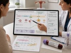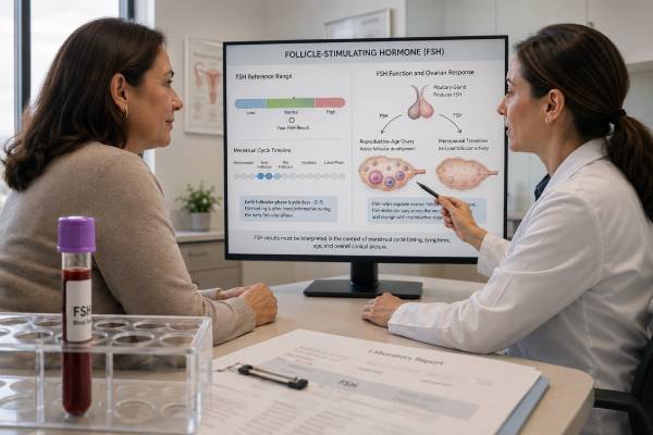
A follicle-stimulating hormone (FSH) test measures a pituitary signal that supports ovarian follicle development and sperm production. In women, FSH changes across the menstrual cycle and rises when ovarian feedback falls, including during menopause and primary ovarian insufficiency. Early-follicular FSH is sometimes used with estradiol to estimate ovarian response, but it varies between cycles and does not measure egg quality or predict natural pregnancy by itself. In men, high FSH can indicate impaired sperm-producing tissue, while low or inappropriately normal FSH can point toward pituitary or hypothalamic suppression. Children require age- and puberty-stage ranges. Hormone therapy, pregnancy, illness, cycle timing, and the laboratory method all affect interpretation. Typical menopause after age 45 is usually diagnosed clinically rather than by repeatedly checking FSH, because levels fluctuate during perimenopause. The result is most useful when paired with estradiol or testosterone, LH, symptoms, and the reason for testing.
- FSH is a pituitary hormone that stimulates ovarian follicles and supports sperm production.
- High FSH with low estradiol or testosterone suggests reduced ovarian or testicular function.
- Low FSH with low sex hormones suggests hypothalamic or pituitary suppression.
- Early-cycle FSH should be interpreted with estradiol; a high E2 can make FSH look falsely reassuring.
- FSH fluctuates in perimenopause and is usually unnecessary for diagnosing typical menopause after age 45.
- A normal FSH does not guarantee fertility, egg quality, open tubes, or normal semen.
Table of Contents
- What the FSH Test Measures
- Why the Test Is Ordered
- Preparation, Timing, and the Testing Process
- Normal Range and How Results Are Interpreted
- What High FSH Results Can Mean
- What Low FSH Results Can Mean
- How Results Fit With Other Hormones and Tests
- Next Steps After the Result
What the FSH Test Measures
GnRH from the hypothalamus stimulates the pituitary to release FSH and LH. In ovaries, FSH promotes follicle growth and estradiol production. In testes, it acts on Sertoli cells and supports the environment needed for sperm production.
Estradiol, inhibin B, and other gonadal signals feed back to the pituitary. When ovarian follicles or testicular tissue are impaired, feedback decreases and FSH rises. When hypothalamic or pituitary signaling is suppressed, FSH may be low or normal despite low sex hormones.
A hormone result is a measurement made under defined laboratory conditions, not a diagnosis by itself. The same concentration can carry different meaning at different ages, cycle stages, pregnancy stages, or times of day. Laboratories also use different analyzers, calibration systems, antibodies, and calculation methods. For that reason, the reference interval printed beside the result should take priority over a range copied from another report or website.
FSH is released in pulses and varies with cycle stage. A single result is a snapshot. It is more dependable when the sample timing and related hormone levels are known and when a repeat confirms an unexpected pattern.
FSH is often combined with other markers in a fertility hormone panel.
Why the Test Is Ordered
FSH is ordered to evaluate menstrual and fertility concerns, menopause or ovarian insufficiency, male gonadal function, and puberty.
- Irregular or absent periods and possible ovarian insufficiency.
- Early-cycle ovarian reserve assessment during fertility evaluation.
- Menopausal symptoms at an unusually young age or an unclear reproductive history.
- Low testosterone, infertility, or suspected impaired sperm production in men.
- Early or delayed puberty.
- Monitoring selected fertility or hormone treatments.
FSH should not be ordered as a stand-alone “fertility age” test. AMH and antral follicle count predict ovarian stimulation response more consistently, while age remains central to egg quality. In men, semen analysis is the primary test of sperm production; FSH helps explain an abnormal result.
Interpretation starts with the reason the test was ordered. A clinician looking for an adrenal enzyme disorder uses different cutoffs and follow-up tests than a clinician evaluating irregular periods, infertility, menopausal symptoms, or androgen deficiency. The result should be read with symptoms, examination findings, age, reproductive stage, and related laboratory values rather than compared with one universal “optimal” number.
The AMH test provides a different ovarian reserve signal and should not be treated as interchangeable with FSH.
Preparation, Timing, and the Testing Process
For ovarian reserve, FSH is commonly collected on cycle days 2–4 with estradiol. Other questions may use random testing.
- Record cycle day, menstrual pattern, pregnancy possibility, and menopause status.
- List hormonal contraception, fertility medicines, estrogen, testosterone, and GnRH medicines.
- Use morning sampling in men when testosterone is measured at the same time.
- Report major illness, undernutrition, heavy exercise, or recent changes in hormone treatment.
- Follow clinic instructions for treatment monitoring rather than using natural-cycle ranges.
Hormonal contraception suppresses FSH and makes ovarian reserve interpretation difficult. During perimenopause, one high result may be followed by a lower value. In children, sensitive assays and puberty-stage intervals are required.
Before collection, provide a complete list of prescription medicines, over-the-counter products, supplements, contraceptives, fertility drugs, and hormone therapy. Do not stop prescribed treatment without instructions. Some products change hormone production, while others alter binding proteins or interfere with an immunoassay. High-dose biotin is a well-known source of interference in several hormone tests, although the direction and size of the error depend on the platform.
Acute illness can temporarily suppress or stimulate reproductive signaling. A test drawn during fever, major surgery, severe calorie restriction, or intense training may describe that temporary state rather than the person’s usual hormone function. When the clinical situation is stable and there is no urgent reason to test immediately, postponing or repeating the measurement can prevent an incorrect label.
Normal Range and How Results Are Interpreted
FSH is usually reported in IU/L or mIU/mL. Approximate adult female values vary by phase, and laboratory cutoffs differ.
| Setting | Approximate pattern | Interpretive note |
|---|---|---|
| Early follicular phase | Often about 3–10 IU/L | Interpret with estradiol and lab range |
| Midcycle | Can rise with the LH surge | Timing-dependent physiologic increase |
| Luteal phase | Often lower than midcycle | Not a reserve-testing window |
| Postmenopause | Frequently above 25–30 IU/L | Fluctuation occurs in transition |
| Adult men | Relatively stable within lab interval | High values may reflect seminiferous damage |
An early-cycle FSH above roughly 10–15 IU/L may suggest reduced ovarian response in some laboratories, but no universal cutoff predicts pregnancy. Primary ovarian insufficiency uses persistent menstrual disturbance and elevated FSH according to guideline criteria, not one borderline result.
A value close to a cutoff deserves more caution than a value that is clearly and repeatedly abnormal. Borderline results may move across the reference limit because of normal biologic variation, recent illness, sleep disruption, exercise, stress, or sample timing. Clinicians often repeat a test under better-controlled conditions before ordering imaging or beginning treatment. The repeat is most useful when the timing, medication list, and assay method are documented.
Trends can be useful when the same question is followed over time, but only when the measurements are reasonably comparable. A change between laboratories may reflect a method difference. A change after starting or stopping hormones may be expected. Record the laboratory, units, collection time, cycle day or pregnancy week, and relevant treatment so later results can be compared in context.
Estradiol should be checked with early-cycle FSH; the estradiol test explains why an elevated E2 can suppress FSH.
What High FSH Results Can Mean
High FSH means the pituitary is increasing stimulation because gonadal feedback is reduced or because a physiologic stage such as menopause is present.
- Natural menopause or the menopausal transition.
- Primary ovarian insufficiency or ovarian damage from surgery, chemotherapy, or radiation.
- Turner syndrome or another ovarian-development condition.
- Primary testicular failure, Klinefelter syndrome, or damage to sperm-producing tissue.
- Normal midcycle rise when sampled near ovulation.
- Incorrect comparison with an age-, sex-, or stage-inappropriate range.
In fertility care, high FSH often predicts a lower egg yield from stimulation, but some cycles and pregnancies still occur. It does not directly measure the genetic quality of eggs. Repeating an unexpectedly high early-cycle result and reviewing estradiol, AMH, and antral follicle count can improve counseling.
In men, high FSH with low sperm concentration suggests primary seminiferous-tubule impairment. Testosterone may remain normal because Leydig-cell function is separate. Genetic evaluation may be indicated with severe oligospermia or azoospermia.
Persistent high FSH with menstrual loss before age 40 needs evaluation for ovarian insufficiency and its causes. In men with infertility, combine FSH with semen analysis, LH, testosterone, examination, and reproductive-urology assessment.
A primary ovarian insufficiency panel is more complete than FSH alone when early ovarian failure is suspected.
What Low FSH Results Can Mean
Low FSH may be normal in some cycle phases or suppressed by hormones. It becomes clinically important when estradiol or testosterone is also low.
- Hypothalamic suppression from low energy availability, intensive exercise, stress, or chronic illness.
- Pituitary disease, surgery, radiation, or other hormone deficiencies.
- High prolactin.
- Pregnancy or hormonal contraception.
- Testosterone, estrogen, or GnRH-based treatment.
- Normal low value during a phase when strong FSH stimulation is not expected.
Low FSH with low LH and estradiol can occur in functional hypothalamic amenorrhea. The history of nutrition, weight change, exercise, stress, and bone health is central. Low FSH with low testosterone in men may reflect obesity, sleep disruption, opioids, severe illness, or pituitary disease.
A normal-range FSH can still be “inappropriately normal” when the gonadal hormone is clearly low. The pituitary should increase its signal in primary gonadal failure, so failure to rise points toward central suppression.
Follow-up may include LH, estradiol or testosterone, prolactin, TSH, other pituitary hormones, nutrition assessment, and MRI when structural disease is possible. Treatment targets the cause and protects bone and reproductive health.
A prolactin test is useful when low gonadotropins occur with cycle changes, galactorrhea, or pituitary symptoms.
How Results Fit With Other Hormones and Tests
FSH interpretation depends on the sex hormone and LH pattern.
| FSH | Related hormones | Possible interpretation |
|---|---|---|
| High | Estradiol low | Menopause or primary ovarian insufficiency |
| High | Testosterone low or normal; semen abnormal | Primary testicular or seminiferous dysfunction |
| Low/normal | Estradiol low | Hypothalamic or pituitary suppression |
| Low/normal | Testosterone low | Central hypogonadism or functional suppression |
| Normal early cycle | Estradiol unexpectedly high | FSH may be masked; reserve interpretation is limited |
Inhibin B can add information about Sertoli-cell or follicular activity in selected cases, but it is not a routine replacement for semen analysis or established ovarian reserve measures. Clinical context determines whether the added test is worthwhile.
Next Steps After the Result
The next step is to place FSH in the correct cycle, age, sex, and treatment context and then identify whether the pattern is gonadal or central.
- Verify timing, units, reference interval, pregnancy status, and hormone exposure.
- Pair FSH with estradiol in women or testosterone in men.
- Repeat a borderline or unexpected result when confirmation changes management.
- Use AMH and ultrasound for ovarian response planning and semen analysis for male fertility.
- Evaluate prolactin, thyroid, nutrition, chronic illness, genetics, or pituitary disease as indicated.
- Discuss treatment, fertility preservation, and bone protection with the appropriate specialist.
In typical menopause, treatment is based on symptoms and health history rather than normalizing FSH. In ovarian insufficiency, hormone replacement often protects bone and other tissues until the usual menopause age unless contraindicated. In central suppression, recovery may require nutrition, disease treatment, medication changes, or pituitary care.
Bring the actual report to the follow-up visit rather than only recalling that the level was “high” or “low.” The report shows units, the laboratory interval, specimen type, and sometimes the assay method. Ask whether the result needs confirmation, whether a related hormone should be measured at the same time, and what finding would change management.
A useful plan defines the next step and its timing. That may be no action, a repeat test, a dynamic stimulation or suppression test, ultrasound, semen analysis, genetic testing, or referral to endocrinology, reproductive endocrinology, urology, or maternal-fetal medicine. It should also identify symptoms that warrant earlier contact instead of waiting for the routine appointment.
Rapidly progressive symptoms deserve faster evaluation than a mild, stable laboratory abnormality. Examples include sudden virilization, severe headache with visual change, signs of adrenal crisis, heavy bleeding with dizziness, or concerning pregnancy symptoms. The laboratory number may guide the workup, but urgent decisions are driven by the whole clinical picture.
Treatment should address the confirmed cause and the person’s goals. Lowering or raising a laboratory value without establishing why it is abnormal can hide an important condition or create new problems. Hormone therapy can affect fertility, blood counts, liver function, clot risk, bone health, and pregnancy, so monitoring plans should be individualized.
Seek prompt assessment for severe headache with visual change, neurologic symptoms, sudden testicular pain, or heavy bleeding with dizziness. FSH cannot rule out urgent pituitary, testicular, pregnancy, or bleeding conditions.
When a repeat measurement is planned, try to reproduce the conditions that matter for this test. Use the same laboratory when practical, note the collection time, and record any change in medicines or reproductive stage. Consistency does not remove all biologic variation, but it makes a true trend easier to distinguish from noise.
Reference intervals describe a population selected by the laboratory; they do not define a treatment target for every person. A result inside the interval can still require attention when symptoms are strong or when another hormone shows a clear mismatch. Conversely, a small deviation may not represent disease when the timing or clinical setting explains it.
A laboratory result can answer only the question built into the test. It may show hormone concentration, but it cannot by itself prove ovulation quality, egg quality, sperm production, placental health, or the cause of a symptom. Those conclusions usually require a combination of history, examination, imaging, and other targeted tests.
When fertility is the concern, age, duration of trying to conceive, menstrual pattern, pregnancy history, semen findings, and tubal or uterine factors often affect the plan as much as a single hormone value. Testing both partners in parallel can prevent months of delay and reduce the chance that an incidental hormone result distracts from a more direct cause.
When symptoms are being monitored rather than fertility, define the outcome that treatment is meant to improve. Examples include restoration of periods, relief of hot flashes, reduction of unwanted hair growth, recovery of sexual function, preservation of bone health, or safe progression of puberty. Laboratory monitoring is most useful when tied to one of these clinical outcomes.
People using nonprescription “hormone balance” products should mention them before testing. DHEA, progesterone creams, testosterone boosters, compounded preparations, and biotin can alter physiology or make results harder to interpret. Product labels may not reliably predict the dose absorbed, so the safest approach is to review the exact container or ingredient list with the clinician.
Results can also differ because laboratories report conventional and SI units. A value expressed in ng/dL cannot be compared directly with one expressed in nmol/L without a hormone-specific conversion. Always compare the number, unit, and interval together. This simple check prevents many apparent contradictions between reports.
An abnormal result can have emotional weight, especially during fertility treatment, pregnancy, or puberty evaluation. Ask the clinician to separate what is known from what remains uncertain. A clear explanation of the likely causes, the confirmation plan, and the time frame for action is more useful than trying to interpret every decimal place independently.
Clinicians also consider whether the test result agrees with the physical findings. A marked biochemical abnormality without expected symptoms may prompt confirmation with a more specific laboratory method. Strong symptoms with a normal result may lead to testing at a different time, measuring a related hormone, or looking for a nonhormonal cause.
The pace of change can be as important as the level itself. Slow changes over years commonly fit physiologic aging or a chronic endocrine condition. Changes over weeks or a few months raise more concern for a new medicine effect, pregnancy-related change, acute illness, or a hormone-producing lesion, especially when symptoms progress quickly.
No single follow-up pathway fits every abnormal value. Mild and explainable results may only need observation. Persistent or substantial abnormalities may require specialist review and tests chosen to locate the source of hormone production or the level of signaling failure. The sequence should be deliberate so that each test answers a specific unresolved question.
Keep a copy of the laboratory report and the order indication. The clinician needs more than the highlighted flag: the numeric value, unit, reference interval, specimen type, collection date and time, and any comment about the assay can change interpretation. A result copied into a patient portal message without those details may be impossible to compare with a later test. This documentation is especially important when care moves between a primary clinician, endocrinologist, fertility clinic, and hospital laboratory.
Before repeating testing, decide what the repeat is meant to resolve. It may confirm persistence, correct a timing error, remove a medication effect, or use a more specific assay. Repeating the same poorly timed test without changing the conditions often reproduces uncertainty rather than solving it. The clinician should also state what result would lead to observation, another laboratory test, imaging, or treatment.
Small deviations from a reference interval are common because the interval usually contains about 95% of a selected comparison population. Some healthy people fall outside it, while some people with disease fall inside it. The clinical importance depends on how far the value is from the limit, whether it persists, whether related hormones agree, and whether symptoms fit the physiology. A reference flag is therefore a prompt for interpretation, not proof of disease.
Hormone results are also affected by changes in binding proteins and metabolism. Liver disease, thyroid disease, kidney disease, pregnancy, body composition, and oral estrogen exposure can change measured concentrations without producing the same change in tissue effect. When a binding issue is suspected, the clinician may use a free or calculated hormone, a related binding protein, or a more specific laboratory method rather than interpreting the total concentration alone.
Fertility decisions should not be reduced to a single endocrine value. A hormone may help choose medication dose, confirm ovulation, or identify a treatable disorder, but it does not measure every step required for pregnancy. Reproductive age, sperm exposure or semen quality, tubal patency, uterine anatomy, timing, prior pregnancies, and the duration of trying all influence the next step. A balanced evaluation prevents overreaction to an incidental result.
Testing can be different during pregnancy, adolescence, menopause, or hormone treatment because physiology is intentionally changing. A range derived from untreated adults may be inappropriate in these settings. Ask whether the laboratory interval matches the patient’s age and reproductive state and whether the clinical team uses a separate treatment or pregnancy target. This is particularly important when a portal automatically marks a result high or low against a generic range.
A laboratory method with greater analytical specificity is useful when the expected concentration is low, the result is surprising, or a major decision depends on the number. Mass-spectrometry methods can separate closely related steroids better than many immunoassays, while dynamic tests can reveal reserve or responsiveness that a basal sample cannot show. The best method depends on the hormone and question; a technically sophisticated test is not automatically necessary for every routine result.
References
- Comprehensive Review of Estradiol, Progesterone, Luteinizing Hormone, and Follicle-Stimulating Hormone Reference Intervals in Transgender, Nonbinary, and Cisgender Individuals 2025 (Review)
- Fertility evaluation of infertile women: a committee opinion 2021 (Committee Opinion)
- The Clinical Utility of Measures of Ovarian Reserve 2025 (Review)
- Current evaluation of amenorrhea: a committee opinion 2024 (Committee Opinion)
- Guideline on premature ovarian insufficiency 2024 (Guideline)
Disclaimer
This article is general information and cannot diagnose infertility, menopause, ovarian insufficiency, or testicular disease. FSH requires cycle-, age-, sex-, and treatment-specific interpretation. Review abnormal results with a qualified clinician.