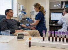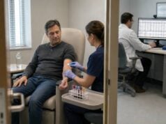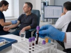
A hypopituitarism blood test panel evaluates whether the pituitary is producing enough signals for the adrenal glands, thyroid, gonads, growth hormone system, and water balance. The panel does not simply look for pituitary hormones below a reference range. In central hormone deficiency, a pituitary result may be low, normal, or slightly elevated but still inappropriate because the target-gland hormone is low. Morning cortisol and ACTH assess the adrenal axis; free T4 and TSH assess the thyroid axis; LH, FSH, estradiol or testosterone assess reproductive function; IGF-1 screens the GH axis; prolactin provides information about pituitary and stalk function; and sodium, serum osmolality, urine osmolality, or copeptin may be needed for vasopressin disorders. Some axes require dynamic testing. Results must be interpreted with symptoms, medications, illness, pregnancy, menstrual status, age, prior pituitary treatment, and imaging.
- Hypopituitarism may affect one hormone or several.
- Target-gland hormones are as important as pituitary hormone values.
- ACTH deficiency is the most urgent deficit to recognize.
- TSH can be “normal” despite central hypothyroidism.
- Random GH is not useful for diagnosing GH deficiency.
- Hormone replacement order matters, with cortisol addressed before thyroid hormone.
Table of Contents
- What Hypopituitarism Means
- Tests in the Panel
- How Each Axis Is Interpreted
- Dynamic Testing and Imaging
- Causes and Result Pitfalls
- Treatment, Monitoring, and Sequence
- Urgent Symptoms and Questions
What Hypopituitarism Means
Hypopituitarism is partial or complete deficiency of one or more anterior or posterior pituitary hormones. Panhypopituitarism usually refers to broad anterior pituitary failure, while hypothalamic disease can produce a similar pattern by disrupting releasing hormones or transport through the pituitary stalk.
The anterior pituitary produces ACTH, TSH, LH, FSH, GH, and prolactin. The posterior pituitary releases vasopressin and oxytocin produced in the hypothalamus. Deficiency patterns depend on the location, size, speed, and cause of damage.
Symptoms are often nonspecific. Fatigue, weakness, low mood, reduced exercise capacity, weight change, low libido, menstrual disturbance, infertility, cold intolerance, low blood pressure, and low sodium can have many causes. The combination, timing, and relation to pituitary risk are more informative than one symptom.
A large or rapidly expanding lesion may cause headache, visual-field loss, double vision, or cranial-nerve symptoms. A slowly evolving lesion may first reduce GH and gonadotropins, while ACTH and TSH are preserved until later. This sequence is not universal.
Congenital hypopituitarism may present with neonatal hypoglycemia, prolonged jaundice, micropenis, undescended testes, poor growth, midline facial features, or visual abnormalities. Acquired disease may follow a pituitary tumor, surgery, radiation, head trauma, stroke, infection, inflammation, infiltrative disease, postpartum hemorrhage, immune-checkpoint therapy, or other medications.
The diagnosis is physiologic. A pituitary hormone should be judged against the hormone made by its target gland. Low free T4 should trigger a rise in TSH; low cortisol should trigger ACTH; low testosterone or estradiol should trigger LH and FSH. Failure to mount that response suggests central dysfunction even when the pituitary value is technically inside the population interval.
Tests in the Panel
A baseline panel is usually collected in the morning because cortisol and testosterone have important diurnal rhythms. The exact set depends on age, sex, symptoms, and treatment.
| Pituitary axis | Baseline tests | Additional tests when needed |
|---|---|---|
| Adrenal | 8–9 a.m. cortisol, ACTH, sodium, glucose | ACTH stimulation, insulin tolerance, metyrapone |
| Thyroid | Free T4, TSH | Repeat by another method if interference suspected |
| Gonadal | LH, FSH, estradiol or morning testosterone | Prolactin, pregnancy test, semen analysis, pelvic or testicular evaluation |
| Growth hormone | IGF-1 | GH stimulation test |
| Prolactin | Prolactin | Dilution for hook effect, macroprolactin testing |
| Water balance | Sodium, plasma osmolality, urine osmolality | Copeptin, water deprivation, desmopressin response |
A complete metabolic panel helps identify sodium, glucose, kidney, and liver abnormalities. Blood count, ferritin, and other tests may evaluate alternative causes of fatigue or menstrual change.
Morning cortisol is a screening value, not a direct measure of ACTH reserve. A clearly low concentration strongly supports adrenal insufficiency, while a clearly high value usually excludes it. Intermediate values require dynamic testing. Cutoffs depend on the cortisol assay.
Free T4 is central to thyroid-axis interpretation. In pituitary disease, TSH may be low, normal, or mildly elevated because secreted TSH can have reduced biologic activity. A central hypothyroidism panel is interpreted from the free T4–TSH relationship.
LH and FSH are paired with estradiol or testosterone. Low sex steroids with low or normal gonadotropins support central hypogonadism after physiologic suppression and medication effects are considered. In a postmenopausal woman, gonadotropins should be high; low or normal values can be a sensitive clue to pituitary failure.
IGF-1 screens for GH deficiency but does not exclude it. Random GH is not useful because secretion is pulsatile. Adults with a strong pituitary history and inconclusive IGF-1 may need stimulation testing.
Prolactin can be low with extensive pituitary destruction or elevated when stalk compression reduces hypothalamic dopamine delivery. A modest elevation therefore does not necessarily indicate a prolactinoma.
Posterior pituitary assessment is symptom-driven. Excessive thirst and hypotonic polyuria prompt paired sodium, plasma osmolality, and urine osmolality, followed by copeptin or other dynamic testing.
How Each Axis Is Interpreted
ACTH–Cortisol Axis
Central adrenal insufficiency results from inadequate ACTH stimulation. Cortisol is low, while ACTH is low or inappropriately normal. Unlike primary adrenal failure, potassium is usually normal because aldosterone is mainly regulated by the renin-angiotensin system. Low sodium can occur through impaired water excretion.
A morning cortisol below roughly 3–5 µg/dL strongly suggests deficiency in many assays, while a value above approximately 14–18 µg/dL often indicates adequate reserve. Modern assays may use lower upper cutoffs. Intermediate values need a validated dynamic test.
Recent pituitary surgery can create a false-reassuring ACTH stimulation result because adrenal glands may still respond before atrophy develops. Timing and pretest probability matter. The insulin tolerance test evaluates the full axis but carries hypoglycemia risk.
TSH–Free T4 Axis
Central hypothyroidism is suggested by low free T4 with TSH that is low, normal, or not appropriately elevated. TSH alone is unsafe for screening known pituitary disease. Severe illness, glucocorticoids, dopamine, and assay interference can mimic this pattern.
During levothyroxine treatment, TSH is not the dosing target. Free T4 is generally maintained in the upper half of the laboratory interval when appropriate, with adjustment for age, cardiac disease, pregnancy, and symptoms.
LH, FSH, and Sex Hormones
In men, low testosterone should be confirmed on morning samples when clinically stable. Low testosterone with low or normal LH and FSH supports central hypogonadism. High gonadotropins suggest primary testicular failure.
In premenopausal women, interpretation depends on menstrual phase, contraception, pregnancy, and ovarian function. Low estradiol with low or normal gonadotropins can indicate central hypogonadism. After menopause, failure of FSH and LH to rise is abnormal.
Hyperprolactinemia can suppress GnRH and create a central pattern. Opioids, glucocorticoids, severe illness, undernutrition, intense exercise, and obesity can also suppress the axis.
GH–IGF-1 Axis
Low IGF-1 supports adult GH deficiency when nutrition, liver disease, diabetes, thyroid status, and age are considered. Most adults require a stimulation test. A known genetic lesion or multiple pituitary deficits with strong structural evidence may permit diagnosis without dynamic testing under selected guidelines.
In children, growth velocity, height SDS, bone age, IGF-1, IGFBP-3, and stimulation testing are integrated. A low IGF-1 alone is insufficient.
Prolactin
A low prolactin can indicate severe lactotroph damage, especially when postpartum lactation fails. A modestly high prolactin can result from stalk effect, medications, pregnancy, hypothyroidism, stress, kidney disease, or macroprolactin. Very high values raise concern for prolactinoma, but the assay should be diluted when a large tumor and unexpectedly modest result suggest the hook effect.
Vasopressin and Water Balance
AVP deficiency produces high-volume dilute urine. Sodium may be normal when thirst and water access compensate, or high when they do not. The diagnosis is based on paired osmolality and dynamic testing rather than a random ADH number. Postoperative patients may pass through changing phases of polyuria and antidiuresis.
Dynamic Testing and Imaging
Basal blood tests diagnose some axes directly, but ACTH and GH reserve often require stimulation. Test selection depends on safety and the clinical question.
The ACTH stimulation test measures cortisol before and after synthetic ACTH. It is convenient and safe but may miss very recent central deficiency. The insulin tolerance test induces hypoglycemia and assesses both cortisol and GH. It is contraindicated in seizure disorders and significant cardiovascular disease.
Glucagon can stimulate both GH and cortisol and is an alternative when ITT is unsuitable. The test lasts several hours and can cause nausea, vomiting, and delayed hypoglycemia. Macimorelin is an oral adult GH test in some settings.
Water-balance testing may use hypertonic saline-stimulated copeptin, arginine-stimulated copeptin, or supervised water deprivation with desmopressin. These tests are not combined casually with anterior pituitary testing because sodium changes require specific monitoring.
Pituitary MRI with contrast evaluates the gland, stalk, hypothalamus, cavernous sinus, optic chiasm, and surrounding structures. Imaging can identify adenoma, craniopharyngioma, inflammation, infiltration, hemorrhage, congenital abnormalities, empty sella, or postoperative change.
Imaging cannot prove a functional deficit. A large lesion may preserve hormone function, while a small lesion or radiation injury can cause major deficiency. Laboratory and imaging findings must be interpreted together.
Visual-field testing is performed when a lesion approaches or compresses the optic pathways. Formal perimetry can detect peripheral loss before the patient notices it.
Genetic testing is considered in congenital cases, family clusters, syndromic features, or combined hormone deficiencies. Results can guide surveillance because some genetic conditions evolve over time.
After cranial radiation, pituitary deficits can appear years later. A normal panel soon after treatment does not end follow-up. The schedule depends on dose, age, tumor type, and field.
Causes and Result Pitfalls
Pituitary neuroendocrine tumors and their treatment are common causes in adults. Nonfunctioning tumors can compress normal tissue or the stalk. Functioning tumors may cause hormone excess while also reducing other axes.
Other acquired causes include:
- pituitary surgery or radiation;
- traumatic brain injury or subarachnoid hemorrhage;
- pituitary apoplexy;
- lymphocytic or immune-checkpoint inhibitor hypophysitis;
- sarcoidosis, hemochromatosis, and Langerhans cell histiocytosis;
- metastasis or infection;
- Sheehan syndrome after severe postpartum hemorrhage;
- chronic glucocorticoids, opioids, or other suppressive medicines.
Laboratory pitfalls are common because endocrine axes are dynamic. Acute illness can lower TSH, free T4, testosterone, and IGF-1 while raising prolactin and cortisol. Elective testing is best performed when clinically stable unless the result will change urgent care.
Exogenous hormones alter feedback. Glucocorticoids suppress ACTH, levothyroxine suppresses TSH, testosterone and estrogen suppress gonadotropins, and GH changes IGF-1. The panel should document every replacement, dose, route, and timing relative to blood collection.
Oral estrogen increases binding proteins and lowers hepatic IGF-1 response. Biotin can interfere with several immunoassays. Heterophile antibodies, macroprolactin, cortisol-binding globulin changes, and free T4 method limitations can produce internally inconsistent results.
Pregnancy changes cortisol-binding globulin, thyroid-binding globulin, prolactin, GH physiology, and gonadotropins. Pregnancy-specific interpretation is essential. The postpartum period raises separate concerns for hypophysitis and Sheehan syndrome.
A “normal” pituitary hormone can be abnormal if the target hormone is low. Conversely, a target hormone near the lower limit may be appropriate for age or medication. Trends, symptoms, and physiologic pairing prevent overdiagnosis.
The panel should be repeated when the result was obtained during acute illness, after a recent medication change, with inconsistent timing, or when assay interference is suspected. Repeating a flawed panel without correcting the issue adds little.
Special situations that can change the panel
Pregnancy changes several pituitary and target-gland hormones at the same time. Cortisol-binding globulin and thyroid-binding globulin rise, total cortisol and total thyroid hormone increase, prolactin becomes physiologically high, and the pituitary enlarges. Standard nonpregnant ranges can therefore mislead. New severe headache, visual symptoms, vomiting, or low blood pressure during pregnancy or after delivery requires prompt assessment for pituitary apoplexy, lymphocytic hypophysitis, or adrenal insufficiency. After major postpartum bleeding, failure to lactate, persistent amenorrhea, fatigue, and low blood pressure can signal postpartum pituitary injury.
Children require age-, sex-, and puberty-specific interpretation. Low IGF-1 may reflect poor nutrition, chronic disease, delayed puberty, or GH deficiency. LH and FSH are naturally low before puberty, so an adult range cannot diagnose deficiency. Growth velocity, bone age, pubertal stage, and family growth pattern help determine whether dynamic GH testing or genetic assessment is justified. Congenital pituitary disorders may present with neonatal hypoglycemia, prolonged jaundice, micropenis, poor growth, or delayed puberty rather than the adult symptom pattern.
Critical illness can temporarily suppress TSH, T3, gonadotropins, testosterone, and IGF-1 while raising cortisol and prolactin. Opioids, glucocorticoids, dopamine, sedatives, and intensive-care treatments add further distortion. Unless an endocrine emergency is suspected, clinicians often repeat nonurgent axes after recovery. A low cortisol in a severely ill person, however, cannot be dismissed solely as illness-related when hypotension, hyponatremia, hypoglycemia, or known pituitary disease is present.
Pituitary surgery and radiation require planned reassessment because hormone status can improve, remain stable, or worsen. Early after surgery, sodium and urine output help detect diabetes insipidus or delayed hyponatremia. Cortisol may need immediate coverage and formal retesting later. TSH can look normal despite low free T4, and gonadal or GH recovery may take months. Radiation-related deficiencies can appear years after treatment, so a normal panel shortly after therapy does not end surveillance.
Result patterns are more informative than isolated flags. Low free T4 with a non-elevated TSH supports central thyroid deficiency; low testosterone or estradiol with low or normal LH and FSH supports central hypogonadism; and a low morning cortisol with a low or normal ACTH raises concern for central adrenal insufficiency. A high prolactin with otherwise low pituitary hormones may reflect stalk compression, while a very large mass with only modest prolactin should prompt dilution testing for the hook effect. A normal random GH does not exclude deficiency or excess because secretion is pulsatile.
Repeat testing should preserve context. Use the same laboratory when possible, record medication timing, collect morning samples for cortisol and testosterone, and compare free T4 rather than TSH alone during central hypothyroidism treatment. When one result conflicts with symptoms or imaging, checking assay interference, binding-protein changes, and recent medication exposure is safer than assuming the pituitary suddenly changed.
Replacement hormones can also alter later test results. Hydrocortisone may lower TSH and change thyroid hormone conversion; estrogen can raise binding proteins; testosterone affects blood counts and reproductive feedback; and GH treatment can uncover or worsen central thyroid or adrenal deficiency. Follow-up panels should therefore state which replacements were taken, the dose, and the interval between the last dose and collection.
A patient carrying emergency steroid instructions should know that routine laboratory timing must never delay stress-dose glucocorticoids during severe illness, vomiting, surgery, or collapse. Similarly, abrupt polyuria after pituitary surgery requires same-day sodium and fluid-balance assessment rather than waiting for a scheduled panel.
Treatment, Monitoring, and Sequence
Treatment replaces target-gland hormones and addresses the cause. Hormones are generally replaced in a safety-conscious order.
Glucocorticoids come first when ACTH deficiency is possible. Starting levothyroxine increases cortisol clearance and metabolic demand and can precipitate adrenal crisis in an untreated patient. When testing is inconclusive and treatment cannot wait, clinicians may provide glucocorticoid coverage before thyroid hormone.
Central adrenal insufficiency is treated with hydrocortisone or another glucocorticoid. Patients need sick-day rules, emergency injection education, medical identification, and plans for surgery, vomiting, fever, or trauma. ACTH and cortisol are not used to titrate routine replacement.
Central hypothyroidism is treated with levothyroxine and monitored with free T4, not TSH. Blood timing relative to the dose should be consistent.
Gonadal hormone replacement depends on age, symptoms, fertility goals, contraindications, and sex. Testosterone replacement suppresses sperm production; men seeking fertility may need gonadotropin therapy instead. Women may use estrogen and progestogen when appropriate, while fertility treatment may require gonadotropins.
GH replacement is considered after other axes are stable and adult deficiency is confirmed. Dosing is titrated by age, sex, estrogen use, symptoms, side effects, and IGF-1. Starting GH can reveal central hypothyroidism or alter glucocorticoid needs.
AVP deficiency is treated with desmopressin. Overreplacement plus excess drinking can cause hyponatremia. Patients need clear fluid and dose instructions and often planned periods of aquaresis.
Monitoring is individualized. Some deficits are permanent; others recover after surgery or inflammation. New deficits can develop after radiation or tumor growth. The panel is repeated according to cause, symptoms, pregnancy, medication changes, and imaging.
Urgent Symptoms and Questions
Adrenal crisis is the most urgent complication. Warning signs include severe weakness, vomiting, abdominal pain, low blood pressure, fainting, confusion, fever, or low glucose. A patient with known ACTH deficiency needs emergency steroid instructions and should not delay treatment while waiting for a cortisol result.
Pituitary apoplexy can cause sudden severe headache, visual loss, double vision, vomiting, altered consciousness, and acute hormone failure. It requires emergency assessment, cortisol treatment when indicated, MRI, and neurosurgical and endocrine care.
Severe hypernatremia from AVP deficiency can cause intense thirst, confusion, weakness, seizures, or coma. Hyponatremia from desmopressin or other causes can produce headache, nausea, confusion, and seizures.
Questions to ask about a hypopituitarism panel include:
- Which axes are clearly deficient, borderline, or normal?
- Were target-gland hormones interpreted with their pituitary signals?
- Was morning cortisol high enough to exclude ACTH deficiency, or is dynamic testing needed?
- Is free T4 low despite a normal TSH?
- Were testosterone or estradiol measured at an appropriate time and physiologic stage?
- Does IGF-1 require a GH stimulation test?
- Could prolactin reflect medication, stalk effect, macroprolactin, or hook effect?
- Do thirst, urine output, sodium, and osmolality suggest AVP deficiency?
- What did MRI and visual-field testing show?
- In what order should hormone replacements begin?
- What are my sick-day and emergency steroid instructions?
The clearest report identifies each axis separately, states whether the result is definitive or requires stimulation, and explains how medication and timing affect interpretation. Hypopituitarism is not one low “pituitary level”; it is a set of target-gland relationships that must be evaluated and replaced safely.
Long-term follow-up remains necessary because pituitary reserve can change after tumor growth, surgery, radiation, pregnancy, or new medication exposure.
Results require longitudinal clinical interpretation.
References
- Hypopituitarism 2024 (Review)
- An Update on Advances in Hypopituitarism: Etiology, Diagnosis, and Current Management 2024 (Review)
- Hypopituitarism 2025 (Clinical Review)
- Pitfalls in the lab assessment of hypopituitarism 2024 (Review)
- Hypopituitarism 2025 (Laboratory Guidance)
- Update on Current Evidence for the Diagnosis and Management of Pituitary Neuroendocrine Tumors in Pregnancy 2023 (Review)
Disclaimer
This article provides general education and cannot diagnose hypopituitarism or determine hormone replacement doses. Cortisol, thyroid, gonadal, GH, prolactin, and water-balance results require axis-specific interpretation with medications, timing, illness, and imaging. Seek emergency care for suspected adrenal crisis, sudden severe headache with visual symptoms, confusion, seizures, or inability to keep steroid medicine down.