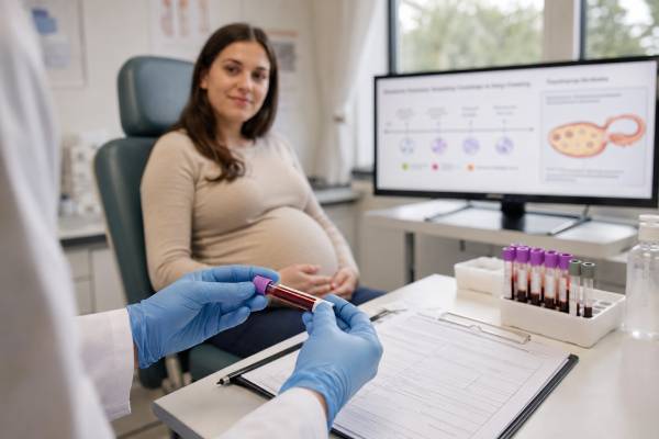
An inhibin A test measures a protein hormone made by ovarian granulosa cells and, during pregnancy, mainly by the placenta. Its most familiar use is as one of four markers in the second-trimester quad screen, where it contributes to an estimate of the chance that a fetus has trisomy 21. Inhibin A can also be measured in selected evaluations of ovarian sex cord-stromal tumors, especially granulosa cell tumors, and in research or specialized assessment of ovarian function.
The meaning of the result depends completely on why the test was ordered. In pregnancy, the laboratory adjusts the value for gestational age and maternal factors and reports a multiple of the median, or MoM, as part of a calculated risk. Outside pregnancy, a concentration is compared with the laboratory’s sex-, age-, and menopausal-status reference interval. A high or low result alone does not diagnose a chromosome condition, prove placental disease, measure egg quality, or confirm an ovarian tumor.
- In pregnancy, inhibin A is usually measured between 15 and 22 weeks as part of the quad screen, not as a stand-alone test.
- A higher inhibin A MoM can increase the calculated screening risk for trisomy 21, but only diagnostic testing can confirm a fetal chromosome condition.
- The quad screen combines inhibin A with AFP, hCG, and unconjugated estriol plus maternal and pregnancy information.
- Elevated inhibin A may also be associated with placental dysfunction and adverse pregnancy outcomes, but it does not predict an outcome with certainty.
- Outside pregnancy, inhibin A may help monitor some granulosa cell tumors when it was elevated before treatment.
- Inhibin A is not a routine ovarian reserve test; AMH, antral follicle count, and early-cycle FSH are generally more useful.
Table of Contents
- What Inhibin A Is
- Inhibin A in Pregnancy Screening
- Preparation, Timing, and the Blood Draw
- Understanding High, Low, and Normal Results
- Ovarian Function and Tumor-Marker Uses
- Follow-Up After an Abnormal Pregnancy Screen
- Limitations and Common Interpretation Mistakes
- When to Contact a Clinician
What Inhibin A Is
Inhibins are protein hormones in the transforming growth factor beta family. They are made of two linked subunits. Inhibin A contains an alpha subunit and a beta-A subunit, while inhibin B contains the same alpha subunit paired with a beta-B subunit. Although the names are similar, the two hormones have different patterns of secretion and different clinical uses.
In reproductive-age women who are not pregnant, granulosa cells in ovarian follicles and the corpus luteum produce inhibin A. It participates in feedback between the ovaries and pituitary gland, helping restrain follicle-stimulating hormone, or FSH. Levels are generally lower in the early follicular phase, rise around ovulation, and are higher during the luteal phase after ovulation. They fall after menopause because active ovarian follicular tissue declines.
During pregnancy, the placenta becomes a major source. Maternal serum inhibin A changes with gestational age, which is why a raw concentration cannot be compared meaningfully without knowing the exact pregnancy dating. Screening laboratories convert the result to a multiple of the median for pregnancies at the same gestational age and then adjust the risk calculation for relevant factors.
Inhibin A has three distinct clinical contexts:
| Context | How the result is used | What it cannot do alone |
|---|---|---|
| Second-trimester prenatal screening | Contributes to calculated risk for trisomy 21 and, with other markers, trisomy 18 and open neural tube defects | Diagnose a fetal chromosome or structural condition |
| Placental-risk assessment | May add information about risk of preeclampsia, fetal growth restriction, preterm birth, or stillbirth in selected settings | Predict exactly whether a complication will occur |
| Ovarian tumor monitoring | May help follow some granulosa cell or other sex cord-stromal tumors | Screen the general population for ovarian cancer |
An inhibin B test is more commonly discussed as a marker of early follicular activity or Sertoli-cell function. Neither inhibin A nor inhibin B should be treated as a direct count of eggs or sperm.
Inhibin A in Pregnancy Screening
The quad screen is a maternal blood screening test usually offered in the second trimester. It measures:
- Alpha-fetoprotein, or AFP
- Human chorionic gonadotropin, usually total hCG
- Unconjugated estriol, or uE3
- Dimeric inhibin A
The four values are combined with gestational age and maternal information to estimate the chance of selected fetal conditions. The test is called screening because it separates pregnancies into higher- and lower-probability groups. It does not examine fetal chromosomes directly.
For trisomy 21, the classic quad-screen pattern often includes higher hCG and inhibin A with lower AFP and unconjugated estriol. A laboratory algorithm calculates a numerical risk, such as 1 in 150 or 1 in 2,000. The cutoff used to label a screen “positive” varies by program, but a common threshold has historically been around 1 in 270 for trisomy 21. The final report and local program criteria should be used rather than applying a cutoff found online.
For trisomy 18, AFP, hCG, and unconjugated estriol may be low, while inhibin A contributes less strongly to the pattern. AFP also helps screen for open neural tube defects. This means the quad screen answers several questions through a combined model; the inhibin A value cannot be separated from the other markers and interpreted as though it were an independent diagnostic test.
The quad screen is commonly performed between 15 and 22 weeks of pregnancy, with many programs preferring 16–18 weeks for optimal performance. It may be used when first-trimester screening was not completed, when cell-free DNA screening is unavailable or declined, or as part of a regional prenatal screening pathway. Cell-free DNA screening is generally more sensitive and specific for common trisomies, but it is still a screening test and does not assess open neural tube defects. A second-trimester ultrasound remains important regardless of the screening method chosen.
The result can be affected by:
- Incorrect gestational age
- Multiple pregnancy
- Maternal weight
- Diabetes treated with insulin
- Smoking status in some algorithms
- Race or ethnicity adjustments used by a particular program
- Conception through assisted reproductive technology
- Fetal demise or a vanishing twin
- Laboratory and assay differences
Because dating errors can change the expected median, ultrasound confirmation of gestational age may materially change the risk calculation. An AFP, hCG, estriol, and inhibin A quad screen should therefore be interpreted from the full laboratory report rather than from one marker copied into a patient portal.
Preparation, Timing, and the Blood Draw
No fasting is usually required for inhibin A testing. The sample is collected from a vein in the arm, and the draw generally takes a few minutes. Temporary soreness, bruising, or lightheadedness can occur.
For prenatal screening, the most important preparation is accurate information. Before collection, the clinic or laboratory may confirm:
- The first day of the last menstrual period
- Ultrasound-based gestational age and estimated due date
- Maternal date of birth and weight
- Whether the pregnancy involves one fetus or multiples
- Whether donor eggs, embryo transfer, or other assisted reproduction was used
- Diabetes status and relevant medicines
- Previous prenatal screening results
A small error in the estimated gestational age can shift marker medians and alter the calculated risk. If ultrasound dating later changes, the laboratory may be able to recalculate the result without repeating the blood draw, provided the sample was collected within the valid screening window.
For a nonpregnancy inhibin A test, the ordering clinician should record menstrual status, cycle phase if known, menopausal status, hormone therapy, prior ovarian surgery, and the reason for testing. Inhibin A varies across the cycle, so a result drawn in the luteal phase should not be compared with an early-follicular reference interval. Pregnancy must also be considered because placental production can make values much higher than nonpregnant levels.
Biotin can interfere with some immunoassays. Patients should report high-dose biotin supplements, but they should not stop prescribed treatment without instructions. The laboratory may recommend withholding biotin for a defined period or using an unaffected assay.
Understanding High, Low, and Normal Results
Pregnancy results and MoM
Prenatal screening reports often express inhibin A as MoM. A value of 1.0 MoM is at the median for the reference population at that gestational age after applicable adjustments. A value of 2.0 MoM is approximately twice the median, while 0.5 MoM is approximately half the median.
“Normal” does not mean that every pregnancy must be close to 1.0 MoM. Screening programs expect a distribution of values. The clinical report uses the complete marker pattern and maternal factors to calculate risk. A high inhibin A value may increase the trisomy 21 risk, but a high value can occur in a chromosomally typical pregnancy. Likewise, a value near the median does not guarantee that the fetus has no chromosome or structural condition.
Some obstetric guidance treats second-trimester inhibin A at or above about 2.0 MoM as a marker associated with increased risk of placental complications, especially when other serum markers are also abnormal. This association may influence later growth assessment or antenatal surveillance, but management depends on the complete pregnancy history, ultrasound findings, blood pressure, other marker values, and local guidance.
High inhibin A outside pregnancy
A high nonpregnancy result may occur with:
- Granulosa cell tumors and some other ovarian sex cord-stromal tumors
- Residual or recurrent tumor in a patient whose cancer previously secreted inhibin A
- Normal luteal-phase ovarian activity
- Exogenous reproductive hormones or ovarian stimulation
- Assay interference or an inappropriate reference interval
A high inhibin A result is not specific for cancer. Imaging, symptoms, physical findings, pathology, other tumor markers, and prior values determine its significance. In someone already treated for a granulosa cell tumor, a rising trend may be more meaningful than a single isolated concentration.
Low inhibin A outside pregnancy
Low or undetectable inhibin A may be expected after menopause and during the early follicular phase. It can also reflect reduced functioning granulosa-cell tissue, but the result is not sufficiently validated as a stand-alone ovarian reserve measure. An ovarian reserve test panel usually relies more on AMH, early-cycle FSH and estradiol, and antral follicle count.
A low inhibin A value in a patient being followed for a previously secreting tumor may be reassuring, but only when interpreted with the treatment history and the same assay over time. A normal marker does not exclude a tumor that does not produce inhibin A.
Ovarian Function and Tumor-Marker Uses
Inhibin A participates in normal ovarian feedback, but routine fertility care rarely uses it alone. Its concentration changes with cycle stage and the corpus luteum, making it less stable than AMH for estimating the pool of small recruitable follicles. It also offers less practical information than ultrasound antral follicle count for predicting response to ovarian stimulation.
For ovarian reserve, the most useful distinctions are:
| Marker | Main value | Main limitation |
|---|---|---|
| AMH | Estimates recruitable follicle quantity and expected stimulation response | Does not measure egg quality or guarantee natural fertility |
| Antral follicle count | Direct ultrasound count of visible small follicles | Operator- and equipment-dependent |
| Early-cycle FSH and estradiol | Shows pituitary-ovarian feedback at baseline | Varies between cycles and may become abnormal relatively late |
| Inhibin B | Reflects early follicular granulosa-cell activity | Assay variability and limited stand-alone predictive value |
| Inhibin A | Reflects luteal and placental activity; selected tumor use | Strong cycle and pregnancy dependence; not routine for reserve |
Inhibin A has a more established role as a tumor marker for selected ovarian sex cord-stromal tumors, especially adult granulosa cell tumors. These uncommon tumors arise from cells involved in sex-hormone production and may secrete inhibin A, inhibin B, estradiol, or anti-Müllerian hormone. Symptoms can include abnormal uterine bleeding, postmenopausal bleeding, pelvic pressure, abdominal swelling, or an adnexal mass.
The marker is most useful when it was elevated at diagnosis and falls after successful treatment. A later rise can prompt imaging and clinical assessment for recurrence. Granulosa cell tumors can recur years after treatment, so long-term follow-up may be recommended. The exact surveillance schedule is individualized.
Inhibin A should not be used as a general ovarian cancer screening test. Most ovarian cancers are epithelial tumors and do not reliably produce inhibin A. Screening people without symptoms or a known tumor using inhibin A can create false alarms and does not have proven benefit.
Follow-Up After an Abnormal Pregnancy Screen
A screen-positive quad test means the calculated probability crossed the program’s threshold. It does not mean the fetus definitely has the condition. Many screen-positive pregnancies have a typical chromosome result.
Follow-up commonly includes:
- Reviewing the dates and pregnancy information. Incorrect gestational age, maternal weight, or fetal number may change the calculation.
- A detailed ultrasound. Ultrasound can confirm dating, assess fetal anatomy, look for structural findings, and evaluate the placenta.
- Genetic counseling. A counselor or clinician explains the numerical risk, test limitations, and available choices in neutral terms.
- Additional screening when appropriate. Cell-free DNA may be offered as a more accurate screening test for common aneuploidies, although using it after a positive serum screen can delay definitive diagnosis and may not answer every concern.
- Diagnostic testing. Amniocentesis can analyze fetal genetic material and provide a diagnostic answer for the chromosomes tested. It is invasive and carries a small procedure-related risk that should be discussed with an experienced clinician.
A result associated with placental risk may lead to additional growth ultrasounds, blood-pressure attention, or antenatal fetal surveillance later in pregnancy. This is not automatic for every elevated value. The decision depends on how high the marker is, whether AFP or hCG is also elevated, maternal medical conditions, fetal growth, and local practice.
An inhibin A pregnancy test result should be discussed promptly enough to preserve all follow-up options. Waiting for the next routine appointment may be reasonable for a mildly abnormal marker with a clearly low final risk, but a screen-positive report deserves timely review.
Limitations and Common Interpretation Mistakes
The test is vulnerable to several common errors.
Treating MoM as a concentration. A value of 2.0 MoM is not 2.0 pg/mL. MoM is a relative value adjusted to a gestational-age median.
Interpreting inhibin A alone. The quad screen is a multivariable calculation. The final risk is more informative than one marker.
Calling a positive screen a diagnosis. Screening identifies increased probability. Chorionic-villus sampling or amniocentesis is needed for a diagnostic chromosome result.
Assuming a negative screen rules out all conditions. The quad screen assesses selected chromosome conditions and open neural tube defects. It does not detect every genetic syndrome, birth defect, or pregnancy complication.
Comparing pregnancy and nonpregnancy ranges. Placental production makes pregnancy values fundamentally different.
Using a single high value to diagnose an ovarian tumor. A tumor evaluation requires imaging, clinical assessment, and often surgery and pathology.
Using inhibin A as a fertility score. It does not measure egg quality or predict natural conception reliably.
Screening performance also depends on population characteristics and laboratory algorithms. Detection rates quoted by one program may not match another. Cell-free DNA has changed prenatal screening pathways, but serum screening remains useful in many settings, particularly when access, timing, cost, or the need for AFP-based neural tube defect screening influences the choice.
When to Contact a Clinician
Contact the prenatal-care team after receiving a screen-positive result, a high inhibin A MoM, or a report that recommends genetic counseling or ultrasound. Ask for the final calculated risk, the exact gestational age used, whether the dating is ultrasound-confirmed, and which follow-up options are available.
During pregnancy, urgent assessment is appropriate for heavy bleeding, severe abdominal pain, severe headache with visual symptoms, shortness of breath, sudden swelling, markedly reduced fetal movement later in pregnancy, or other warning signs provided by the obstetric team. An inhibin A result should never delay evaluation of symptoms.
Outside pregnancy, arrange clinical review for persistent abnormal uterine bleeding, postmenopausal bleeding, increasing abdominal size, pelvic pressure, unexplained early satiety, or a new adnexal mass. These symptoms have many noncancerous causes, but they warrant evaluation. A rising inhibin A level during surveillance after granulosa cell tumor treatment should be discussed with the oncology team rather than interpreted independently.
The safest way to use inhibin A is to keep the testing context visible. In pregnancy, it is one input in a prenatal risk model. In oncology, it is a trend marker for selected tumors. In fertility care, it is usually secondary to better-validated ovarian reserve measures.
Patients can make the follow-up visit more productive by bringing the complete laboratory report rather than only the highlighted abnormal value. Useful details include the raw concentration, MoM, final calculated risk, gestational age used by the laboratory, other quad-screen markers, and ultrasound dating. For tumor surveillance, bring prior inhibin A values, the assay laboratory, treatment dates, imaging reports, and the value measured before treatment. Trends are easiest to interpret when the same assay is used over time.
References
- Screening for Fetal Chromosomal Abnormalities 2026 (Guideline)
- Prenatal Genetic Screening Tests 2025 (Official Page)
- Indications for Outpatient Antenatal Fetal Surveillance 2021 (Guideline)
- Biochemical Screening for Fetal Trisomy 21 2023 (Review)
- Predictive value of quad serum markers for adverse pregnancy outcomes 2021 (Cohort Study)
- Maternal Serum Activin A, Inhibin A and Follistatin-Related Protein Concentrations in Preeclampsia: A Systematic Review and Meta-Analysis 2023 (Systematic Review)
Disclaimer
This article provides general information and does not interpret an individual prenatal screen, diagnose a fetal condition, or evaluate an ovarian mass. Pregnancy screening and tumor-marker results require review by the ordering clinician using the laboratory method, gestational age or menstrual status, imaging, and medical history. Seek urgent obstetric care for severe pain, heavy bleeding, severe headache with visual changes, or reduced fetal movement when instructed by your care team.