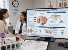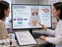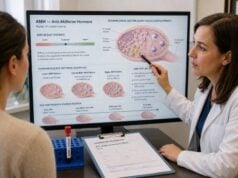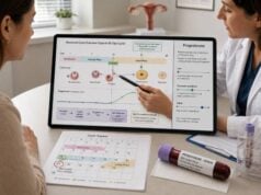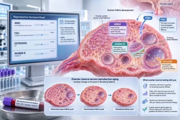
An ovarian reserve test panel estimates how the ovaries may respond to fertility treatment by measuring anti-Müllerian hormone (AMH), follicle-stimulating hormone (FSH), estradiol, and sometimes inhibin B. These tests mainly reflect the number of recruitable follicles—not egg quality, the chance of natural conception in a specific month, or whether pregnancy is possible. Age remains the strongest routine predictor of egg quality and chromosome-related pregnancy risk. AMH can usually be checked on any cycle day, while FSH, estradiol, and inhibin B are commonly measured early in the menstrual cycle. Results must be interpreted together with age, cycle history, medications, prior ovarian surgery or chemotherapy, and an ultrasound antral follicle count. A low result can help a fertility specialist plan treatment sooner or choose a medication dose, but it does not prove infertility. A high AMH level may indicate a strong follicle response or polycystic ovary syndrome (PCOS), and it can increase the risk of ovarian hyperstimulation during treatment.
- Ovarian reserve tests estimate egg quantity and expected treatment response, not egg quality.
- AMH and antral follicle count are generally the most useful modern reserve markers.
- FSH, estradiol, and inhibin B are usually interpreted from cycle days 2–4.
- A low AMH or high FSH does not mean pregnancy is impossible.
- A high AMH level can occur with PCOS and may predict an excessive stimulation response.
- Reference intervals vary by age, assay, laboratory, and fertility clinic.
Table of Contents
- What Ovarian Reserve Testing Measures
- Who May Need an Ovarian Reserve Panel
- When and How the Tests Are Done
- AMH, FSH, Estradiol, and Inhibin B Explained
- How to Interpret Result Patterns
- Normal Ranges and Important Limitations
- Additional Tests and Next Steps
What Ovarian Reserve Testing Measures
Ovarian reserve describes the remaining pool of eggs and, more practically, the number of follicles that may be recruited during a menstrual cycle or fertility treatment. A blood panel cannot count all remaining eggs directly. Instead, it measures hormones produced by small ovarian follicles or by the pituitary gland in response to ovarian feedback.
The most important distinction is between egg quantity and egg quality. AMH, FSH, estradiol, inhibin B, and antral follicle count can help estimate quantity and likely response to injectable fertility medications. They do not directly measure whether an egg has the correct number of chromosomes. Egg quality declines mainly with age, especially in the late 30s and 40s, even when AMH is normal or high.
Ovarian reserve results are most useful for treatment planning. A specialist may use them to estimate whether the ovaries are likely to produce few, average, or many follicles during in vitro fertilization (IVF). The findings can influence medication dosing, counseling about expected egg yield, and discussion of alternatives. They may also support evaluation for diminished ovarian reserve or primary ovarian insufficiency when menstrual cycles become irregular or stop.
Reserve testing is less accurate as a screening test for natural fertility in people who have not tried to conceive. Studies show that low AMH or elevated FSH does not reliably identify who will or will not become pregnant naturally over the next several months. A person with low reserve may ovulate one egg regularly and conceive, while someone with reassuring reserve may have blocked fallopian tubes, endometriosis, sperm-related infertility, or age-related egg-quality problems.
For that reason, an ovarian hormone assessment should be treated as one part of a broader fertility evaluation rather than a pass-or-fail fertility score. Results are most meaningful when interpreted with reproductive age, pregnancy goals, time trying to conceive, medical history, and ultrasound findings.
Who May Need an Ovarian Reserve Panel
A clinician may order ovarian reserve testing when someone has infertility, is planning fertility treatment, or has a condition that could reduce the follicle pool. Testing is especially useful before ovarian stimulation because it helps predict response and reduce the chance of either an inadequate dose or excessive stimulation.
Common reasons for testing include:
- Difficulty becoming pregnant after 12 months when younger than 35, or after 6 months at age 35 or older
- Planning IVF, egg freezing, embryo freezing, or fertility preservation
- Irregular, infrequent, or absent periods
- A family history of early menopause or primary ovarian insufficiency
- Previous ovarian surgery, ovarian torsion, severe endometriosis, or removal of one ovary
- Previous chemotherapy, pelvic radiation, or another gonadotoxic treatment
- A known genetic condition associated with ovarian insufficiency
- Concern about an unexpectedly weak response to prior fertility medication
- A need to estimate the risk of a very high response, particularly with PCOS
Testing may also be discussed before delaying pregnancy, but the limitations matter. A normal AMH result does not guarantee future fertility, preserve egg quality, or predict the exact age of menopause. A low result may cause unnecessary anxiety if it is interpreted without context. Decisions about elective egg freezing should therefore consider age, family-building plans, expected egg yield, cost, and the fact that no test can guarantee a future live birth.
People younger than 40 with several months of missed periods may need a broader evaluation for primary ovarian insufficiency. In that setting, clinicians usually rely on menstrual history and repeated elevation of FSH rather than AMH alone. Pregnancy, thyroid disease, high prolactin, hypothalamic causes, and medication effects must also be considered. The primary ovarian insufficiency panel is different from routine IVF-response testing because the clinical question is whether ovarian function has become persistently impaired.
Routine reserve screening is generally not recommended as the only test in people without infertility risk factors. It is also not a substitute for semen analysis, assessment of ovulation, or evaluation of the uterus and fallopian tubes when pregnancy has not occurred.
When and How the Tests Are Done
AMH is relatively stable across the menstrual cycle and can usually be measured on any day. FSH, estradiol, and inhibin B change substantially during the cycle, so they are commonly drawn in the early follicular phase—usually cycle day 2, 3, or 4. Cycle day 1 is the first day of full menstrual bleeding, not light spotting.
A typical testing plan may include:
| Test | Usual timing | Main purpose |
|---|---|---|
| AMH | Any cycle day in most cases | Estimates the pool of small growing follicles and expected stimulation response |
| FSH | Cycle days 2–4 | Measures pituitary effort needed to recruit follicles |
| Estradiol | Cycle days 2–4 with FSH | Checks baseline ovarian activity and whether FSH may be artificially suppressed |
| Inhibin B | Cycle days 2–4 | Reflects activity of early developing follicles; used less often than AMH |
| Antral follicle count | Early follicular ultrasound | Counts visible small follicles in both ovaries |
Fasting is usually not required for these hormone tests unless other labs, such as fasting glucose or insulin, are ordered at the same time. Blood can generally be drawn at any time of day, although following the laboratory’s instructions improves consistency.
Tell the clinician about hormonal contraception, fertility medications, testosterone therapy, supplements, recent pregnancy, breastfeeding, and any medication that affects the ovaries or pituitary gland. Combined hormonal contraceptives and some other suppressive methods can temporarily lower AMH and antral follicle count. A clinician may recommend testing after a break from contraception when an accurate treatment-planning estimate is important, but contraception should not be stopped without discussing pregnancy prevention and medical risks.
Recent ovarian surgery, an active ovarian cyst, or a fertility-treatment cycle can also affect interpretation. If FSH and estradiol are drawn outside the intended early-cycle window, the numbers may be physiologically higher or lower and should not be compared with early-follicular reference ranges.
The blood draw itself takes only a few minutes. Ultrasound antral follicle count may be performed transvaginally in adults because that approach gives a clearer view of the ovaries. No single test is perfect; agreement between AMH and ultrasound generally increases confidence in the estimate.
AMH, FSH, Estradiol, and Inhibin B Explained
Anti-Müllerian Hormone (AMH)
AMH is produced by granulosa cells in preantral and small antral follicles. Because these follicles represent the recruitable pool, AMH usually declines as ovarian reserve decreases with age. It is widely used to predict the number of eggs that may be obtained during ovarian stimulation.
A low AMH level suggests a lower expected response, but it does not prove that no eggs remain and should not be used to deny fertility treatment. A high AMH level often predicts a larger response. It may occur in people with PCOS because they have a greater number of small follicles. The result is best interpreted with age-specific laboratory data and an AMH test interpretation, not a single internet cutoff.
Follicle-Stimulating Hormone (FSH)
FSH is released by the pituitary gland to stimulate follicle growth. When the ovaries provide less estradiol and inhibin feedback, the pituitary may release more FSH. An elevated early-follicular FSH level can therefore suggest diminished ovarian reserve.
FSH varies from cycle to cycle and may not rise until reserve is already significantly reduced. A single normal value does not rule out diminished reserve. Very high FSH combined with irregular or absent periods raises concern for ovarian insufficiency, but diagnosis usually requires clinical context and repeat testing.
Estradiol (E2)
Estradiol is produced by growing follicles. Early in the cycle, it should generally be relatively low. An elevated baseline estradiol level may indicate early follicle recruitment or a functional ovarian cyst. It can also suppress FSH, creating a normal-looking FSH result despite reduced reserve. This is why clinicians often order FSH and estradiol together.
Estradiol alone is not a reliable reserve test. It rises and falls across the cycle and is strongly influenced by fertility medications, pregnancy, and exogenous estrogen.
Inhibin B
Inhibin B is produced by granulosa cells of small developing follicles and suppresses FSH. Lower early-follicular levels may occur when fewer follicles are active. However, inhibin B has greater cycle-to-cycle variability and generally adds less predictive value than AMH or antral follicle count. Many clinics no longer include it in a standard reserve panel.
An inhibin B test may still be useful in selected cases or research settings, but it should not override more established markers when results conflict.
How to Interpret Result Patterns
Clinicians interpret the pattern rather than labeling one number as good or bad. Age and ultrasound findings can change what the same hormone value means for two different people.
| Common pattern | Possible interpretation | Important caution |
|---|---|---|
| Low AMH, high early FSH, low inhibin B | Diminished ovarian reserve or lower expected stimulation response | Does not prove infertility or measure egg quality directly |
| Low AMH with normal FSH | Early decline in recruitable follicle pool | FSH may remain normal until reserve is more reduced |
| High FSH with high baseline estradiol | Reduced reserve or abnormal early follicle recruitment | Repeat testing and ultrasound may be needed |
| Normal FSH with elevated baseline estradiol | FSH may be falsely reassuring | Check cycle timing and consider an ovarian cyst |
| High AMH and high antral follicle count | High-response pattern, often seen with PCOS | High reserve does not guarantee egg quality or pregnancy |
| Low AMH after hormonal contraception | Possible temporary suppression | Interpret with medication history and consider repeat testing |
| Discordant AMH and follicle count | Assay, ultrasound, timing, or biologic variation | Use the overall clinical picture rather than averaging blindly |
A diminished-reserve pattern may lead a fertility specialist to discuss earlier treatment, a higher or individualized medication dose, realistic egg-yield expectations, or more than one retrieval cycle. It does not automatically mean donor eggs are required. The person’s age, prior response, sperm factors, embryo goals, and personal preferences remain central.
A high-response pattern can be clinically important because it increases the likelihood of many developing follicles and ovarian hyperstimulation syndrome. Clinics may use lower gonadotropin doses, a different trigger medication, or a freeze-all strategy to reduce risk. High AMH is common in PCOS, but PCOS diagnosis requires clinical criteria rather than AMH alone. A PCOS hormone evaluation may include androgen testing and metabolic assessment when symptoms support it.
When menstrual periods have stopped, very low AMH is not enough by itself to diagnose menopause or primary ovarian insufficiency. FSH, estradiol, pregnancy testing, thyroid testing, prolactin, symptoms, and the duration of cycle changes may all be relevant.
Normal Ranges and Important Limitations
There is no universal ovarian reserve “normal range.” Laboratories use different AMH assays, calibration methods, units, and age groups. Fertility clinics may also use their own response categories based on treatment outcomes. The reference interval printed on the report should be the starting point.
Broad examples sometimes used in adult fertility practice include AMH below about 1 ng/mL as lower reserve, approximately 1–3 ng/mL as an intermediate range, and above about 3–4 ng/mL as a higher-response range. These are not diagnostic boundaries. AMH naturally falls with age, so a value that is below average at 25 may be expected at 42. Results may be reported in ng/mL or pmol/L; 1 ng/mL is approximately 7.14 pmol/L.
Early-follicular FSH below roughly 10 IU/L is often considered reassuring, 10–20 IU/L may suggest reduced reserve, and values above 20 IU/L may indicate marked reduction. Cutoffs differ substantially, and FSH fluctuates. Estradiol above approximately 60–80 pg/mL early in the cycle can make a normal FSH less reassuring, but the laboratory’s range and ultrasound findings matter. Inhibin B cutoffs are even less standardized and should be interpreted by the ordering specialist.
Key limitations include:
- AMH and antral follicle count predict egg yield better than natural pregnancy or live birth.
- No reserve marker directly measures chromosome competence or embryo quality.
- A normal panel cannot rule out age-related decline, endometriosis, tubal disease, uterine factors, or sperm problems.
- Low values may still be compatible with regular ovulation and spontaneous conception.
- High AMH can reflect PCOS and excessive stimulation risk rather than superior fertility.
- Hormonal contraception, pregnancy, recent ovarian procedures, and laboratory differences can shift results.
- Results can vary between assays, so trend testing is most useful when the same laboratory and method are used.
Because of these limitations, repeating AMH frequently to watch it decline usually does not improve decision-making. Testing is most valuable when the result will change a concrete plan.
Additional Tests and Next Steps
The next step depends on why the panel was ordered. In fertility treatment, the most useful companion test is usually a transvaginal ultrasound for antral follicle count. AMH and follicle count together provide a better estimate of ovarian response than either alone.
A complete infertility evaluation may also include:
- Menstrual and ovulation history
- Semen analysis for the sperm-producing partner
- Assessment of fallopian tube openness, often with hysterosalpingography
- Pelvic ultrasound for fibroids, polyps, endometriomas, or ovarian cysts
- Thyroid-stimulating hormone and prolactin when cycle disturbance is present
- Progesterone testing when ovulation is uncertain
- Genetic testing when primary ovarian insufficiency is suspected
- Review of medications, cancer treatment, surgery, and family history
Seek specialist review promptly when periods stop before age 40, FSH is repeatedly in a menopausal range, AMH is extremely low for age, there is a history of gonadotoxic treatment, or pregnancy has not occurred within the recommended evaluation window. Sudden severe pelvic pain, heavy bleeding, fainting, or a positive pregnancy test with pain requires urgent medical care and is not an ovarian reserve issue.
When results suggest reduced reserve, practical decisions may include trying to conceive sooner, proceeding with fertility treatment, considering egg or embryo freezing, or discussing donor eggs only when appropriate. The right choice depends on age, desired family size, finances, values, and the probability of success with each option. A reserve panel is a planning tool—not a verdict on whether someone can become a parent.
When results are borderline, repeating the panel immediately is rarely the best first response. A clinician may instead confirm that the blood was drawn at the correct cycle stage, review contraception or supplements, and compare the result with antral follicle count. If repeat testing is appropriate, using the same laboratory reduces method-related differences. Counseling should also address emotional impact: reserve labels can sound final even though they describe probability and treatment response. Ask what the result changes, what uncertainty remains, and which next decision is time-sensitive. That conversation is more useful than focusing on a single percentile or comparing numbers with another person of a different age.
References
- Guideline for the prevention, diagnosis and treatment of infertility 2025 (Guideline)
- Guideline on Ovarian Stimulation for IVF/ICSI 2025 (Guideline)
- The best ovarian reserve marker to predict ovarian response following controlled ovarian hyperstimulation: a systematic review and meta-analysis 2024 (Systematic Review and Meta-analysis)
- Guideline on premature ovarian insufficiency 2024 (Guideline)
- Fertility evaluation of infertile women: a committee opinion 2021 (Committee Opinion)
- Testing and interpreting measures of ovarian reserve: a committee opinion 2020 (Committee Opinion)
Disclaimer
This article is for general educational purposes and does not replace individualized medical advice, diagnosis, or treatment. Ovarian reserve results vary by age, assay, cycle timing, medication use, and clinical circumstances; discuss your report with a qualified reproductive health professional.