
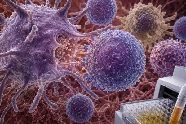
An interleukin-12 test measures a cytokine that connects early innate immune sensing with a strong cellular immune response. Interleukin-12, or IL-12, is produced mainly by activated dendritic cells, macrophages, and other antigen-presenting cells. It acts on natural killer cells and T cells, promotes type 1 helper T-cell development, increases interferon-gamma production, and supports killing of infected or abnormal cells. The biologically active form is IL-12p70, a two-part protein made from p35 and p40 subunits. This detail matters because the p40 subunit is also part of IL-23, and some laboratory methods or treatment discussions refer to p40 rather than active IL-12p70. Direct serum IL-12 testing is used mainly for research and specialist investigation of immune, infectious, inflammatory, or treatment-related questions. A high value is nonspecific, while a low blood level does not prove defective T-cell immunity. Evaluation of the IL-12–interferon-gamma pathway often requires stimulated cell assays, receptor-signaling studies, pathogen history, and genetic testing rather than one resting cytokine concentration.
- IL-12p70 is the active heterodimer; IL-12p40 alone is not the same analyte.
- IL-12 promotes IFN-γ release from T cells and natural killer cells and helps macrophage-centered defense.
- High serum IL-12 may reflect infection, immune activation, inflammation, or an immune-stimulating therapy.
- Low or undetectable IL-12 is common in resting blood and does not diagnose an IL-12 pathway defect.
- In suspected inherited susceptibility to mycobacteria or Salmonella, functional and genetic tests are more informative than a simple cytokine level.
Table of Contents
- IL-12 Biology and Why the Subunits Matter
- Types of IL-12 Tests
- Why a Clinician May Order IL-12 Testing
- Interpreting a High IL-12 Result
- Interpreting a Low or Undetectable Result
- IL-12–IFN-γ Pathway Defects and Infection Susceptibility
- IL-12 Versus IL-23, and Why the Distinction Changes Meaning
- Practical Interpretation, Follow-Up, and Limitations
IL-12 Biology and Why the Subunits Matter
IL-12 is part of a structurally unusual cytokine family whose members are made from paired protein chains. Active IL-12p70 combines a p35 chain encoded by IL12A with a p40 chain encoded by IL12B. The two chains join to form the heterodimer that binds the IL-12 receptor and drives the best-known biological effects.
Dendritic cells and macrophages produce IL-12 after recognizing microbial material and receiving additional activation signals. The cytokine acts especially on natural killer cells and activated T cells. Its receptor contains IL-12Rβ1 and IL-12Rβ2 chains. Signaling through TYK2, JAK2, and STAT4 increases interferon-gamma production and helps naive CD4 T cells develop toward a type 1 helper phenotype. IL-12 also enhances natural killer and CD8 T-cell cytotoxic activity in appropriate settings.
The resulting IL-12–IFN-γ circuit is a two-way partnership. Antigen-presenting cells release IL-12; lymphocytes respond by releasing IFN-γ; IFN-γ then activates macrophages and can further shape antigen presentation. This circuit is particularly important for controlling pathogens that survive inside macrophages, including mycobacteria and Salmonella.
IL-12 is not simply a marker of “strong immunity.” Too little pathway activity can impair control of intracellular infection. Excessive or persistent type 1 activation can contribute to inflammatory tissue injury. The desired response is targeted and time-limited.
Subunit terminology causes frequent confusion. IL-12p40 can circulate alone, as a homodimer, or as part of active IL-12p70. The same p40 chain also joins with p19 to form IL-23. Therefore, an assay that detects p40 may reflect potential contributions from IL-12, IL-23, free p40, and related complexes. An assay specific for IL-12p70 gives a narrower result.
Treatment names can create the same problem. A drug that targets p40 blocks signaling from both IL-12 and IL-23. A drug that targets IL-23p19 is more selective for IL-23. The biological and clinical implications are different, so the exact target should always be identified.
Types of IL-12 Tests
The simplest format is a direct protein assay in serum or plasma. Many laboratories specifically report IL-12p70. Others may report IL-12 without clarifying whether the method detects p70, p40, or both. The assay description, analyte name, and manufacturer information should be checked before interpretation.
Direct measurement may be performed by enzyme immunoassay, chemiluminescent methods, multiplex bead arrays, or ultrasensitive digital platforms. IL-12 concentrations are often very low, and results may fall below the detection limit. An assay’s lower limit, calibration, and antibody specificity strongly influence the number.
A cytokine panel may include IL-12p70 alongside IFN-γ, IL-2, IL-6, IL-10, TNF-α, and chemokines. This can show whether a broader type 1 or inflammatory pattern is present. It cannot identify the disease by itself because overlapping patterns occur in infection, autoimmunity, malignancy, immune therapy, and critical illness.
Stimulated cell testing examines production capacity. Patient monocytes or dendritic cells can be exposed to microbial ligands, then IL-12 release is measured. This asks whether cells can make the cytokine under defined conditions. It may be paired with tests of IFN-γ production after IL-12 stimulation or receptor signaling after adding recombinant cytokine.
Flow cytometry can measure intracellular IL-12 in defined cell populations or downstream STAT phosphorylation. Gene-expression methods can quantify IL12A, IL12B, or pathway-related transcripts. These methods reveal different aspects of the pathway and should not be treated as direct serum equivalents.
Genetic testing is used when an inborn error of immunity is suspected. Variants in IL12B can reduce functional IL-12 production. Variants in IL12RB1 can impair cellular response to IL-12 and also affect IL-23 signaling because the receptor chain is shared. TYK2, IFNGR genes, STAT1, and other pathway genes may be relevant depending on the phenotype. A molecular finding needs expert classification and functional correlation.
Finally, IL-12 may be measured during investigational cancer therapy. Local or tumor-targeted IL-12 delivery is designed to activate T cells and natural killer cells while limiting systemic toxicity. In that setting, serum IL-12, IFN-γ, chemokines, liver tests, blood counts, and clinical adverse events may serve as pharmacodynamic or safety markers.
Why a Clinician May Order IL-12 Testing
Direct serum IL-12 testing is uncommon in routine primary care. Laboratory directories often describe it as a research-oriented test that may help investigate the pathogenesis of immune, infectious, allergic, or inflammatory disorders. The test has the greatest value when ordered for a precise question.
One question is whether a patient has evidence of broad immune activation. In severe infection, autoimmune inflammation, cytokine release, or unusual multisystem disease, IL-12 can be included in a panel to characterize the signaling pattern. The result is supportive rather than diagnostic.
Another question concerns host defense against intracellular organisms. A child or adult with disseminated Bacillus Calmette-Guérin infection, recurrent nontuberculous mycobacterial disease, extraintestinal Salmonella, or an unusually severe pattern may need evaluation of the IL-12–IFN-γ axis. A resting IL-12 level is usually not enough; specialists use functional stimulation and receptor-response studies.
Cancer immunology is another setting. IL-12 promotes IFN-γ, T-cell activity, natural killer-cell function, and chemokines that can recruit immune cells into tumors. Systemic recombinant IL-12 has substantial toxicity, so modern strategies aim for targeted, local, gene-based, viral, or antibody-linked delivery. Measurement during trials can confirm exposure and downstream immune activation.
Autoimmune and inflammatory research also uses IL-12 testing. Type 1 immune activity has been studied in inflammatory bowel disease, psoriasis, inflammatory arthritis, and other conditions. However, much early literature attributed effects to IL-12 when p40 blockade was actually suppressing both IL-12 and IL-23. Modern interpretation must distinguish these pathways.
Clinicians may also evaluate a patient receiving a biologic that blocks IL-12/23p40. Direct cytokine levels are not routinely required to determine whether the drug is working. Symptoms, disease-specific activity measures, imaging, endoscopy, skin assessment, laboratory safety monitoring, and drug-specific guidance are more important.
An isolated IL-12 test is generally not appropriate as a wellness screen, a measure of overall T-cell strength, or a way to decide whether a person needs “immune boosting.” Cytokine concentrations are dynamic and context-dependent. A result has meaning only when connected to symptoms, exposures, medications, and a plausible pathway question.
Interpreting a High IL-12 Result
A high IL-12p70 result indicates increased immunoreactive cytokine in the specimen. It does not establish whether the cytokine is the main driver of illness or a secondary response.
Intracellular infection is a biologically plausible cause. Antigen-presenting cells can release IL-12 after detecting bacteria, parasites, fungi, or viruses, particularly when type 1 cellular immunity is needed. The blood level varies with timing and tissue compartment. Pathogen-specific culture, molecular testing, serology, microscopy, imaging, or biopsy is required for diagnosis.
General inflammatory activation can raise IL-12 with other cytokines. Tissue injury and innate immune stimulation may activate dendritic cells and macrophages. A high value can occur in severe illness without indicating a unique pathway disorder. CRP, ferritin, blood counts, organ tests, and the clinical course usually provide more immediate information.
Autoimmune or immune-mediated disease may involve IL-12-driven type 1 responses. Yet elevated concentrations overlap across diagnoses and may not correlate tightly with disease activity. A result cannot replace disease-specific autoantibodies, imaging, pathology, endoscopy, or established scoring systems.
Immune therapy may deliberately increase IL-12 or its downstream signals. In an IL-12-based cancer trial, a high level may reflect drug exposure. IFN-γ, CXCL9, CXCL10, activated T cells, fever, liver-enzyme changes, and other effects may be monitored. Interpretation must follow the protocol because treatment-associated concentrations are not compared with ordinary diagnostic ranges in the same way.
Malignant or tumor-associated immune activity can also alter IL-12, but a high value is not a cancer screening test. Tumors may suppress IL-12 locally even when systemic inflammation is present, or surrounding antigen-presenting cells may produce it. Tissue context is more informative than serum alone.
Assay cross-reactivity is important. A method that detects p40 may report a high result because of IL-23 or free p40 rather than active IL-12p70. The laboratory should clarify what the capture and detection antibodies recognize.
Unexpectedly high values should be reviewed for preanalytic artifacts. Delayed separation can permit ongoing cytokine release. Heterophile antibodies, rheumatoid factor, high-dose hook effects, or matrix interference can distort immunoassays. A repeat specimen, dilution study, or alternate platform may resolve a discordant result.
The magnitude of elevation should be judged only against the laboratory’s method-specific range. Cross-platform numerical comparisons are unreliable even when units are identical.
Interpreting a Low or Undetectable Result
Low circulating IL-12 is common in healthy people because production is stimulus-dependent and often local. An undetectable result means the amount was below the assay’s detection limit, not that the pathway is absent.
Timing can explain a low result during infection. IL-12 may have peaked before collection, may be produced mainly in tissue, or may be consumed rapidly through receptor binding. Other cytokines can sustain the response after circulating IL-12 falls.
A low resting value does not prove that dendritic cells or macrophages cannot produce IL-12. Stimulated testing is needed to assess production capacity. Cells must be viable and present in adequate numbers, and the assay must include controls.
Medications can reduce production. Glucocorticoids, calcineurin inhibitors, antimetabolites, JAK or TYK2 inhibitors, biologics, and cytotoxic therapies may suppress antigen-presenting cells or downstream signaling. Anti-IL-12/23p40 treatment can also change measured free and drug-bound analyte differently depending on assay design.
A true IL-12 production defect is rare. IL12B deficiency impairs the p40 subunit and can reduce IL-12 and IL-23 function. A receptor defect may leave the cytokine level normal or high because cells cannot respond. Thus, concentration alone cannot localize the abnormal step.
A normal IL-12 result also does not exclude T-cell dysfunction. T cells may be reduced in number, fail to express receptors, have impaired STAT4 signaling, or respond abnormally to a different stimulus. A T-cell count test measures quantity, while proliferation, cytokine release, and signaling assays measure aspects of function.
Low IL-12 should not be treated with supplements or cytokine products outside specialist care. Increasing a proinflammatory cytokine indiscriminately could cause fever, liver injury, cytopenias, or other toxicity. The underlying diagnosis must come first.
IL-12–IFN-γ Pathway Defects and Infection Susceptibility
The IL-12–IFN-γ axis is central to a group of inborn errors sometimes grouped under Mendelian susceptibility to mycobacterial disease. Affected people have selective difficulty controlling weakly virulent mycobacteria, environmental mycobacteria, BCG vaccine strains, Salmonella, and sometimes other intracellular organisms.
The exact infection pattern depends on the gene and whether the defect is partial or complete. Complete interferon-gamma receptor defects can present early and severely. IL-12 receptor beta 1 deficiency is among the more common genetic defects in this pathway and can have variable penetrance. Some people remain well for years, while others develop disseminated infection.
Clinical clues include persistent lymphadenitis, unusual bone lesions, hepatosplenomegaly, recurrent bloodstream Salmonella, disseminated nontuberculous mycobacteria, or severe disease after BCG vaccination. Routine immunoglobulins and lymphocyte counts can be normal, which is why the pathogen pattern matters.
Evaluation is stepwise. Cultures and molecular identification establish the organism. HIV testing and common acquired causes of immunosuppression are assessed. Blood cells may be stimulated to measure IL-12 and IFN-γ production. Recombinant IL-12 or IFN-γ can be added to determine whether the pathway responds. Flow cytometry may examine receptor expression and STAT phosphorylation. Genetic testing then targets plausible genes or uses a broader inborn-error panel.
The result of an interferon-gamma test must also be understood precisely. A tuberculosis IGRA measures antigen-specific T-cell release and does not directly diagnose an inherited IL-12 pathway defect. A serum IFN-γ level is also different from a controlled functional response assay.
Treatment includes prolonged pathogen-directed antimicrobial therapy and specialist management of the immune defect. Recombinant IFN-γ can help selected patients whose cells retain receptor responsiveness, but it would not correct a complete IFN-γ receptor defect. Hematopoietic stem-cell transplantation may be considered for severe disorders. Decisions require immunology and infectious-disease expertise.
Acquired neutralizing anti-IFN-γ autoantibodies can mimic part of this pathway failure in adults. Testing for those antibodies is distinct from measuring IL-12 and may be appropriate when disseminated intracellular infections begin later in life.
IL-12 Versus IL-23, and Why the Distinction Changes Meaning
IL-12 and IL-23 share the p40 subunit but lead to different dominant immune programs. IL-12 pairs p35 with p40 and supports type 1 T-cell and natural killer-cell responses with IFN-γ production. IL-23 pairs p19 with p40 and helps maintain and expand IL-17-producing lymphocytes in several inflammatory settings.
Because the IL12B gene encodes p40, variants affecting it can disrupt both cytokines. Likewise, the IL-12Rβ1 receptor chain participates in both the IL-12 and IL-23 receptor complexes. A patient with IL12RB1 deficiency may therefore show impaired IFN-γ responses and altered IL-17-related immunity.
The distinction is also therapeutic. Ustekinumab targets p40 and blocks both IL-12 and IL-23. Other biologics target IL-23p19 and preserve IL-12 signaling. These agents have different indications, efficacy profiles, and theoretical infection effects.
Older experimental conclusions sometimes assigned inflammatory disease to IL-12 because blocking p40 improved the model. Once IL-23 was discovered, some effects were reattributed to IL-23. Current articles and test reports should state whether they measured p70, p40, p35, or a shared pathway signal.
A direct IL-23 test is itself specialized and not a routine diagnostic test for psoriasis or inflammatory bowel disease. Measuring both cytokines does not automatically reveal which one is driving tissue inflammation, because receptor expression and local cellular responses matter.
This shared-subunit biology is a good example of why cytokine names cannot be interpreted as isolated molecules. Structure, receptor use, and downstream cell type determine meaning.
Practical Interpretation, Follow-Up, and Limitations
Begin with the report details. Does it say IL-12p70 or IL-12p40? Was the specimen serum, plasma, or cell-culture fluid? Was the result direct or stimulated? What were the detection limit, reference interval, and controls? Without those details, “high IL-12” is incomplete information.
Next, reconstruct the clinical timing. Note fever or active infection, recent vaccination, autoimmune flare, immune therapy, antimicrobial treatment, and immunosuppressive medication. Cytokine levels can change within hours, so a sample drawn during an attack is not equivalent to one drawn after recovery.
If acute infection is possible, diagnosis relies on pathogen-specific testing and organ assessment. Serious symptoms such as breathing difficulty, confusion, low blood pressure, persistent high fever, severe abdominal pain, or rapidly worsening condition require urgent care. Specialized cytokine results should not delay antibiotics or other emergency treatment.
For broader immune evaluation, clinicians may order a complete blood count, immunoglobulins, lymphocyte subsets, vaccine antibodies, inflammatory markers, cultures, and imaging. Natural killer-cell and T-cell function can be assessed separately. The natural killer cell function test evaluates cytotoxic performance, not IL-12 production.
When intracellular infection susceptibility is the concern, referral to clinical immunology and infectious disease is appropriate. Functional studies should ideally be arranged with the performing laboratory before collection because live-cell handling requirements are strict. Genetic counseling may be needed before and after sequencing.
For serial testing, use the same laboratory, specimen type, and method. A change across platforms may be analytical rather than biological. Even on one platform, small shifts near the lower detection limit should not be overinterpreted. Ask whether the laboratory validated the assay for the patient’s age group and clinical setting, whether the reported range is a healthy reference interval or a disease-specific cutoff, and whether biologic therapy can interfere with antibody detection. When a stimulated study is performed, review the negative and positive controls, cell counts, incubation time, and transport conditions. A failed control makes the experiment uninterpretable rather than proving that IL-12 immunity is absent. Borderline findings are strongest when they are reproducible and agree with a characteristic infection pattern or a downstream functional abnormality.
The most responsible conclusion is often that IL-12 supports a pattern but cannot establish cause. Its value increases when it is linked to a defined stimulus, paired downstream response, characteristic infection history, or treatment protocol. It decreases when ordered as an isolated marker without a clinical hypothesis. Patients should also know that an abnormal research cytokine does not automatically require treatment; the actionable target is the confirmed disease, infection, or treatment toxicity, not the laboratory signal in isolation.
References
- Serum cytokine panels in pediatric clinical practice 2024 (Clinical review)
- Interleukin 12, Serum 2026 (Laboratory test guidance)
- Preclinical and clinical studies of a tumor targeting IL-12 immunocytokine 2024 (Translational review)
- Emerging interleukin targets in the tumour microenvironment 2023 (Review)
- Revitalizing Cytokine-Based Cancer Immunotherapy through Innovative Delivery Strategies 2023 (Review)
- Cytokine-armed oncolytic herpes simplex viruses for cancer immunotherapy 2024 (Review)
Disclaimer
This article is for general educational purposes and does not diagnose infection, immune deficiency, autoimmune disease, or cancer. IL-12 results are specialized, method-dependent, and should be interpreted by a qualified clinician with the exact analyte, assay controls, medications, and clinical history. Seek urgent medical care for severe infection symptoms, breathing difficulty, confusion, shock, or rapid deterioration.





