
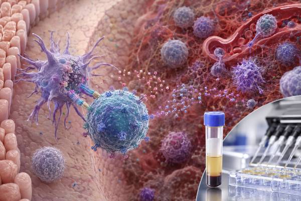
An interleukin-23 (IL-23) test measures a cytokine involved in type 17 immunity, a defense system that helps protect skin and mucosal surfaces from selected bacteria and fungi but can also drive chronic inflammatory disease. IL-23 is strongly linked to psoriasis, psoriatic arthritis, and inflammatory bowel disease, which is why medicines that block its p19 subunit are used for several immune-mediated conditions. A blood IL-23 level, however, is not a routine diagnostic test for those diseases and is not the same as measuring pathway activity in affected tissue. Results vary with the assay, specimen, disease site, treatment, timing, and the molecular form detected. Many commercial cytokine panels are laboratory-developed or research-oriented, and there is no universal “optimal” IL-23 range. A high result may support evidence of immune activation in the right context, while a low result may be normal or reflect treatment. The most useful interpretation combines the exact method with symptoms, examination, established disease markers, imaging or endoscopy when needed, and response to therapy.
- IL-23 is a two-subunit cytokine made mainly by activated antigen-presenting cells.
- It sustains pathogenic Th17 and other type 17 immune responses rather than acting as a simple inflammation score.
- Serum IL-23 does not reliably diagnose psoriasis, arthritis, or inflammatory bowel disease by itself.
- The p19 subunit is specific to IL-23; the p40 subunit is shared with IL-12.
- IL-23 inhibitor treatment is monitored primarily with clinical and disease-specific measures, not a target serum IL-23 value.
Table of Contents
- IL-23 Biology and the Th17 Pathway
- What an IL-23 Test Measures
- Why IL-23 Testing May Be Ordered
- Specimens, Preparation, and Procedure
- How to Interpret IL-23 Results
- What High IL-23 Can Mean
- What Low IL-23 Can Mean
- Related Tests and IL-23-Targeted Treatment
IL-23 Biology and the Th17 Pathway
IL-23 belongs to the IL-12 cytokine family. It is a heterodimer, meaning it is built from two different protein subunits: p19 and p40. The p19 subunit is encoded by IL23A and is specific to IL-23. The p40 subunit is encoded by IL12B and is shared with IL-12. This shared architecture matters clinically because a test or medicine directed at p40 affects both IL-12 and IL-23 pathways, while a p19-directed product is more selective for IL-23.
Dendritic cells, macrophages, monocytes, and other antigen-presenting cells can produce IL-23 after sensing microbes, tissue damage, or inflammatory signals. The cytokine binds a receptor composed of IL-23R and IL-12Rβ1. Receptor signaling activates JAK2, TYK2, STAT3, and related pathways. Susceptibility variants in genes such as IL23R, IL12B, TYK2, and downstream regulators have helped establish the pathway’s role in immune-mediated disease, although genetic risk does not mean that a person’s blood IL-23 concentration will be high.
IL-23 is often discussed with T helper 17 cells. Early Th17 differentiation is initiated by a broader cytokine environment that can include IL-1, IL-6, and transforming growth factor-beta. IL-23 is especially important for expansion, persistence, tissue accumulation, and pathogenic programming of IL-23-responsive cells. These cells can produce IL-17A, IL-17F, IL-22, granulocyte-macrophage colony-stimulating factor, and other mediators. IL-23 also acts on gamma-delta T cells, innate lymphoid cells, and selected natural killer T-cell populations, so the pathway is not limited to conventional CD4 Th17 cells.
Type 17 immunity is protective at epithelial barriers. IL-17-family cytokines recruit and activate neutrophils, stimulate antimicrobial peptides, and strengthen responses against extracellular bacteria and fungi. Excessive or poorly controlled signaling can instead produce keratinocyte activation, intestinal inflammation, enthesitis, synovitis, and tissue remodeling. This protective-versus-pathogenic balance explains why blocking the pathway can improve inflammatory disease but may also alter infection risk.
IL-23 is not the only driver of IL-17 production. Some type 17 responses can persist with limited IL-23 dependence, and the importance of the pathway differs by organ and disease. This helps explain why IL-23 inhibitors are highly effective for plaque psoriasis and useful in selected bowel and joint diseases but are not interchangeable with IL-17 inhibitors across every condition. It also means a measured IL-23 level cannot predict the full downstream response.
What an IL-23 Test Measures
The phrase “IL-23 test” usually refers to an immunoassay that detects IL-23 protein in serum, plasma, cell-culture supernatant, tissue fluid, or a research specimen. Methods include enzyme-linked immunosorbent assays, electrochemiluminescent platforms, bead-based multiplex assays, and newer high-sensitivity systems. The report should state the specimen, units, analytical method, lower limit of detection, and whether the assay targets intact IL-23 or a particular subunit.
An assay intended to measure intact IL-23 should recognize the biologically relevant p19-p40 heterodimer. A p19 measurement may detect material related to IL-23, but analytical specificity depends on the antibody pair. A p40 assay is not IL-23-specific because free p40, p40 homodimers, and IL-12 can contribute. This distinction is central when comparing studies or interpreting a result from a broad panel.
Serum and plasma are not interchangeable. Clotting, anticoagulants, platelet and leukocyte activation, processing delays, storage temperature, and freeze-thaw cycles can affect cytokine recovery. IL-23 is often present at low concentrations, where minor preanalytical differences and assay background have a large relative effect. A value from one laboratory should not be compared with a reference interval from another platform.
Some laboratories stimulate peripheral blood mononuclear cells with microbial products, receptor ligands, or other reagents and then measure IL-23 released into culture. That approach evaluates the sample’s capacity to produce IL-23 under defined conditions rather than the concentration circulating in the person. It can be useful in mechanistic research but does not directly reproduce the environment of skin, intestine, or joint tissue.
Tissue studies may measure IL23A or IL12B messenger RNA, protein staining, spatial expression, or downstream gene signatures. These methods can demonstrate pathway activity near the disease site more clearly than serum testing, but they are usually research tools or part of specialized pathology. An IL-23 gene-expression result is not numerically comparable with a protein concentration.
IL-23 can also appear in a cytokine panel. A multiplex panel may help characterize a pattern, but adding many analytes increases the chance that at least one falls outside a statistical reference interval. A mild isolated abnormality should not be treated as proof of disease without clinical validation.
Why IL-23 Testing May Be Ordered
Direct IL-23 testing is most common in clinical research, translational immunology, drug-development studies, and selected specialist evaluations. It is not a standard screening test for autoimmune disease and is not required before prescribing an IL-23 inhibitor. The ordering question should be explicit.
Researchers may measure IL-23 to study psoriasis, psoriatic arthritis, inflammatory bowel disease, hidradenitis suppurativa, axial or peripheral spondyloarthritis, uveitis, or other inflammatory conditions. The goal may be to compare disease groups, define tissue pathways, assess pharmacodynamic change, or search for response biomarkers. An association at the group level does not necessarily create a clinically useful cutoff for an individual patient.
In psoriasis, diagnosis is usually clinical and sometimes supported by skin biopsy. Disease severity is assessed through body surface area, plaque characteristics, symptoms, nail and joint involvement, and validated scores. Serum IL-23 is not needed to confirm that a rash is psoriasis or to distinguish it reliably from eczema, fungal infection, or another dermatosis.
In inflammatory bowel disease, diagnosis and monitoring rely on symptoms, stool inflammatory markers, blood tests, imaging, endoscopy, and histology. Cytokine-pathway research has guided effective treatment, but a serum IL-23 concentration does not replace fecal calprotectin, C-reactive protein, endoscopic findings, or tissue assessment. A normal blood value does not exclude active Crohn disease or ulcerative colitis.
In inflammatory arthritis, clinicians assess swollen and tender joints, entheses, dactylitis, skin and nail disease, imaging, and conventional inflammatory markers. IL-23 may be biologically important even when the circulating concentration is low or undetectable. Treatment selection is based on the diagnosed condition, manifestations, previous therapies, comorbidities, safety factors, and guideline recommendations—not on a stand-alone cytokine result.
Testing may be included in a clinical trial of an IL-23 inhibitor or an upstream signaling drug. Samples are then collected at protocol-defined times and interpreted against baseline, drug exposure, receptor occupancy, downstream proteins, gene expression, and clinical outcomes. Such research results should not be used outside the protocol as though they were routine diagnostic laboratory values.
A direct IL-23 test is generally not useful for unexplained fatigue, generalized pain, or a desire to measure “autoimmune inflammation” without a focused evaluation. These symptoms deserve a history, examination, and targeted testing selected for the likely causes.
Specimens, Preparation, and Procedure
For a serum or plasma IL-23 test, fasting is usually unnecessary unless another ordered test requires it. Patients should tell the clinician and laboratory about acute infections, recent vaccination, corticosteroids, conventional immune suppressants, biologic drugs, JAK or TYK2 inhibitors, antibiotics, pregnancy, and recent surgery. These factors can affect immune signaling or the interpretation of an inflammatory result. Medicines should not be stopped without instructions from the prescribing clinician.
A phlebotomist collects blood into the tube specified by the laboratory. Serum requires clotting before separation; plasma uses an anticoagulant. Prompt centrifugation and controlled storage are especially important for low-level cytokines. Research protocols may require blood to be processed within a fixed number of minutes and frozen at a defined temperature. Deviations can make longitudinal samples incomparable.
The ordinary risks are those of venipuncture: short-lived discomfort, bruising, lightheadedness, and rarely infection or persistent bleeding. The test itself does not expose the person to IL-23 or alter immune function.
For cell-stimulation testing, blood must often arrive while immune cells remain viable. The laboratory isolates mononuclear cells, counts them, places a standardized number in culture, applies a stimulus, and measures IL-23 in the supernatant after a defined incubation. Negative controls estimate spontaneous release, while positive controls check whether the cells and assay can respond. Poor viability, delayed transport, or a failed control may invalidate the result.
Tissue research requires a different procedure. A skin biopsy, intestinal biopsy, synovial sample, or other material may be analyzed for protein or RNA. The clinical reason for obtaining tissue should stand on its own; invasive sampling is not justified solely to obtain an IL-23 value in most care settings.
Before interpreting the report, verify five items: the exact analyte, whether intact IL-23 or a subunit was measured, the specimen, the units, and the laboratory’s reference or decision limits. Results in pg/mL from serum cannot be compared with normalized gene-expression values, tissue staining intensity, or culture concentrations.
How to Interpret IL-23 Results
There is no universal normal range for IL-23. Some healthy reference samples have undetectable concentrations on one platform and measurable concentrations on another. Laboratories may establish their own interval, provide only an analytical detection limit, or mark the test as research use only. The report’s method-specific information takes priority over ranges found online.
A result below the detection limit means the assay could not quantify IL-23 above its threshold in that specimen. It does not mean the pathway is absent. IL-23 may be produced locally, bind receptors, degrade, or fluctuate outside the sampling time. Active tissue disease can coexist with a low blood concentration.
A measurable result should be considered alongside assay precision near the low end. When a value is close to the limit of quantification, small numerical differences may not represent a meaningful biological change. Repeating the sample on another platform can create a different number because calibration and antibody specificity differ.
Trend testing is most informative when the same specimen type, collection timing, processing protocol, and assay are used. Even then, a fall in serum IL-23 does not automatically equal remission, and a rise does not automatically predict a flare. Clinical endpoints and established biomarkers should lead interpretation.
The p19-p40 distinction can change the conclusion. An elevated p40-related signal may reflect IL-12 biology, free subunit, or other molecular forms rather than intact IL-23. A report that simply says “IL-23” without sufficient method detail may need clarification from the laboratory.
Patterns may be more useful than isolated numbers in research. IL-23 may be examined with IL-17A, IL-17F, IL-22, IL-6, tumor necrosis factor, GM-CSF, and chemokines. Still, blood cytokines do not map neatly onto tissue pathways. Downstream markers can be normal despite active disease, or elevated for unrelated reasons.
The result should answer a defined question. For diagnosis, ask whether the assay has validated sensitivity and specificity for the suspected disease. For prognosis, ask whether a cutoff predicts an outcome in comparable patients. For treatment monitoring, ask whether changing the number has been shown to improve care. In many current clinical settings, the answer is no, which limits the value of direct IL-23 testing.
What High IL-23 Can Mean
A high IL-23 result indicates more detectable analyte than the laboratory’s comparison limit. It may be consistent with activation of monocytes, macrophages, dendritic cells, or tissue inflammatory pathways, but it is nonspecific. The same finding can occur in different immune-mediated diseases, infections, malignancies, or research cohorts, and not every patient with those conditions has an elevated blood level.
Psoriasis is the clearest example of a disease with a central IL-23 pathway. IL-23-producing cells in skin help maintain pathogenic type 17 responses, and p19 inhibitors can produce major clinical improvement. Nevertheless, serum concentration does not measure plaque severity reliably enough to replace examination. A high value also does not prove that a particular IL-23 inhibitor will work.
Psoriatic arthritis and related spondyloarthritides involve complex inflammation at joints, entheses, skin, and gut. IL-23 can be important in initiation or selected cellular compartments, while established disease may use downstream or partly independent pathways. This helps explain why treatment effects differ by manifestation and why a circulating cytokine level is an incomplete guide.
Crohn disease and ulcerative colitis can involve increased IL-23 pathway activity within intestinal tissue. A high serum level may accompany activity in some people, but stool calprotectin, endoscopy, imaging, symptoms, anemia, albumin, and C-reactive protein provide more established information. Infection must also be excluded when bowel symptoms worsen because immune-suppressing treatment can alter the presentation.
Infections can stimulate IL-23 as antigen-presenting cells respond to microbial signals. Because the IL-23/IL-17 axis supports neutrophil recruitment and barrier defense, an elevated result during infection may be protective rather than evidence of autoimmunity. Clinical microbiology and source evaluation are needed before attributing inflammation to an immune-mediated disorder.
Technical explanations include cross-reactivity, interference, specimen handling, or measurement of a shared subunit. A surprising extreme value should prompt review of units, dilution, assay range, and laboratory comments. The appropriate response is to investigate the underlying clinical problem, not to try to lower IL-23 directly based on one test.
What Low IL-23 Can Mean
A low or undetectable resting IL-23 level is often normal. Cytokines are not continuously abundant hormones; they may be produced for short periods near activated cells and rapidly consumed. A low value does not prove weak Th17 immunity, immune deficiency, or absence of autoimmune disease.
Treatment is an important cause of reduced pathway activity. Selective p19 antibodies bind IL-23, and assays may detect free cytokine, drug-bound cytokine, both, or neither depending on design. This can produce an apparent decrease or, paradoxically, altered measured concentrations. Upstream corticosteroids, conventional immunosuppressants, JAK/TYK2 pathway drugs, and control of the underlying disease can also lower production.
A low stimulated result may reflect reduced monocyte or dendritic-cell responsiveness, medication effects, poor cell viability, inadequate stimulation, or sample delay. It should be interpreted with positive controls and other cytokines. An isolated low response does not diagnose a receptor or signaling defect.
Rare inherited abnormalities in IL-23-related pathways may change susceptibility to infection or inflammation, but direct serum testing is not the diagnostic approach. Clinical immunology evaluation may include cell phenotyping, functional studies, targeted signaling assays, and genetic testing based on the phenotype.
Because IL-23 shares p40 with IL-12 and receptor components with related pathways, one low analyte does not describe the entire network. A person can have normal IL-23 but abnormal IL-17 production, or low circulating protein with strong tissue gene expression. The phrase “low IL-23” should therefore remain a laboratory description, not a disease label.
Related Tests and IL-23-Targeted Treatment
Related testing depends on the organ involved. Suspected psoriasis may require dermatologic examination and occasionally biopsy. Joint symptoms may prompt inflammatory markers, rheumatoid factor or anti-CCP antibodies when appropriate, ultrasound, radiographs, or MRI. Bowel symptoms may require complete blood count, metabolic and liver tests, C-reactive protein, fecal calprotectin, stool infection studies, imaging, and endoscopy.
An IL-17 test measures a downstream cytokine but does not substitute for IL-23 testing, and neither test is routinely necessary to diagnose pathway-associated diseases. A chemokine panel may describe cell-recruitment signals in research. Conventional markers such as C-reactive protein are less pathway-specific but usually have clearer clinical roles.
Biologic medicines can target IL-23 p19 or the shared p40 subunit. Selective p19 inhibitors include agents used for plaque psoriasis, psoriatic arthritis, and inflammatory bowel disease, with indications varying by product and country. Ustekinumab and related products target p40 and therefore inhibit both IL-12 and IL-23 signaling. These distinctions affect efficacy, safety, and disease-specific use.
Before treatment, clinicians evaluate for active infection and usually screen for tuberculosis according to the product label and local guidance. Vaccinations should be reviewed, and live vaccines are generally avoided during significant biologic immunosuppression. Liver tests may be required for selected products and indications. Patients should report fever, persistent cough, painful urination, severe diarrhea, or other infection symptoms.
Treatment response is monitored through the affected organ: skin clearance and symptoms, joint and enthesis findings, bowel symptoms, biomarkers, imaging, or endoscopic healing. Drug levels and anti-drug antibodies are being studied or used selectively for some biologics, but direct serum IL-23 is not a standard therapeutic target. A normal value does not justify stopping effective treatment, and a high value does not automatically justify dose escalation.
The most accurate interpretation is modest: an IL-23 result can describe one measured part of a complex pathway. It becomes useful only when the assay is analytically sound, the clinical question is defined, and more established evidence supports the conclusion.
References
- IL-23 past, present, and future: a roadmap to advancing IL-23 science and therapy 2024 (Review)
- The Role of IL-23 in the Development of Inflammatory Diseases 2025 (Review)
- The Role of Interleukin 23/17 Axis in Psoriasis Management 2024 (Review)
- Type 17 immunity: novel insights into intestinal homeostasis and inflammatory bowel disease 2024 (Review)
- Targeting IL-23 for IBD: Rationale and Progress to Date 2023 (Review)
- The Role of IL-23 in the Pathogenesis and Therapy of Inflammatory Bowel Disease 2023 (Review)
Disclaimer
This article provides general education and is not a diagnosis or treatment recommendation. IL-23 testing is specialized, assay-dependent, and usually insufficient to diagnose or monitor an immune-mediated disease by itself. Discuss results and medication decisions with the clinician who knows the symptoms, examination, established biomarkers, and treatment history.





