
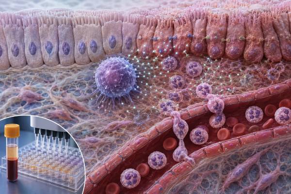
An interleukin-17 test measures a cytokine involved in barrier defense, neutrophil recruitment, and several immune-mediated inflammatory diseases. The name “IL-17” usually refers to IL-17A, but the IL-17 family contains six members, and IL-17F has overlapping functions. T helper 17 cells are a major source, yet gamma-delta T cells, innate lymphoid cells, natural killer T cells, and other populations can also release IL-17 rapidly. The cytokine stimulates epithelial cells, fibroblasts, and other tissue cells to produce chemokines, antimicrobial proteins, granulocyte growth factors, and additional inflammatory mediators. This protects mucosal surfaces against Candida and some extracellular bacteria, but excessive or misdirected signaling contributes to psoriasis, psoriatic arthritis, axial spondyloarthritis, and other disorders. Direct serum IL-17 testing is primarily research-oriented and is not a stand-alone diagnostic test for autoimmunity or a routine guide to biologic treatment. A value must be interpreted according to the exact family member, assay method, specimen, disease activity, medications, and whether the laboratory measured soluble cytokine, IL-17–producing cells, or a broader Th17 signature.
- “IL-17” usually means IL-17A, but IL-17F and other family members are separate analytes.
- Th17 cells are important producers, but a serum IL-17 value is not a Th17 cell count.
- High IL-17 may fit neutrophilic or autoimmune inflammation, yet it cannot diagnose one disease.
- Low or undetectable blood IL-17 does not exclude active IL-17 signaling inside skin, joints, bowel, or lung.
- Weak IL-17 immunity is clinically associated with chronic mucocutaneous Candida susceptibility, especially in selected inborn errors or during IL-17 blockade.
Table of Contents
- The Th17–IL-17 Axis and Barrier Defense
- Which IL-17 Form Is Being Tested?
- Why IL-17 Testing Is Ordered
- What a High IL-17 Result May Mean
- What a Low or Undetectable Result May Mean
- IL-17 in Psoriasis, Arthritis, Bowel Disease, and Treatment
- IL-17 Defects, Candida, and Immune Evaluation
- Assay Limitations and the Most Useful Follow-Up
The Th17–IL-17 Axis and Barrier Defense
IL-17A is a proinflammatory cytokine best known for activating tissue cells rather than directly killing microbes. When it binds receptors on epithelial cells, keratinocytes, fibroblasts, endothelial cells, and other targets, those cells release chemokines such as CXCL1 and CXCL8, granulocyte colony-stimulating factor, antimicrobial peptides, and cytokines. The result is recruitment, production, and activation of neutrophils at a threatened surface.
This pathway is especially useful at the skin, mouth, airways, and gastrointestinal tract. Candida species and some extracellular bacteria are controlled partly through IL-17-dependent barrier responses. IL-17 can work with IL-22 to strengthen antimicrobial protein production and epithelial defense.
Th17 cells develop from naive CD4 T cells under a cytokine environment involving signals such as IL-6, IL-1, TGF-β, and IL-23, although human differentiation is more complex than one fixed recipe. STAT3 and the transcription factor RORγt are central. IL-23 is particularly important for maintaining and expanding pathogenic or inflammatory Th17 programs. A direct IL-23 test therefore examines an upstream cytokine, not the same analyte as IL-17.
Th17 cells are not the only source. Gamma-delta T cells, mucosal-associated invariant T cells, invariant natural killer T cells, innate lymphoid cells, and some myeloid or tissue populations can release IL-17 rapidly. This is why a serum concentration cannot reveal the source or prove expansion of conventional Th17 cells.
The IL-17 family includes IL-17A through IL-17F. IL-17A and IL-17F are most closely related and can form homodimers or an IL-17A/F heterodimer. They share receptor components and biological functions but differ in potency and tissue contribution. IL-17E is better known as IL-25 and promotes type 2 immunity, making it functionally distinct from IL-17A despite the family name.
Physiologic IL-17 is protective. Pathologic IL-17 becomes a problem when the response is persistent, occurs without a useful microbial target, or amplifies self-sustaining inflammation. In psoriasis, for example, IL-17 acts directly on keratinocytes and participates in a loop involving dendritic cells, IL-23, inflammatory T cells, and activated skin cells. Successful IL-17-targeted therapies confirm the pathway’s importance, but a therapeutic target is not automatically a useful serum diagnostic marker.
Which IL-17 Form Is Being Tested?
The test report should state whether the analyte is IL-17A, IL-17F, total IL-17, or another family member. An assay labeled only “IL-17” often targets IL-17A, but this should not be assumed. Antibodies used in one platform may recognize homodimers differently from heterodimers.
A direct serum or plasma assay quantifies soluble protein. Common technologies include enzyme-linked immunoassays, electrochemiluminescence, multiplex bead arrays, and ultrasensitive digital methods. Concentrations are often low, and many healthy or treated samples fall below the assay’s detection limit.
Multiplex testing places IL-17 beside IL-6, IL-8, TNF-α, IL-10, interferons, and other cytokines. This may help describe an inflammatory pattern, particularly in research. It also increases complexity: different analytes have different stability, dynamic ranges, and susceptibility to matrix effects. One borderline result among many measurements is weak evidence unless it is reproducible and clinically coherent.
Cell-based tests answer different questions. Intracellular cytokine staining uses flow cytometry after stimulation to identify the percentage and phenotype of cells capable of producing IL-17. ELISpot counts IL-17-secreting cells under particular conditions. Gene-expression panels measure IL17A, IL17F, RORC, or a larger Th17 signature. Tissue biopsy can localize IL-17-related transcripts or proteins in an affected organ.
A flow cytometry immune panel that reports T-cell subsets usually does not include functional Th17 measurement unless specially designed. Standard CD4 counts cannot be converted into a Th17 count.
Functional immune studies may stimulate cells with Candida antigens, mitogens, or cytokines and measure IL-17 production. These tests can support evaluation of rare immune defects but require viable cells, appropriate controls, and specialist interpretation. A low stimulated response can result from lymphopenia, medication, specimen delay, or a true pathway abnormality.
Genetic tests evaluate genes affecting Th17 development or IL-17 signaling. They do not measure the current cytokine concentration. A pathogenic STAT3, IL17RA, IL17F, ACT1, CARD9, or related variant has different consequences and must be interpreted with the infection and clinical phenotype.
Why IL-17 Testing Is Ordered
Direct serum IL-17 testing is primarily used to study disease mechanisms. Commercial laboratories commonly describe it as research-oriented support for investigating immune, infectious, allergic, or inflammatory disorders rather than as a validated diagnostic test.
In psoriasis and psoriatic arthritis research, IL-17A may be measured to explore pathway activation, compare tissue and blood, or study treatment response. Clinical diagnosis still depends on skin and joint findings, imaging when needed, and exclusion of mimics. A normal serum result does not argue strongly against an IL-17-driven skin lesion.
In axial spondyloarthritis, IL-17 is biologically important and targeted therapies can be effective. Yet no serum IL-17 cutoff establishes the diagnosis. Inflammatory back-pain features, imaging of the sacroiliac joints, HLA-B27 in an appropriate context, inflammatory markers, and examination are more useful.
In inflammatory bowel disease, researchers examine Th17-related cytokines because the pathway participates in mucosal inflammation. The effect of blocking IL-17 differs from the effect of blocking IL-23, and direct IL-17 inhibition can worsen or trigger bowel inflammation in some patients. This illustrates that cytokine roles depend on tissue and cannot be predicted from a blood value alone.
In severe asthma, chronic obstructive lung disease, neutrophilic airway disease, and infection research, IL-17 may be measured in sputum or bronchoalveolar lavage rather than blood. Local samples may better reflect airway signaling, but they have their own collection and normalization problems.
In suspected immune deficiency, direct serum IL-17 is usually less useful than history and functional testing. Recurrent or persistent oral thrush, esophageal candidiasis, chronic nail or skin Candida infection, or unusual fungal disease can prompt assessment of Th17 development and IL-17 signaling.
Testing may also appear in studies of rheumatoid arthritis, multiple sclerosis, lupus, hidradenitis suppurativa, bullous skin disease, transplant rejection, cancer, and severe infection. An association in a research cohort does not mean an isolated patient result has validated sensitivity, specificity, or a treatment threshold.
Before ordering, the clinician should be able to explain what decision a high or low result would change. If no action follows, established disease-specific tests may offer more value.
What a High IL-17 Result May Mean
A high direct IL-17A result indicates that more immunoreactive protein was present than in the laboratory’s reference group. It does not identify the producing cell or the affected tissue.
Psoriatic disease is a well-established IL-17-associated condition. Activated T cells and innate lymphocytes produce IL-17 in skin and entheses, while keratinocytes and stromal cells amplify inflammation. Serum levels may be elevated in some studies but overlap substantially with controls and may not track every disease domain.
Spondyloarthritis can involve IL-17 at the enthesis, joint, skin, and bowel. A high concentration may be compatible with pathway activity but cannot distinguish axial spondyloarthritis from psoriatic arthritis or another inflammatory condition.
Infection and barrier inflammation can raise IL-17 as the body recruits neutrophils against extracellular bacteria or fungi. The cytokine does not identify the organism. Cultures, molecular tests, microscopy, imaging, and examination are required.
Neutrophilic inflammatory disease may produce a high value through IL-17-driven chemokines and growth factors. Conditions affecting airways, skin, joints, or bowel can share this pattern. A neutrophil count test shows circulating cell quantity, while IL-17 reflects one signal that can promote neutrophil recruitment and production.
Autoimmune or immune dysregulation can involve Th17 cells, but IL-17 is not a universal autoimmune marker. Some diseases show mixed Th1, Th2, interferon, B-cell, and innate pathways. The dominant pathway can differ among patients with the same diagnosis.
Obesity and metabolic inflammation, malignancy, recent immune stimulation, and certain therapies may alter IL-17 in research settings. These associations are too nonspecific for screening.
An unexpected isolated elevation may be analytical. Heterophile antibodies, rheumatoid factor, delayed processing, platelet or leukocyte activation, and multiplex matrix effects can change results. Repeat testing on a fresh specimen or a different method may be considered when the clinical picture is inconsistent.
A high number is not a reason to start an IL-17 inhibitor by itself. Biologic treatment is prescribed for a confirmed condition using approved criteria, disease severity, comorbidities, infection screening, and prior treatment response. Direct cytokine levels are not routinely used to choose among agents.
What a Low or Undetectable Result May Mean
Undetectable serum IL-17 is common. The cytokine is often produced locally, binds receptors rapidly, and may be present below the assay’s lower limit. The result does not mean the person lacks Th17 cells or cannot recruit neutrophils.
A low value during treated autoimmune disease may reflect reduced pathway activity, but it can also reflect assay timing, tissue confinement, or drug interference. Antibodies that bind IL-17 may change whether an immunoassay detects free, total, or drug-bound cytokine. The test must be validated for patients receiving that therapy.
Glucocorticoids, JAK inhibitors, conventional immunosuppressants, lymphocyte-depleting treatment, and biologics targeting IL-23 or IL-17 can reduce production or signaling. A low result should be considered in relation to dose and interval since treatment.
True impaired IL-17 immunity is evaluated through the clinical pattern. Chronic mucocutaneous candidiasis is the classic clue. Patients may have persistent thrush, recurrent Candida infection of skin and nails, or esophageal disease. Some inborn errors also cause bacterial skin or respiratory infections, autoimmunity, aneurysms, endocrine disease, or other features depending on the gene.
A receptor defect can exist despite a normal or high ligand concentration. If IL-17RA or downstream ACT1 signaling is impaired, measuring serum IL-17 does not show whether target cells respond. Functional stimulation and signaling assays are needed.
A low stimulated IL-17 response can be caused by too few CD4 cells, poor viability, inadequate stimulation, delayed transport, or medication. Controls must demonstrate that cells were capable of responding. An invalid assay is not evidence of deficiency.
Low IL-17 is also not necessarily desirable in health. The pathway protects barrier surfaces. Excessive suppression can increase mucocutaneous Candida risk. The goal is appropriate activity, not the lowest possible value.
IL-17 in Psoriasis, Arthritis, Bowel Disease, and Treatment
The strongest clinical evidence for the IL-17 pathway comes from treatment response rather than serum testing. Monoclonal antibodies can target IL-17A, both IL-17A and IL-17F, or the IL-17 receptor. These therapies are used for selected inflammatory skin and musculoskeletal diseases.
In plaque psoriasis, blocking IL-17 can rapidly reduce keratinocyte activation and skin inflammation. In psoriatic arthritis, treatment can improve peripheral joints, enthesitis, dactylitis, and skin disease. In axial spondyloarthritis, IL-17 inhibition can reduce spinal symptoms and inflammation. Treatment selection depends on the full disease phenotype, including bowel and eye disease.
Candida infection is a mechanism-related risk because IL-17 supports mucosal antifungal defense. Most reported infections are mucocutaneous and treatable, but recurrent symptoms need evaluation. Patients should report persistent mouth soreness, white plaques, painful swallowing, genital symptoms, or unusual skin and nail infection.
Inflammatory bowel disease requires caution. Although Th17-related signals are present in bowel inflammation, direct IL-17 blockade has not produced the same benefit as IL-23 blockade and may worsen bowel disease in susceptible patients. A history of chronic diarrhea, blood in stool, abdominal pain, weight loss, or known Crohn disease affects drug choice.
Other safety considerations vary by product and patient. Clinicians review vaccination status, infection history, tuberculosis risk according to local guidance, other immunosuppressants, pregnancy plans, and comorbid disease. The serum IL-17 concentration is not used as a universal safety screen.
Response monitoring uses symptoms, examination, skin scores, joint counts, function, inflammatory markers, and imaging when appropriate. A patient may improve clinically without a measurable baseline IL-17 level. Conversely, a reduced serum level does not prove control of all tissue disease.
This difference between pathway validation and biomarker validation is important. A cytokine can be an excellent drug target yet a poor blood diagnostic test because it acts locally, changes rapidly, and overlaps among conditions.
IL-17 Defects, Candida, and Immune Evaluation
Chronic mucocutaneous candidiasis describes persistent or recurrent Candida infection of mucous membranes, skin, and nails. It can result from several defects that reduce the number, development, production, or signaling of IL-17-producing cells.
Autosomal dominant STAT3 loss-of-function disease impairs Th17 development and is associated with eczema, characteristic bacterial infections, very high IgE, skeletal or dental findings, and mucocutaneous candidiasis in some patients. Gain-of-function STAT1 variants can suppress Th17 programs and cause chronic candidiasis with variable bacterial, viral, autoimmune, vascular, or endocrine complications.
Variants directly affecting IL-17F, IL-17RA, IL-17RC, or ACT1 can produce a more focused susceptibility to chronic mucocutaneous Candida and selected bacterial infections. CARD9 deficiency can cause invasive or deep fungal infection through a related but broader antifungal pathway. APECED can involve neutralizing antibodies against IL-17 family cytokines along with endocrine autoimmunity.
Evaluation begins with confirming the organism and site. Recurrent white oral plaques are not always Candida, and culture or microscopy may be needed. The clinician documents onset age, infection frequency, response to antifungals, bacterial and viral infections, growth, eczema, fractures, retained primary teeth, endocrine symptoms, family history, and medication exposure.
Laboratory work may include complete blood count, immunoglobulins, lymphocyte subsets, HIV testing, vaccine responses, Th17-cell or cytokine-production assays, autoantibodies, and genetic sequencing. The exact panel is guided by the phenotype. A standard CD4 count can be normal despite a selective Th17 defect.
Treatment combines antifungal management with care directed at the underlying immune disorder. Long-term antifungal exposure can select resistant organisms, so species identification and susceptibility testing may be important. Immune-directed therapy is individualized and should not be based on a low serum IL-17 value alone.
Assay Limitations and the Most Useful Follow-Up
Specimen type is a major source of variation. Serum forms after clotting, during which cells and platelets can release mediators. Plasma contains anticoagulant that can affect assay chemistry. Sputum, tissue, and lavage samples require normalization because dilution and cellular content vary.
Processing should follow the performing laboratory’s instructions. Delayed centrifugation, temperature changes, hemolysis, and repeated freeze-thaw cycles can affect low-concentration cytokines. Samples for live-cell functional testing require stricter timing than frozen serum.
Reference ranges are platform-specific. An IL-17A value from a multiplex bead assay cannot be compared directly with a single-analyte ultrasensitive result. The lower detection limit, quantification limit, subtype specificity, and handling of values below range should be reviewed.
Interpretation becomes stronger when several independent observations agree: compatible tissue disease, a reproducible cytokine or cellular signature, response to pathway-targeted treatment, and exclusion of infection or another mimic. It remains weak when based on one borderline serum result. When a trend is requested, specimens should be collected at comparable points in the treatment cycle and analyzed by the same laboratory. A numerical fall can reflect lower production, greater drug binding, fewer circulating producer cells, or simple variation near the detection limit. Clinical improvement should therefore be documented independently rather than inferred from the cytokine number.
For suspected autoimmune inflammation, follow-up may include CRP, erythrocyte sedimentation rate, complete blood count, disease-specific antibodies, imaging, skin or bowel assessment, and specialist examination. For recurrent Candida, functional and genetic immune testing is more relevant.
Urgent medical attention is needed for difficulty breathing, rapidly spreading skin infection, high fever with deterioration, confusion, severe dehydration, painful swallowing with inability to drink, or signs of sepsis. The cytokine result should never delay infection treatment.
Useful questions for the ordering clinician include: Was IL-17A or another family member measured? Is the assay validated for this clinical use? Was the sample collected before or after immune therapy? Does the result match tissue findings? Will repeating it change management? The answers determine whether the test is a meaningful pathway clue or a nonspecific research observation. In most patients, the confirmed diagnosis and organ-level response matter more than achieving any particular serum IL-17 target. That distinction prevents unnecessary repeat testing and overtreatment.
References
- Th17 cell pathogenicity in autoimmune disease 2025 (Review)
- Role of interleukin 17 and T helper cells 17 cells as a new pathway of biomarkers in autoimmune diseases 2025 (Review)
- Short-term risk and long-term incidence rate of infection and malignancy with IL-17 and IL-23 inhibitors in adult patients with psoriasis and psoriatic arthritis: a systematic review and meta-analysis 2023 (Systematic review and meta-analysis)
- Bimekizumab: A Review in Psoriatic Arthritis 2024 (Drug review)
- Homozygosity for the Common IL23R R381Q Variant Underlies Chronic Mucocutaneous Candidiasis 2025 (Genetic study)
- Interleukin 17, Serum 2026 (Laboratory test guidance)
Disclaimer
This article is for general educational purposes and does not diagnose autoimmune disease, fungal infection, immune deficiency, or another condition. IL-17 assays are specialized and method-dependent, and results should be interpreted by a qualified clinician with symptoms, medications, infection history, and disease-specific testing. Seek urgent care for severe infection, breathing difficulty, confusion, or rapid clinical worsening.





