
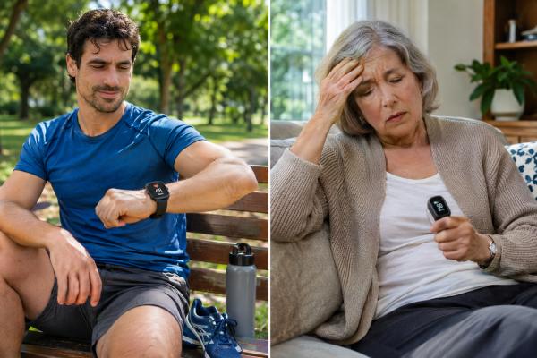
A low heart rate is not automatically a heart problem. Bradycardia usually means a resting heart rate below 60 beats per minute, but many healthy people—especially endurance-trained athletes and people during sleep—regularly fall below that number without symptoms or danger. What matters is whether the heart rate is appropriate for the situation and whether the heart is pumping enough blood to the brain and body.
A pulse in the 40s can be normal in a fit, symptom-free person. The same rate can be concerning in someone with fainting, chest pain, severe weakness, shortness of breath, confusion, or low blood pressure. Medications, thyroid disease, electrolyte problems, sleep apnea, aging of the heart’s electrical system, sinus node dysfunction, and heart block can all slow the pulse.
The useful question is not simply “How low is too low?” It is “Why is it low, and is it causing symptoms or an abnormal rhythm?” This guide explains common causes, warning signs, how doctors evaluate bradycardia, when athletes should be checked, and when emergency care is needed.
Table of Contents
- What Is Bradycardia?
- When a Low Heart Rate Can Be Normal
- Common Causes of Bradycardia
- Symptoms and Warning Signs
- How Bradycardia Is Evaluated
- Treatment, Medication Changes, and Pacemakers
- When to Seek Urgent Care
What Is Bradycardia?
Bradycardia is a heart rate that is slower than expected, commonly defined as fewer than 60 beats per minute in adults. That definition is useful for describing a number, but it does not tell you whether the rhythm is healthy or harmful.
Your heart rate is controlled by electrical signals that usually start in the sinus node, the heart’s natural pacemaker. The sinus node speeds the heart up during exercise, stress, fever, or dehydration and slows it during rest and sleep. Electrical signals then travel through the atria, the atrioventricular node, and the ventricles.
A slow rate can arise because:
- the sinus node is firing slowly;
- the sinus node pauses or fails to fire at times;
- electrical signals are delayed or blocked between the atria and ventricles;
- a medication suppresses the sinus or AV node;
- the body is in a high-vagal-tone state, as can occur during sleep or in trained athletes.
If you want to compare your resting pulse with typical ranges across daily activities, see normal heart-rate ranges at rest and during exercise.
The number alone is not enough
A resting pulse of 55 beats per minute in a healthy person who feels well may need no treatment. A pulse of 55 in someone with fainting and advanced heart block is a different situation. Likewise, a pulse of 42 in a competitive runner may be expected, while a new pulse of 42 in an older adult taking several heart-rate-slowing medications warrants review.
Clinicians therefore focus on four questions:
- Is the rhythm truly slow, and what rhythm is it?
- Is the person having symptoms because of it?
- Is there a reversible cause?
- Is there evidence of significant conduction disease that could worsen?
When a Low Heart Rate Can Be Normal
A low resting heart rate can be normal in physically fit people, during sleep, and in some healthy adults with naturally slower sinus rates. Normal bradycardia is typically symptom-free and the heart rate rises appropriately with activity.
Athletes and endurance training
Regular endurance training changes the heart and autonomic nervous system. Many runners, cyclists, swimmers, rowers, and other well-trained athletes develop resting sinus bradycardia. Rates in the 40s are common, and some highly trained people dip into the 30s during sleep.
Recent athlete studies show that low rates and short pauses can occur in healthy endurance athletes without adverse outcomes. Still, athletic status should not be used to dismiss every slow rhythm. Symptoms, age, ECG findings, and the heart-rate response to exercise matter.
A trained person deserves further evaluation if bradycardia is accompanied by:
- fainting or near-fainting;
- exercise intolerance that is new or unexplained;
- unusual shortness of breath;
- chest pain;
- persistent dizziness;
- an inability to raise heart rate appropriately during exertion;
- long pauses or higher-grade heart block;
- a family history of sudden cardiac death or inherited rhythm disease.
Sleep
Heart rate normally falls during sleep, especially during deeper sleep stages. The sleeping pulse can be substantially lower than the daytime resting rate. This is usually normal if the rhythm is otherwise healthy.
Sleep apnea is an important exception. Repeated airway obstruction can produce oxygen drops and large swings in autonomic activity, sometimes causing nighttime bradycardia or pauses. If a low nighttime heart rate occurs with loud snoring, witnessed breathing pauses, daytime sleepiness, or resistant hypertension, ask about sleep apnea. The article on sleep apnea symptoms and cardiovascular effects explains the common clues.
Naturally low resting heart rate
Some people simply have a lower baseline heart rate. If it has been stable for years, you feel well, your ECG is normal, and the heart rate rises with activity, it may be your normal physiology.
Common Causes of Bradycardia
Bradycardia can come from medication effects, electrical conduction disease, metabolic problems, sleep-related changes, or damage to the heart’s conduction system. Identifying the cause is more important than treating the number.
Medications
Many medicines intentionally slow heart rate. Common examples include:
- beta blockers;
- non-dihydropyridine calcium-channel blockers such as verapamil or diltiazem;
- digoxin;
- some antiarrhythmic drugs;
- ivabradine;
- certain sedatives, opioids, or other drugs in specific circumstances.
A medication can become more likely to cause bradycardia after a dose increase, during kidney or liver dysfunction, or when combined with another rate-slowing medicine. Do not stop a prescribed drug abruptly without medical guidance; some drugs can cause rebound problems when discontinued suddenly.
Sinus node dysfunction
Sinus node dysfunction, sometimes called sick sinus syndrome, occurs when the heart’s natural pacemaker cannot reliably generate an appropriate rate. It becomes more common with age as fibrosis and other changes affect the conduction system.
Patterns may include persistent sinus bradycardia, sinus pauses, or alternating slow and fast rhythms. Some people have “tachy-brady syndrome,” in which atrial fibrillation or another fast atrial rhythm alternates with slow rates or pauses.
Atrioventricular block
Heart block means electrical signals from the atria are delayed or fail to conduct to the ventricles. The seriousness depends on the type.
- First-degree AV block is delayed conduction and is often benign by itself.
- Mobitz I second-degree block can occur in healthy people and athletes, especially at rest or during sleep, but context matters.
- Mobitz II second-degree block is more concerning because it can progress to complete heart block.
- Third-degree, or complete, heart block means atrial signals are not conducting to the ventricles and usually requires urgent specialist assessment.
An ECG is needed to distinguish these rhythms; a smartwatch heart-rate number cannot diagnose heart block.
Thyroid and electrolyte problems
Severe hypothyroidism can slow the heart. Abnormal potassium, calcium, magnesium, or other metabolic disturbances can also affect conduction. These are especially important when bradycardia develops suddenly or in a person with kidney disease, vomiting, diarrhea, or medication changes.
Heart disease or injury
A heart attack can cause bradycardia when it affects blood supply to the conduction system. Inflammation, infiltrative disease, prior heart surgery, congenital heart disease, and other structural problems can also damage electrical pathways.
Hypothermia and severe illness
Low body temperature can slow the heart significantly. Severe illness, neurological injury, and certain toxic exposures can also produce bradycardia. These situations require treatment of the underlying cause.
Symptoms and Warning Signs
Bradycardia becomes clinically important when the slow rate reduces cardiac output enough to cause symptoms or unstable circulation. The brain is often the first organ to show reduced blood flow.
Possible symptoms include:
- dizziness or lightheadedness;
- near-fainting or fainting;
- unusual fatigue;
- exercise intolerance;
- shortness of breath;
- chest discomfort;
- confusion or difficulty concentrating;
- weakness;
- low blood pressure;
- palpitations, especially when slow and fast rhythms alternate.
Symptoms can be intermittent. Someone may feel normal during a clinic visit yet have pauses or marked bradycardia at night or during an episode of dizziness. That is why ambulatory rhythm monitoring is often useful when symptoms are episodic.
Chronotropic incompetence
A heart rate does not have to be extremely low at rest to cause problems. Some people have chronotropic incompetence, meaning the heart does not speed up enough during exercise. They may describe poor stamina, breathlessness, or feeling as if they cannot “get going” despite effort.
Because many factors affect exercise heart rate—including age, fitness, medications, temperature, and illness—the pattern should be evaluated rather than judged against a simple formula. For background, see target heart rate by age and exercise intensity.
How Bradycardia Is Evaluated
The key test is an electrocardiogram, but evaluation often also includes medication review, blood tests, and longer rhythm monitoring. The goal is to connect symptoms with the actual rhythm and identify reversible causes.
A clinician may ask:
- What is your usual resting heart rate?
- Is the low rate new?
- Do you faint, become dizzy, or lose exercise capacity?
- Does the rate rise during activity?
- Are you an endurance athlete?
- Do you take beta blockers, calcium-channel blockers, digoxin, or antiarrhythmics?
- Do symptoms occur after medication doses, during sleep, or with exertion?
- Is there a history of thyroid disease, heart attack, cardiac surgery, sleep apnea, or infection?
ECG and rhythm monitors
A standard ECG shows the rhythm at one moment. If symptoms are intermittent, a Holter monitor, patch monitor, event monitor, or implantable loop recorder may be considered depending on how often episodes occur.
Patch monitors can be useful when symptoms do not happen every day. The guide to Zio patch heart monitoring explains how longer ambulatory monitoring is commonly used.
Blood tests
Depending on the situation, testing may include:
- thyroid-stimulating hormone;
- electrolytes;
- kidney function;
- medication levels for selected drugs;
- tests for infection, ischemia, or other causes when indicated.
Exercise testing
An exercise test can show whether heart rate increases appropriately with exertion and whether symptoms correlate with an inadequate response. This can be useful when a person mainly reports reduced stamina rather than fainting.
Echocardiography
An echocardiogram may be ordered if there is concern for structural heart disease, cardiomyopathy, valve disease, prior heart damage, or conduction abnormalities that could be associated with structural problems.
What an ECG can reveal
An electrocardiogram does more than confirm that the pulse is slow. It shows where each heartbeat starts and how the electrical signal travels through the heart. Sinus bradycardia means the normal sinus node is still starting the beats, just at a slower rate. In sick sinus syndrome, the sinus node may pause, alternate between slow and fast rhythms, or fail to increase the rate appropriately with activity. Atrioventricular, or AV, block means conduction from the upper to the lower chambers is delayed or interrupted.
These distinctions matter because the same heart rate can have very different implications. A resting rate of 45 beats per minute with normal conduction in a well-trained, symptom-free athlete can be physiologic. A rate of 45 caused by high-grade AV block in someone who is dizzy or fainting is a different problem and may require urgent treatment.
Wearables are useful clues, not a diagnosis
Smartwatches and fitness trackers can reveal a recurring low-rate pattern, but wrist sensors can misread motion, poor skin contact, cold hands, or irregular rhythms. If a device repeatedly reports unusually low rates, confirm the pattern by checking your pulse manually or with a validated device and discuss it if symptoms are present. A single overnight alert in an otherwise well person is usually less informative than a consistent pattern tied to fainting, exercise intolerance, or daytime fatigue.
When symptoms are intermittent, a standard office ECG may look normal. A Holter monitor, patch monitor, or event monitor can connect symptoms with the rhythm over hours to weeks. For example, a longer-term patch heart monitor can be useful when episodes are too infrequent for a short ECG.
Treatment, Medication Changes, and Pacemakers
Asymptomatic physiologic bradycardia often needs no treatment, while symptomatic or high-risk conduction disease may require a pacemaker. Treatment depends on the rhythm, symptoms, and cause—not on one heart-rate cutoff.
Correct reversible causes first
When possible, clinicians address factors such as:
- excessive doses or combinations of rate-slowing medicines;
- hypothyroidism;
- electrolyte abnormalities;
- sleep apnea;
- acute ischemia;
- infection or inflammation;
- hypothermia.
Medication changes need to balance competing needs. For example, a beta blocker may be important after a heart attack, for heart failure, or for an arrhythmia, so the answer may be dose adjustment rather than simply stopping it.
When a pacemaker may be needed
A permanent pacemaker is designed to prevent the heart rate from dropping below a programmed level and to maintain appropriate timing when the conduction system fails.
Pacemaker decisions are especially likely when there is:
- symptomatic sinus node dysfunction with a clear link between symptoms and bradycardia;
- certain forms of Mobitz II second-degree AV block;
- high-grade AV block;
- complete heart block;
- persistent symptomatic bradycardia when an essential medication cannot reasonably be withdrawn.
There is no single resting pulse at which every person needs pacing. A symptom-free athlete at 42 beats per minute is not treated the same way as an older adult with repeated syncope and intermittent complete heart block.
Modern pacing strategies increasingly include conduction-system pacing in selected patients, but the exact device and pacing approach depend on the underlying conduction problem and the expected amount of ventricular pacing.
When to Seek Urgent Care
Seek emergency care for a slow heart rate accompanied by fainting, chest pain, severe shortness of breath, confusion, signs of shock, or severe ongoing weakness. These symptoms can indicate that the heart is not maintaining adequate circulation or that a serious condition is causing the bradycardia.
Urgent warning signs include:
- loss of consciousness or repeated near-fainting;
- chest pressure or suspected heart attack symptoms;
- severe breathing difficulty;
- new confusion or inability to stay awake;
- very low blood pressure with pale, cool, or clammy skin;
- a slow pulse after a suspected medication overdose;
- a new slow pulse with severe weakness after a heart procedure or recent heart attack;
- known high-grade heart block with worsening symptoms.
If a wearable shows a heart rate in the 30s or 40s but you feel completely well, confirm the reading by checking your pulse or using a validated device. Wearables can misread heart rate because of poor skin contact, motion artifact, or missed beats. Persistent unexpected readings still deserve medical review, particularly if you are not highly trained.
For a new low resting pulse without emergency symptoms, schedule a clinical assessment rather than trying to raise the heart rate with caffeine or supplements. Bring a list of medicines and supplements and, if possible, a log of heart rate, blood pressure, symptoms, sleep, and exercise.
The most useful rule is this: slow can be normal when it is appropriate, stable, and symptom-free; it becomes concerning when it is new, symptomatic, caused by conduction disease, or prevents the heart from meeting the body’s needs.
Exercise response is another useful clue. A healthy heart rate should usually rise with physical activity and fall during recovery. If the rate barely increases despite exertion and you develop unusual fatigue, shortness of breath, or poor exercise tolerance, clinicians may consider chronotropic incompetence—the inability to raise heart rate appropriately for demand. An exercise test can sometimes help document this pattern. By contrast, a fit person may start from a low resting rate yet increase the rate normally during activity and feel completely well. That difference is one reason symptoms and functional response matter more than a resting number alone.
Sleep deserves attention too. Heart rate normally slows overnight, but marked bradycardia or pauses can be associated with sleep apnea in some people. Loud snoring, witnessed breathing pauses, morning headaches, or significant daytime sleepiness are reasons to discuss possible sleep apnea with a clinician rather than assuming every nighttime low-rate alert is simply athletic conditioning.
References
- Electrocardiogram essentials: Bradycardia 2025 (Review)
- Symptomatic bradyarrhythmias in the athlete-Underlying mechanisms and treatments. 2024 (Review)
- Wide Spectrum of Bradyarrhythmias and Supraventricular Tachyarrhythmias in Sportsmen: Run Forrest, Run?! 2024 (Review)
- Appropriate application of diagnostics for identifying patients in need of pacemaker therapy. 2023 (Review)
- The Athlete’s Heart-Challenges and Controversies: JACC Focus Seminar 4/4 2022 (Review)
Disclaimer
This article is for general education and does not replace medical evaluation. Seek urgent care for a slow heart rate with fainting, chest pain, severe breathing difficulty, confusion, or signs of poor circulation. Do not stop beta blockers, antiarrhythmics, or other prescribed heart medicines without guidance from your clinician.