
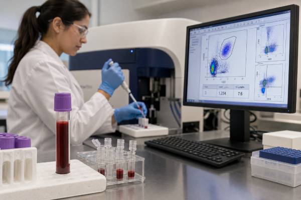
A natural killer cell count test measures the number and percentage of circulating NK cells, usually by flow cytometry. Most laboratories identify NK cells as lymphocytes that lack CD3 and express CD16, CD56, or both. The result can help evaluate unusual or severe viral infections, selected inborn errors of immunity, immune recovery after transplantation, treatment effects, or persistent lymphocytosis. It does not directly show whether NK cells can recognize, degranulate against, or kill target cells. A person may have a normal count but impaired NK-cell function, while a low count during acute illness, corticosteroid use, chemotherapy, or temporary cell redistribution may not represent a primary NK-cell deficiency. Interpretation requires the absolute count, percentage, age-specific reference interval, other lymphocyte subsets, infection history, medicines, and often repeat testing. True natural killer cell deficiency is rare and is considered most strongly when low NK-cell numbers or defective function persist and match a characteristic clinical pattern, especially severe, recurrent, or difficult-to-control herpesvirus or papillomavirus disease.
- NK cells are generally CD3-negative and CD16-positive, CD56-positive, or both.
- The absolute count and percentage should be reviewed together because either can be misleading alone.
- NK-cell quantity is not the same as cytotoxicity, degranulation, cytokine production, or antibody-dependent killing.
- Acute infection, stress, medicines, age, and specimen handling can change the result.
- Persistent abnormalities require confirmation and evaluation for secondary causes before a rare genetic deficiency is diagnosed.
Table of Contents
- NK Cells and Their Role in Immune Defense
- How an NK Cell Count Is Measured
- When the Test Is Useful
- What a Low NK Cell Count Can Mean
- What a High NK Cell Count Can Mean
- Why Count and Function Are Different
- Age, Timing, Medicines, and Testing Pitfalls
- Follow-Up Testing and Practical Interpretation
NK Cells and Their Role in Immune Defense
Natural killer cells are innate lymphocytes that respond quickly to stressed, infected, or transformed cells. Unlike conventional T cells, they do not require one unique rearranged antigen receptor to recognize a specific target. Instead, they integrate signals from many activating and inhibitory receptors. Healthy cells normally display molecules that engage inhibitory NK receptors, while infected or malignant cells may lose those protective signals or gain stress ligands that favor activation.
When activation outweighs inhibition, NK cells can release perforin and granzymes. Perforin helps create access to the target cell, and granzymes initiate programmed cell death. NK cells also express death-inducing ligands and release cytokines such as interferon-gamma, which helps coordinate macrophages and other immune cells. These mechanisms are important early in infection, before a fully expanded antigen-specific T-cell response develops.
CD16 allows many NK cells to perform antibody-dependent cellular cytotoxicity. CD16 binds the Fc portion of antibodies already attached to a target. This connects antibody recognition to NK-cell killing and is one reason NK cells participate in responses to some infections, therapeutic monoclonal antibodies, and antibody-coated tumor cells.
NK cells are especially associated with defense against herpesviruses, including herpes simplex virus, varicella-zoster virus, cytomegalovirus, and Epstein–Barr virus. They also contribute to control of human papillomaviruses and other pathogens. However, viral defense is shared with T cells, antibodies, interferons, macrophages, and tissue barriers. Recurrent viral infection does not automatically mean an NK-cell disorder.
NK cells also influence pregnancy, tissue remodeling, inflammation, autoimmunity, and cancer surveillance. Uterine NK cells have a specialized phenotype and role in placental development; a routine blood count does not measure them. Similarly, circulating NK-cell numbers do not directly describe NK cells inside the liver, lungs, lymph nodes, tumors, or other tissues.
Most blood NK cells are CD56-dim and usually express CD16 strongly. They are commonly associated with cytotoxic activity and antibody-dependent killing. A smaller CD56-bright population produces cytokines efficiently and often has lower or absent CD16. Expanded phenotyping can separate these populations, but a basic NK count usually combines them.
How an NK Cell Count Is Measured
The test is performed on anticoagulated whole blood using flow cytometry. Fluorescent antibodies bind surface markers, and the instrument analyzes individual cells as they pass through a laser. Laboratories first identify viable leukocytes and the lymphocyte region, often using CD45 and light-scatter characteristics, then exclude T cells with CD3 and sometimes exclude B cells with CD19.
A common definition is CD45-positive, CD3-negative, CD19-negative lymphocytes expressing CD16, CD56, or both. Other laboratories use CD3-negative CD16/CD56-positive gates without CD19 exclusion. The report should state the marker combination because small differences can affect the population included.
Results are usually reported as the percentage of lymphocytes that are NK cells and the absolute number per microliter. The percentage describes composition; the absolute count estimates how many NK cells are present in a volume of blood. Absolute numbers may be measured directly with counting beads or calculated by combining the flow-cytometry percentage with the absolute lymphocyte count from a complete blood count.
Both values matter. A patient with broad lymphopenia can have a normal NK percentage but a low absolute NK count. A high NK percentage may simply reflect loss of T or B cells while the absolute NK number remains normal. For evaluation of deficiency, a persistently low absolute count generally carries more direct meaning than percentage alone, but the complete pattern must be reviewed.
Expanded panels can report CD56-bright/CD16-low and CD56-dim/CD16-positive populations, NKT-like cells, activating and inhibitory receptors, maturation markers, or intracellular perforin and granzyme. The label “NKT cell” is used inconsistently in clinical reports. Some laboratories report CD3-positive CD16/CD56-positive cells, which are T cells expressing NK-associated markers and are not conventional CD3-negative NK cells.
Quality depends on proper gating, adequate cell events, specimen viability, and timely transport. Delayed processing can change CD16 or CD56 expression and reduce cell recovery. Clots, extreme temperatures, old specimens, or severe leukopenia may make enumeration unreliable. A precise-looking number does not remove uncertainty when only a small number of NK events were collected.
An NK enumeration panel is not designed to diagnose NK-cell leukemia or lymphoma. Persistent unexplained lymphocytosis, abnormal large granular lymphocytes, cytopenias, organ enlargement, or a mass requires diagnostic hematopathology with morphology, broader immunophenotyping, molecular studies, and often tissue evaluation.
When the Test Is Useful
Clinicians may order an NK cell count when the infection pattern suggests a cellular immune problem. The most characteristic concern is unusually severe, recurrent, persistent, or treatment-resistant herpesvirus or papillomavirus infection. Examples include disseminated varicella, severe recurrent herpes, uncontrolled cytomegalovirus, extensive warts, or complications that are disproportionate to the person’s age and known medical conditions.
The test is also part of a lymphocyte subset panel used to evaluate suspected inborn errors of immunity. T-, B-, and NK-cell patterns help narrow pathways involved in severe combined immunodeficiency and related disorders. In newborns or infants with opportunistic infection, chronic diarrhea, poor growth, absent thymic output, or an abnormal newborn screen, the entire lymphocyte profile is more informative than an isolated NK count.
After hematopoietic cell transplantation, NK cells often reappear earlier than T and B cells. Enumeration may help describe immune reconstitution, although a recovered number does not prove normal receptor diversity or function. Similar monitoring may be used after solid-organ transplantation, chemotherapy, cellular therapy, or selected immunosuppressive regimens.
In HIV, cancer, autoimmune disease, and chronic viral illness, research and specialized clinical panels may include NK counts or subsets. These results are usually supportive rather than decisive. They should not be used as a general wellness score, a measure of “immune strength,” or a stand-alone explanation for fatigue, frequent common colds, infertility, or nonspecific inflammatory symptoms.
Some laboratories offer NK counts in reproductive immunology. Peripheral-blood NK cells are biologically different from uterine NK cells, and a blood result does not directly measure implantation-site immunity. Major reproductive decisions should not be based on an isolated peripheral NK percentage without evidence-based clinical guidance.
High or low counts may also be discovered incidentally on flow cytometry performed for another reason. The next step depends on whether the finding is transient, whether other blood-cell lines are abnormal, and whether symptoms suggest infection, inflammation, marrow disease, treatment effect, or clonal lymphoproliferation.
Testing is most useful when it answers a defined question: Are NK cells persistently absent or reduced? Is the abnormality isolated or part of broader lymphopenia? Has the population recovered after treatment? Does the phenotype justify a function assay or genetic evaluation? Without such a question, minor deviations are likely to generate confusion rather than useful care.
What a Low NK Cell Count Can Mean
A low NK count has many secondary causes. Acute viral infection, severe bacterial illness, critical illness, surgery, stress hormones, malnutrition, protein loss, bone marrow suppression, chemotherapy, radiation, corticosteroids, and other immunosuppressive medicines can reduce circulating numbers. Cells can also leave the blood and enter tissues, producing temporary lymphopenia without permanent loss of the total NK-cell pool.
Broad lymphopenia points toward a different process from isolated NK-cell reduction. The complete blood count, total lymphocyte count, CD3 T cells, CD4 and CD8 subsets, and CD19 B cells show whether multiple populations are affected. Cytopenias in platelets, neutrophils, or red cells raise concern for marrow failure, severe infection, malignancy, medication toxicity, or systemic disease.
True natural killer cell deficiency is rare. It is generally considered only when the numerical or functional defect is stable over time, secondary causes have been excluded, and the clinical phenotype is compatible. Historically, “classical” or developmental NK-cell deficiency describes very low or absent NK cells, while “functional” NK-cell deficiency describes normal numbers with impaired killing. Modern genetic classification is more specific because several disorders affect NK development, maturation, signaling, or cytotoxic machinery.
GATA2-related disease can cause marked loss of NK cells along with monocytes, B cells, or dendritic cells and can be associated with viral infections, myelodysplasia, leukemia risk, lymphedema, and other features. Other genetic conditions affect NK-cell development more selectively or occur within broader combined immunodeficiencies. The phenotype and associated cell abnormalities guide gene selection.
Low NK cells are also seen in severe combined immunodeficiency patterns. For example, some cytokine-signaling defects produce low T and NK cells with preserved B-cell numbers, while recombination defects may produce low T and B cells with preserved NK cells. These patterns are not diagnoses, but they help prioritize urgent functional and genetic studies.
A single low count during illness should usually be repeated after recovery when clinically safe. Persistent severe infection, an abnormal newborn screen, or profound combined lymphopenia should not wait for routine repetition. The degree of urgency depends on age, organism, organ involvement, other lymphocyte populations, and whether the patient is clinically stable.
Low blood NK numbers do not predict every infection. Some people with very low counts remain relatively well, while others with normal counts have defective degranulation or receptor signaling. Vaccination history, antiviral treatment, immunoglobulin levels, T-cell responses, and the full infection record are essential context.
What a High NK Cell Count Can Mean
A high NK percentage or absolute count is often reactive. Viral infections can expand or redistribute NK populations, and the pattern may persist during recovery. Inflammation, smoking, physiologic stress, and immune reconstitution after treatment can also alter circulating numbers. The timing of collection relative to symptoms matters.
A high percentage does not necessarily mean increased NK-cell mass. If T cells fall, NK cells can occupy a larger share of the remaining lymphocytes while the absolute NK count stays normal. Reports should therefore be read with the total lymphocyte count and all major subsets.
Persistent NK-cell lymphocytosis deserves a different evaluation from a transient rise. Large granular lymphocytes include cytotoxic T cells and NK cells. An expanded NK-like population may be polyclonal or reactive, but chronic lymphoproliferative disorders of NK cells and aggressive NK-cell malignancies are possible in the correct clinical setting.
Warning features include persistent marked lymphocytosis, anemia, neutropenia, thrombocytopenia, constitutional symptoms, liver or spleen enlargement, abnormal liver tests, hemophagocytic features, or an atypical population on smear. A routine enumeration test cannot determine whether cells are clonal or malignant. Diagnostic evaluation may require a broader flow panel, killer-cell immunoglobulin-like receptor patterns, Epstein–Barr virus testing, STAT3 or other molecular studies, marrow examination, and tissue biopsy.
High NK counts do not prove enhanced antiviral or anticancer protection. Expanded cells may be immature, chronically stimulated, exhausted, dysfunctional, or clonal. Their receptor repertoire and ability to degranulate can matter more than quantity.
Treatment context is also important. Some cytokines and cellular therapies can expand NK cells, while recovery after transplantation may produce a temporary numerical predominance. A value that is expected under a protocol may not represent disease, but it still needs to be interpreted with symptoms and other blood findings.
Why Count and Function Are Different
Enumeration asks how many cells match a surface-marker definition. Function testing asks what those cells do after stimulation. The two results can disagree because NK activity depends on receptors, signaling proteins, cytotoxic granules, cell maturation, metabolic state, and the target-cell interaction—not simply the number of cells present.
An NK cell function test may measure direct cytotoxicity against standardized target cells. Other assays assess degranulation by measuring surface CD107a after stimulation, intracellular perforin or granzyme, interferon-gamma production, or receptor expression. Each test examines a different step and has its own specimen and transport requirements.
A low cytotoxicity result can reflect too few NK cells in the tested sample rather than poor function per cell. Laboratories may normalize or interpret activity in relation to the NK-cell percentage, but methods differ. Conversely, a normal bulk cytotoxicity result can sometimes mask a small abnormal subset or a defect that appears only with a specific stimulus.
Functional assays are especially vulnerable to delays, temperature changes, acute illness, corticosteroids, and other treatments. Fresh blood is often required, and shipping over a weekend can invalidate the result. A low value should be confirmed under appropriate conditions and interpreted with controls before a genetic conclusion is made.
Familial HLH and related disorders can involve cytotoxic pathways shared by NK cells and CD8 T cells. Screening may include perforin expression and CD107a degranulation rather than a general NK count alone. The clinical question—developmental NK deficiency, impaired killing, degranulation defect, or hyperinflammation—determines the correct assay.
This distinction prevents two common errors. A normal count does not rule out functional NK deficiency, and a low count does not automatically prove defective killing. Both quantity and quality may need assessment when the infection history is compelling.
Age, Timing, Medicines, and Testing Pitfalls
NK-cell reference intervals vary by age and laboratory. Infants and children have changing lymphocyte numbers as the immune system develops, so adult ranges should not be applied. Older age also changes subset composition, receptor expression, and function. The report’s age-specific interval is the correct starting point, not a number found on another laboratory’s website.
Time of day, recent exercise, acute stress, sleep, fever, and smoking can shift circulating lymphocytes. Pregnancy changes immune distribution, and blood NK cells do not represent uterine NK cells. Minor deviations are often best interpreted as trends rather than fixed traits.
Medicines can suppress production, induce cell death, block signaling, or change lymphocyte trafficking. Corticosteroids may rapidly lower circulating lymphocytes. Chemotherapy, radiation, antithymocyte globulin, calcineurin inhibitors, JAK inhibitors, and other agents can affect NK numbers or activity. Growth factors and cytokine therapies may produce different patterns.
Monoclonal antibodies can also complicate marker detection. CD16 can be shed after activation, and therapy or immune complexes may change receptor expression. A gate requiring one marker too strictly could undercount cells whose phenotype has shifted. Laboratories commonly use CD16 and CD56 together to reduce this problem.
Specimen age is crucial. NK cells remain measurable longer than some functions remain intact, so enumeration may be acceptable when a cytotoxicity assay is no longer valid. However, prolonged delay still reduces viability and changes surface markers. The collection tube, temperature, transport schedule, and laboratory acceptance criteria must be followed.
Reference intervals describe most healthy people, not a diagnostic boundary. Some healthy individuals fall slightly outside them. Conversely, a value within range can be abnormal for a patient whose previous count was much higher or whose function is defective. Clinical significance depends on persistence, magnitude, associated infections, and other immune findings.
Commercial “immune profiling” panels may attach broad wellness claims to NK numbers. A circulating count cannot establish chronic viral reactivation, explain nonspecific symptoms, predict cancer, or determine whether supplements are working. Results should be tied to a recognized clinical indication and interpreted by a clinician familiar with flow cytometry and immune deficiency.
Follow-Up Testing and Practical Interpretation
Start by confirming what the report measured. Check whether NK cells were defined as CD3-negative CD16/CD56-positive cells, whether CD19 was excluded, whether absolute counts and percentages are both provided, and whether a subset panel shows T and B cells. Review sample viability and any laboratory comments.
Next, place the result in time. Was the person acutely ill, taking corticosteroids, receiving chemotherapy, recovering from transplantation, or sampled soon after strenuous exercise? Compare with earlier complete blood counts and flow results. A repeat sample after recovery can distinguish a transient shift from a persistent defect when the clinical situation allows.
The infection history should be specific. Record organisms, sites, severity, age at onset, recurrence, need for hospitalization, response to antiviral treatment, vaccine complications, extensive warts, and family history. Ordinary childhood respiratory infections without unusual severity are less suggestive than disseminated herpesvirus or persistent papillomavirus disease.
Companion testing may include complete blood count and smear, quantitative immunoglobulins, vaccine antibody responses, T- and B-cell subsets, HIV testing when appropriate, viral PCR, liver and kidney studies, and evaluation for protein loss or marrow disease. If HLH is suspected, ferritin, triglycerides, fibrinogen, soluble CD25, cytotoxic-protein expression, and degranulation testing may be urgent.
Persistent isolated or combined abnormalities can lead to expanded NK phenotyping, cytotoxicity and CD107a assays, perforin/granzyme studies, chromosomal testing, targeted gene panels, exome or genome sequencing, or marrow assessment. Genetic counseling is valuable when an inherited disorder is possible.
For high counts, determine whether the rise is reactive or persistent. A smear and diagnostic flow cytometry are appropriate when lymphocytosis, cytopenias, organ enlargement, or constitutional symptoms suggest a clonal process. Routine NK enumeration should not be repeatedly ordered as a substitute for hematopathology.
The most useful interpretation is a pattern statement rather than “high” or “low”: the absolute NK count is persistently reduced or increased; the percentage is or is not distorted by other lymphocytes; T and B cells are preserved or affected; the result was or was not obtained during illness or treatment; and the infection phenotype does or does not match an NK-cell disorder. That approach directs the next test while avoiding a diagnosis based on one number.
References
- Clinical, immunologic, and genetic characteristics of 148 patients with natural killer cell deficiency 2025 (Research Article)
- Natural Killer Cell Assays: Clinical Applications and Future Perspectives 2026 (Review)
- Comprehensive snapshots of natural killer cells functions, molecular mechanisms and therapeutics 2024 (Review)
- NK cell subsets and dysfunction during viral infection 2023 (Review)
- Natural Killer Cells Enumeration 2026 (Laboratory Test Guide)
- Natural Killer (NK)/Natural Killer T-Cell Subsets, Blood 2026 (Laboratory Test Guide)
Disclaimer
This article is for general education and does not diagnose natural killer cell deficiency, infection, HLH, leukemia, or lymphoma. NK-cell results require age-specific ranges, repeat confirmation when appropriate, and interpretation with the infection history, medicines, other lymphocyte subsets, and functional testing. Severe or disseminated viral infection, rapidly worsening illness, or profound combined lymphopenia requires prompt specialist care.





