
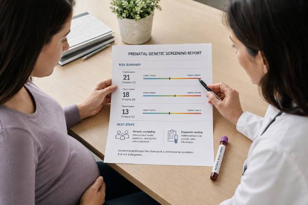
Prenatal genetic screening estimates the chance that a pregnancy is affected by selected chromosome conditions; it does not confirm whether the fetus has one. Screening options include cell-free DNA testing, first-trimester combined screening, second-trimester serum screening, and ultrasound. They differ in timing, conditions assessed, accuracy, cost, and the information they provide. Cell-free DNA screening is the most accurate screening method for common trisomies, but it analyzes DNA fragments released mainly by the placenta and can still produce false-positive, false-negative, or no-result outcomes. Serum tests combine pregnancy-related markers with maternal factors to calculate risk, while ultrasound can identify structural findings and markers that no blood test replaces. Every pregnant patient should have the opportunity to discuss screening and diagnostic testing, regardless of age or baseline risk, and may accept or decline either. The most useful choice depends on what the patient wants to know and what they would do with the information. A high-chance result should be confirmed with chorionic villus sampling or amniocentesis before irreversible decisions.
- Prenatal screening provides a probability, not a diagnosis.
- Cell-free DNA is the most sensitive and specific screening test for trisomies 21, 18, and 13, but positive results still require diagnostic confirmation.
- Patients should generally choose one screening strategy rather than layering multiple independent blood screens.
- Ultrasound remains necessary even after a low-chance cell-free DNA result.
- The positive predictive value varies by condition, population prevalence, and individual circumstances.
- A no-result cell-free DNA test is not automatically reassuring and needs clinical follow-up.
Table of Contents
- What prenatal screening can and cannot tell you
- Main screening options and timing
- How cell-free DNA screening works
- Understanding risk and test performance
- Interpreting low-, high-, and no-result reports
- What happens after an abnormal screen
- Choosing a screening strategy
What prenatal screening can and cannot tell you
Prenatal genetic screening asks whether a pregnancy has a higher or lower chance of selected chromosome conditions than a defined threshold. The conditions most consistently assessed are trisomy 21, trisomy 18, and trisomy 13. Some screening programs also include sex chromosome aneuploidies, open neural tube defects, or selected placental and pregnancy risks. Expanded commercial panels may report microdeletions, rare autosomal trisomies, or genome-wide copy-number changes, but evidence and professional recommendations are not equally strong for every add-on.
A screening result is not a direct fetal diagnosis. Even cell-free DNA, sometimes marketed as noninvasive prenatal testing or NIPT, is more accurately described as screening. Most DNA fragments measured in maternal plasma come from the placenta. The placenta and fetus usually share the same chromosome complement, but they can differ because of confined placental mosaicism. Maternal chromosome variation, a vanished twin, organ transplant, maternal malignancy, laboratory factors, and low fetal fraction can also affect results.
Screening has two legitimate outcomes: it may help a patient avoid invasive testing after a reassuring result, or it may identify a pregnancy for which diagnostic testing deserves consideration. It can also help plan the timing of detailed ultrasound or consultation. It should not be presented as a compulsory step toward diagnosis.
Diagnostic tests answer a different question. Chorionic villus sampling and amniocentesis obtain placental or fetal cells for chromosome or DNA analysis. They can diagnose the conditions for which testing is performed, although every laboratory method has limits. The distinction between prenatal screening and diagnosis is crucial: a high-chance screen changes probability, while a diagnostic result evaluates the sampled cells directly.
A low-chance screen does not guarantee a chromosomally typical or healthy baby. It reduces the chance of the specific conditions assessed. It does not exclude all chromosome abnormalities, single-gene disorders, birth defects, developmental conditions, pregnancy complications, or problems that appear later. The test menu should be read carefully because “genetic screening” can imply a broader assessment than the laboratory actually performs.
Ultrasound is complementary rather than redundant. A first-trimester scan can establish viability, gestational age, fetal number, and major early abnormalities. A detailed mid-trimester anatomy scan evaluates structures that blood screening cannot. A low-chance cell-free DNA result should not be used to skip recommended ultrasound.
Main screening options and timing
Cell-free DNA screening can generally be performed from about 10 weeks of pregnancy. A maternal blood sample is analyzed for chromosome representation. It has the best screening performance for trisomies 21, 18, and 13 and is available in singleton and many twin pregnancies. The exact panel, eligibility, and performance in higher-order multiples, donor-egg pregnancies, vanishing twins, or other complex situations vary.
First-trimester combined screening typically uses maternal age, a blood test measuring pregnancy-associated plasma protein A and free beta-human chorionic gonadotropin, and an ultrasound measurement of nuchal translucency. It is performed within a defined first-trimester window. It provides a risk estimate for common trisomies and may offer broader clues about placental function or fetal development, depending on the program.
Second-trimester serum screening may be a triple or quad screen. The quad screen measures alpha-fetoprotein, human chorionic gonadotropin, unconjugated estriol, and inhibin A. It estimates risk for trisomy 21 and trisomy 18 and can identify increased risk for open neural tube defects through alpha-fetoprotein. It is less accurate for common trisomies than cell-free DNA but remains useful when first-trimester screening was not performed, cfDNA is unavailable, or neural tube defect screening is needed.
Integrated or sequential screening combines first- and second-trimester information under a specific protocol. In an integrated strategy, a final risk may not be released until the second-trimester component is complete. Sequential strategies may release an early result and refine risk later. These programs should not be confused with independently ordering multiple screens and trying to reconcile conflicting reports.
Ultrasound screening includes nuchal translucency assessment and the second-trimester anatomy scan. Ultrasound can identify structural anomalies, soft markers, and growth concerns. Certain findings may lead directly to an offer of diagnostic testing even when blood screening is low chance.
Carrier screening is separate. It evaluates whether prospective parents carry variants for inherited conditions such as cystic fibrosis, spinal muscular atrophy, or hemoglobin disorders. Carrier screening does not estimate the same chromosome risks as cfDNA or serum screening. A pregnancy can have low-chance aneuploidy screening while still being at risk for a single-gene condition inherited from the parents.
Timing matters because each option has a testing window and because later results leave less time for confirmatory testing or decision-making. Prenatal counseling is most useful early, but patients presenting later should still receive options appropriate to gestational age.
How cell-free DNA screening works
During pregnancy, short DNA fragments circulate in maternal blood. The placental contribution is often called fetal DNA, although the fragments primarily arise from trophoblast cells in the placenta. The proportion of placental fragments in the total sample is the fetal fraction.
Laboratories use several methods. Massively parallel sequencing counts fragments from each chromosome. Targeted sequencing focuses on selected chromosomes or regions. SNP-based approaches examine patterns of genetic markers and may estimate fetal fraction or distinguish maternal and placental contributions differently. These methods are not interchangeable, and their capabilities in twins, donor conceptions, triploidy, and vanishing-twin pregnancies differ.
For common trisomies, the laboratory looks for a small excess of fragments from a chromosome. If chromosome 21 material is overrepresented beyond the model’s threshold, the result may be reported as high chance for trisomy 21. Because the signal is statistical and placental, it is not equivalent to counting chromosomes in fetal cells.
Adequate fetal fraction is important. It generally rises with gestational age but is influenced by maternal weight, medications, sample handling, placental biology, and fetal chromosome status. A low fraction may lead to a no-call result. Some laboratories use proprietary risk algorithms when fetal fraction is low, while others do not report. The report should state whether a result was generated and, when available, the fetal fraction.
Test performance is strongest for trisomy 21, followed by trisomy 18 and trisomy 13. Performance for sex chromosome conditions is more variable because maternal X-chromosome mosaicism, age-related X loss, placental mosaicism, and biological diversity can contribute to discordance. Screening for 22q11.2 deletion and other microdeletions has lower population prevalence and more challenging validation; professional recommendations differ on whether selected screening should be offered, but routine population-wide microdeletion screening is not uniformly recommended.
Cell-free DNA may incidentally reveal a maternal chromosome finding or a pattern suggesting maternal disease. Such results are rare and are not a cancer screening program. An unusual multiple-aneuploidy pattern may prompt maternal evaluation after specialist review, but it should not be interpreted without clinical context.
The phrase “99% accurate” is incomplete and potentially misleading. A detection rate near 99% for trisomy 21 does not mean that 99% of positive results are true. The probability that a positive result is correct depends on the condition’s prevalence and the patient’s prior chance, as well as test sensitivity and specificity.
Understanding risk and test performance
Several measures describe screening performance, and each answers a different question.
Sensitivity, or detection rate, is the proportion of affected pregnancies that receive a high-chance result. Specificity is the proportion of unaffected pregnancies that receive a low-chance result. False-positive rate is the proportion of unaffected pregnancies classified as high chance. These are characteristics of the test in a defined population.
Positive predictive value, or PPV, is the probability that a pregnancy with a high-chance result is actually affected. PPV rises when the condition is more common in the tested population and falls when it is rare. A high-chance result for trisomy 21 generally has a higher PPV than a high-chance result for a rare microdeletion, even if both laboratory reports use similar wording.
Negative predictive value, or NPV, is the probability that a pregnancy with a low-chance result is unaffected by the screened condition. NPV is usually very high for common aneuploidies because these conditions are uncommon and cfDNA detection is strong, but it is not 100%.
A result may be presented as a ratio, such as 1 in 50, or as a category above or below a cutoff. “High risk” does not necessarily mean more likely affected than unaffected. For example, a 1-in-20 result is high compared with a screening cutoff, yet 19 of 20 pregnancies with that individual calculated risk would be expected not to have the condition. Cell-free DNA reports may use “high probability,” “aneuploidy detected,” or a PPV estimate; the wording should not replace the underlying concept.
Maternal age affects the baseline chance of common trisomies, but age should not determine whether screening is offered. Current guidance supports offering both screening and diagnostic options to all pregnant patients. Age can influence PPV because it changes prior prevalence, but a younger patient can have an affected pregnancy and an older patient can have an unaffected one.
Twins require separate counseling. CfDNA performs well for common trisomies in many twin pregnancies, but fetal fraction and the contribution of each placenta complicate interpretation. If a high-chance result occurs, the screen may not identify which fetus is affected. Vanishing twins can contribute DNA for weeks and cause discordant results.
Risk estimates are not interchangeable across laboratories or methods. Comparing “low risk” from one test with “negative” from another without understanding their panels and thresholds can create false certainty. The result should be interpreted with the test name, gestational age, ultrasound, pregnancy type, and indication.
Interpreting low-, high-, and no-result reports
A low-chance result substantially reduces the probability of the conditions screened. Routine prenatal care and recommended ultrasound should continue. Diagnostic testing remains available if later ultrasound findings, family history, or personal preference justify it. A patient who wants near-definitive chromosome information may choose CVS or amniocentesis even after a low-chance screen.
A high-chance result means the screening signal exceeded the laboratory threshold. The next steps are genetic counseling, detailed ultrasound when appropriate, and an offer of diagnostic testing. The choice between CVS and amniocentesis depends on gestational age, the suspected condition, ultrasound findings, and the possibility of placental mosaicism. For some results, amniocentesis may better distinguish a placental finding from the fetal chromosome complement.
A no-result, no-call, or test failure means the laboratory could not issue a reliable classification. Common reasons include low fetal fraction, early gestational age, sample problems, technical quality, or an unusual genomic pattern. A no-result is not the same as low chance. Some aneuploidies and placental abnormalities are associated with test failure, so current guidance supports counseling, ultrasound evaluation, and an offer of diagnostic testing. Repeating cfDNA may succeed, but whether to repeat depends on gestational age, ultrasound, maternal factors, and how urgently an answer is needed.
An atypical or outside-the-scope result means the data do not fit the test’s standard model. It may reflect maternal or placental variation, mosaicism, a chromosome change not targeted by the assay, or technical noise. The laboratory and genetics team may recommend maternal testing, diagnostic testing, or no further action based on the pattern. Atypical results should not be translated into a specific fetal diagnosis without evidence.
A sex discrepancy can occur when reported sex chromosome information differs from ultrasound or expectations. Causes include gestational dating, fetal or placental sex chromosome variation, vanishing twin, maternal variation, differences of sex development, sample error, or an ultrasound misclassification. The appropriate response is confidential, nonjudgmental review and confirmatory evaluation—not assumptions.
Serum screening reports use calculated risk and may separately report increased alpha-fetoprotein. Elevated AFP can be associated with open neural tube defects, abdominal wall defects, incorrect dating, multiple gestation, placental problems, or other causes. Follow-up usually begins with a targeted ultrasound and may include amniotic fluid testing depending on the situation.
What happens after an abnormal screen
The first step is to confirm exactly what was screened and what the report says. “Positive prenatal test” is too vague. The clinician should review the condition, method, fetal fraction or analyte values, PPV if supplied, gestational age, fetal number, ultrasound, and relevant maternal history.
Genetic counseling converts the population result into an individualized discussion. It explains the chance that the result is true, possible reasons for discordance, diagnostic options, procedure timing, and the range of outcomes associated with the condition. Counseling should be nondirective and should include the option to decline additional testing.
CVS samples placental tissue, usually earlier in pregnancy. It can provide an earlier diagnostic result but may occasionally detect confined placental mosaicism. Amniocentesis samples amniotic fluid, usually later, and more directly reflects fetal cells. The choice is not simply “earlier versus safer”; it depends on the chromosome condition and clinical context. The CVS diagnostic test and amniocentesis diagnostic test have different sampling considerations.
The laboratory test on the invasive sample should match the question. Rapid aneuploidy testing can address common trisomies quickly. Karyotype can show chromosome number and structure. Chromosomal microarray can detect smaller deletions and duplications. A targeted assay may be required for a suspected microdeletion or familial variant. Sometimes more than one method is appropriate.
Ultrasound can support counseling but cannot confirm or exclude most chromosome conditions by itself. Some affected fetuses have no detectable structural anomaly, while some unaffected fetuses have soft markers. A normal ultrasound after a high-chance cfDNA result lowers concern for certain severe phenotypes but does not erase the screening signal.
Patients should not be pressured to make irreversible decisions from a screening result alone. Professional guidance consistently recommends diagnostic confirmation when results would influence pregnancy termination or other major management. If diagnostic testing is declined, care can still be tailored with imaging, pediatric consultation, delivery planning, and postnatal testing.
A discordant diagnostic result may require review. Potential explanations include placental mosaicism, maternal findings, vanishing twin, sample mix-up, or a false-positive screen. The genetics team may communicate with the screening laboratory and recommend additional maternal or placental studies.
Choosing a screening strategy
There is no single best screening test for every patient. A useful decision starts with goals. Does the patient primarily want the most accurate screen for common trisomies? Is early timing important? Is information about open neural tube defects needed? Would uncertain findings from expanded panels create more harm than benefit? Would the patient choose diagnostic testing regardless of screening?
For many singleton and twin pregnancies, cfDNA is the preferred screen for trisomies 21, 18, and 13 because of its performance. First-trimester combined or serum screening remains a valid option where available. Patients should generally avoid simultaneous independent aneuploidy screens, because multiple tests can increase false-positive results and produce confusing discordance. Serum AFP or ultrasound for neural tube defects can still complement cfDNA because it addresses a different clinical question.
Expanded cfDNA menus deserve careful scrutiny. A laboratory’s ability to report a condition does not prove that population screening provides net benefit. Rare-condition PPVs can be modest, clinical validation may be limited, and confirmatory testing can be complex. SMFM’s 2025 guidance recommends against routine population screening for microdeletions and advises that patients interested in fetal copy-number variants be offered diagnostic testing rather than relying on cfDNA screening.
Cost and access affect choice but should not be confused with medical superiority. Insurance coverage, public programs, laboratory contracts, and local ultrasound availability vary. Patients deserve transparent information about out-of-pocket cost, test scope, and what follow-up would cost if a result is abnormal.
Personal values are central. Some people want information for preparation but would not consider invasive testing. Others want diagnostic certainty early. Some prefer not to know about sex chromosome conditions or adult-onset implications. Screening can be declined entirely without abandoning good prenatal care.
A screening plan should include the response to each possible result before blood is drawn. Knowing whether a high-chance, low-chance, or no-result outcome would lead to diagnostic testing helps determine whether screening is the right first step. This anticipatory approach turns prenatal genetic screening from a routine checkbox into an informed choice.
References
- Screening for Fetal Chromosomal Abnormalities (2026), ACOG practice advisory.
- Society for Maternal-Fetal Medicine Consult Series #74: Cell-Free DNA Screening for Aneuploidies: Updated Guidance (2025), clinical guideline.
- Noninvasive Prenatal Screening for Fetal Chromosome Abnormalities in a General-Risk Population: An Evidence-Based Clinical Guideline of the ACMG (2023), evidence-based guideline.
- Screening and Diagnosis of Fetal Structural Anomalies and Chromosome Conditions (C-Obs 35) (2024), clinical guideline.
- Prenatal Screening and Diagnostic Testing for Fetal Chromosomal Abnormalities (2024), health-system guideline.
- Prenatal Genetic Screening Tests (2025), patient guidance.
Disclaimer
This article provides general educational information and does not replace personalized prenatal care or genetic counseling. Screening availability, test panels, thresholds, follow-up recommendations, and laws vary by location. A qualified clinician should interpret any result using the specific laboratory report, gestational age, ultrasound findings, pregnancy history, and patient preferences.





