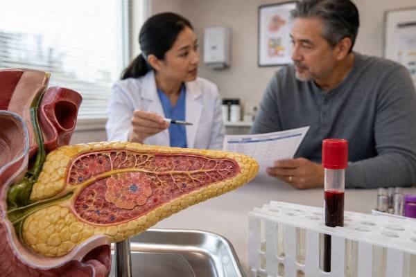
A proinsulin blood test measures the precursor molecule that pancreatic beta cells process into insulin and C-peptide. Healthy beta cells convert most proinsulin before secretion, so only a small amount circulates. Higher levels can occur when beta cells are under pressure from insulin resistance, when kidney clearance is reduced, with rare defects in prohormone processing, or when an insulin-secreting pancreatic neuroendocrine tumor releases incompletely processed hormone. The test has its clearest diagnostic role during confirmed hypoglycemia: proinsulin should be strongly suppressed when glucose is low, so an inappropriately detectable or elevated result can support endogenous hyperinsulinism. It must be interpreted with plasma glucose, insulin, C-peptide, beta-hydroxybutyrate, kidney function, and a sulfonylurea or meglitinide screen. A fasting high value obtained while glucose is normal is not proof of insulinoma, and a normal result does not exclude every tumor. Reference intervals, specimen requirements, and cutoffs differ by assay. Proinsulin is a specialized test, not a routine substitute for glucose, A1C, or standard diabetes evaluation.
- Proinsulin is the intact precursor that is normally split into insulin and C-peptide inside beta cells.
- During true hypoglycemia, proinsulin should be low; failure to suppress supports continued endogenous beta-cell secretion.
- High fasting proinsulin with normal or high glucose may reflect insulin resistance or beta-cell stress rather than a tumor.
- Kidney failure can raise proinsulin, insulin, and C-peptide and complicate interpretation.
- Insulinoma evaluation requires a complete critical sample and medication screen, not a proinsulin result alone.
Table of Contents
- What Proinsulin Is
- Why the Test Is Ordered
- Preparation and Specimen Collection
- Normal Range and Cutoffs
- High Proinsulin Levels
- Proinsulin in Hypoglycemia and Insulinoma
- Proinsulin in Diabetes and Beta-Cell Health
- Limitations, Follow-Up, and Next Steps
What Proinsulin Is
Insulin production begins as preproinsulin, a single protein made in the rough endoplasmic reticulum of pancreatic beta cells. A signal peptide is removed to create proinsulin. The molecule folds and forms disulfide bonds that align the future A and B chains of insulin. It then moves through the Golgi apparatus into secretory granules.
Inside those granules, prohormone convertases and carboxypeptidase enzymes cut proinsulin at two sites. This processing produces mature insulin and C-peptide. The beta cell stores the products and releases them when glucose and other signals stimulate secretion.
Most proinsulin is processed before release, but a small fraction enters the circulation intact or as partially split intermediates. A proinsulin assay may be designed to measure intact proinsulin specifically or may cross-react to some degree with intermediates. The report should be interpreted according to the exact method.
Proinsulin differs from insulin in several important ways:
- It has much lower glucose-lowering biological activity than mature insulin.
- It remains in circulation longer.
- Its concentration is usually much lower than insulin after a meal.
- Disproportionate release can indicate inefficient beta-cell processing.
- It is produced only through endogenous beta-cell synthesis; pharmaceutical insulin does not contain proinsulin.
C-peptide is another product of the same processing pathway. Unlike insulin, it undergoes little first-pass liver extraction and is cleared mainly by the kidneys. Measuring all three peptides can reveal whether insulin activity comes from the pancreas and whether processing appears normal.
Beta cells may release more proinsulin when demand is high. In insulin resistance, the cells increase production to maintain glucose. Stress in the endoplasmic reticulum, rapid granule turnover, impaired convertase activity, or loss of mature granule stores can increase the proportion of immature hormone entering blood. This is why proinsulin is studied as a marker of beta-cell strain.
A tumor can disrupt processing more dramatically. Insulinomas often secrete proinsulin and insulin inappropriately even when glucose is low. Some rare tumors secrete predominantly proinsulin and may cause hypoglycemia with only modest measured insulin. The proinsulin result can therefore expose endogenous secretion that an insulin assay alone might miss.
Why the Test Is Ordered
The most important clinical indication is unexplained hypoglycemia, particularly when an insulinoma or another endogenous hyperinsulinemic disorder is suspected. The test is collected as part of a critical sample during low laboratory plasma glucose, not simply as a routine morning hormone panel.
A clinician may order proinsulin for:
- Recurrent fasting, overnight, or exercise-related hypoglycemia
- Hypoglycemia with confusion, unusual behavior, seizure, or loss of consciousness
- Suspected insulinoma despite a borderline insulin result
- Differentiating endogenous insulin secretion from injected insulin exposure
- Evaluating a positive or unclear C-peptide pattern
- Monitoring selected patients after treatment of an insulin-secreting tumor
- Investigating rare prohormone-processing disorders
The test may also be used in metabolic research or selected diabetes evaluations. Elevated fasting proinsulin and a high proinsulin-to-insulin ratio have been associated with insulin resistance, beta-cell dysfunction, and future type 2 diabetes. However, these measures are not standard diagnostic criteria for prediabetes or diabetes and do not have universal treatment targets.
Proinsulin is not usually needed when hypoglycemia is clearly caused by prescribed insulin or a known medication error. It is also not a screening test for pancreatic cancer. Most pancreatic adenocarcinomas do not create a proinsulin syndrome, and many pancreatic neuroendocrine tumors are nonfunctional.
For unexplained hypoglycemia, clinicians first confirm Whipple’s triad:
- Symptoms or signs compatible with hypoglycemia
- A reliably measured low plasma glucose
- Relief when glucose is raised
A home meter or continuous glucose monitor can identify timing, but low readings should be confirmed with laboratory plasma glucose when safely possible. Sensor lag and reduced low-range accuracy can produce false alarms.
When spontaneous sampling is not possible, a supervised fast may reproduce fasting hypoglycemia. The test can last up to 72 hours under close observation with defined stopping rules. A mixed-meal test may be used for reproducible post-meal symptoms. Neither procedure should be attempted at home.
The ordering question should be explicit. “Proinsulin during hypoglycemia to assess endogenous hyperinsulinism” is different from “fasting proinsulin as a research marker of beta-cell processing.” The same numerical result can have different meaning in those settings.
Preparation and Specimen Collection
For an ordinary fasting measurement, the laboratory may require an overnight fast, usually 8–12 hours. Water is generally allowed. The patient should follow the exact instructions because meals stimulate insulin and proinsulin secretion.
For hypoglycemia evaluation, the essential preparation is a plan to capture the episode safely. The sample should be drawn while plasma glucose is low and before carbohydrate, intravenous dextrose, or glucagon changes the hormone pattern, provided that drawing blood does not delay emergency treatment.
A complete critical sample usually includes:
- Laboratory plasma glucose
- Insulin
- C-peptide
- Proinsulin
- Beta-hydroxybutyrate
- Sulfonylurea and meglitinide screen
Additional tests may include insulin antibodies, cortisol, free fatty acids, liver and kidney tests, and other toxicology. The proinsulin result cannot replace these companion measurements.
The medication history is crucial. Sulfonylureas and meglitinides stimulate the pancreas to secrete insulin, C-peptide, and proinsulin, creating a pattern that can look identical to insulinoma. The drug screen must be collected at the same episode and must include the agents available to the patient or household.
Record all insulin products, even when use is denied or uncertain. Injected insulin ordinarily suppresses proinsulin and C-peptide, but some insulin assays detect analogs poorly. The combination of low proinsulin and low C-peptide may still reveal pancreatic suppression when insulin appears unexpectedly low.
Specimen requirements vary. Proinsulin may be measured in plasma or serum, with rapid separation and freezing required by some laboratories. Samples collected at multiple times should be clearly labeled. Hemolysis, delay, thawing, or use of an incorrect tube can invalidate results.
Glucose must also be processed promptly. Blood cells continue consuming glucose after collection, potentially causing pseudohypoglycemia if plasma is not separated or preserved. A false low glucose paired with ordinary hormone values can create a misleading workup.
Provide the laboratory with the glucose concentration and collection time. Interpretation is much stronger when the report shows that the proinsulin measurement corresponded to actual hypoglycemia.
Normal Range and Cutoffs
Proinsulin is commonly reported in picomoles per liter. One current specialty laboratory gives a fasting reference interval of 3.6–22 pmol/L. Other assays use different intervals, and some report intact proinsulin in picograms per milliliter. The performing laboratory’s units and range take priority.
A fasting reference interval describes a population under ordinary conditions. Hypoglycemia interpretation uses a different physiological expectation: proinsulin should be suppressed below the usual upper limit when glucose is low.
One laboratory reports that most people without insulinoma have proinsulin below 22 pmol/L when blood glucose is below 60 mg/dL, whereas more than 80% of insulinoma patients exceed 22 pmol/L. At that cutoff, specificity during hypoglycemia is very high but sensitivity is incomplete. The same laboratory notes that a lower threshold of 5 pmol/L can increase sensitivity above 95% but substantially reduces specificity.
This tradeoff explains why different sources cite different cutoffs. A high threshold minimizes false positives but can miss tumors. A low threshold catches more cases but labels many non-tumor results abnormal. Clinicians do not choose between them in isolation; they interpret glucose, insulin, C-peptide, proinsulin, ketones, drug testing, organ function, symptoms, and assay characteristics together.
Traditional adult guidance has used proinsulin of at least 5 pmol/L during plasma glucose below about 55 mg/dL as one biochemical criterion for endogenous hyperinsulinemia. It is not a universal rule. Assay calibration, sample timing, and clinical context matter.
For fasting metabolic assessment at normal glucose, a value above the reference range may indicate increased beta-cell demand or impaired processing but does not diagnose a specific disease. Ratios such as proinsulin-to-insulin have been used in research; lack of standardization and different insulin clearance make universal personal targets unreliable.
A “normal” result must also be evaluated for timing. A proinsulin of 10 pmol/L may be normal on an ordinary fasting report but inappropriately detectable during severe hypoglycemia. Conversely, 25 pmol/L after a meal may have less diagnostic significance than the same value during low glucose.
| Context | General interpretation |
|---|---|
| Low glucose with proinsulin suppressed | Appropriate beta-cell shutdown; consider non-endogenous-insulin causes |
| Low glucose with proinsulin inappropriately detectable or high | Supports endogenous beta-cell secretion |
| Normal or high glucose with mildly high proinsulin | May reflect insulin resistance, beta-cell stress, kidney dysfunction, or nonfasting collection |
| Marked fasting proinsulin with multiple hormone abnormalities | Consider rare processing defects or a secreting tumor |
High Proinsulin Levels
High proinsulin means more precursor is circulating than expected for the assay and collection context. It does not automatically mean insulin is high, glucose is low, or a tumor is present.
Insulin resistance and type 2 diabetes
In insulin resistance, beta cells must produce more hormone to maintain glucose. Increased synthesis and rapid secretory demand can release a larger amount and proportion of proinsulin. Early type 2 diabetes may show hyperinsulinemia and hyperproinsulinemia. As beta-cell function deteriorates, insulin output may later fall while glucose rises.
Fasting proinsulin has been associated with future diabetes and cardiovascular risk in population studies, but it is not a standard diagnostic test. Prediabetes and diabetes are diagnosed with fasting plasma glucose, A1C, oral glucose tolerance testing, or random glucose with symptoms. Treatment decisions rely on those validated criteria and overall risk.
Kidney dysfunction
The kidneys contribute to clearance of proinsulin, insulin, and especially C-peptide. Chronic kidney failure can raise all three. Elevated values in kidney disease usually occur without the profound suppressed glucose characteristic of insulinoma, but kidney failure also increases hypoglycemia risk through medication accumulation, poor nutrition, and reduced glucose production. Interpretation requires the paired glucose and estimated glomerular filtration rate.
Insulinoma and related beta-cell disorders
Insulinomas often release excess proinsulin. Some have inefficient processing and a particularly high proinsulin proportion. The finding is diagnostically meaningful only when secretion fails to suppress during hypoglycemia. A random high value without Whipple’s triad should not trigger immediate pancreatic surgery.
Other endogenous hyperinsulinemic conditions include post-bariatric hypoglycemia, non-insulinoma pancreatogenous hypoglycemia syndrome, and rare congenital hyperinsulinism. The clinical timing, imaging, surgical history, age, and genetics help distinguish them.
Insulin secretagogues
Sulfonylureas and meglitinides cause endogenous release, so proinsulin, C-peptide, and insulin can all be high during hypoglycemia. A negative or absent drug screen can lead to a false insulinoma diagnosis. The screen must be broad and timed with the event.
Rare processing disorders
Prohormone convertase 1/3 deficiency impairs processing of multiple peptide hormones. Proinsulin can be strikingly high while mature insulin is low or undetectable. Affected people may have obesity, adrenal insufficiency, infertility, gastrointestinal problems, and characteristic pigmentation or hair findings depending on the genetic disorder. Diagnosis requires specialized endocrine and genetic evaluation.
Variants in the insulin molecule can reduce cleavage efficiency or alter metabolism, producing marked hyperproinsulinemia. Some individuals have few symptoms and no diabetes. A persistent extreme result that does not fit common causes warrants laboratory confirmation and genetic consultation.
Nonfasting and analytical causes
Food stimulates secretion, so a nonfasting sample can exceed a fasting range. Heterophile antibodies, biotin effects in susceptible assays, or cross-reactivity with split proinsulin intermediates can also alter results. The laboratory should investigate when proinsulin is incompatible with insulin, C-peptide, glucose, and the clinical picture.
Proinsulin in Hypoglycemia and Insulinoma
Proinsulin is most powerful as part of a physiological pattern during documented hypoglycemia. When glucose falls, insulin secretion should nearly stop. C-peptide and proinsulin should also fall, and beta-hydroxybutyrate should rise as the body turns to fat and ketones.
Endogenous hyperinsulinemic hypoglycemia typically shows:
- Low plasma glucose
- Insulin that is detectable or high when it should be suppressed
- C-peptide that is detectable or high
- Proinsulin that is detectable or high
- Low beta-hydroxybutyrate
- A substantial glucose rise after glucagon in some protocols
- Negative testing for sulfonylureas and meglitinides
An insulinoma is one possible cause of this pattern. The tumor may be small and invisible on initial imaging. Biochemical confirmation should come first, followed by localization with CT, MRI, endoscopic ultrasound, somatostatin-receptor imaging in selected cases, or specialized functional studies.
Proinsulin helps when insulin is borderline. Modern insulin assays are specific and may report low concentrations even while biologically meaningful secretion continues. A high proinsulin and C-peptide during hypoglycemia can support a beta-cell source despite an insulin value that does not look dramatic.
Proinsulin also helps distinguish injected insulin. Exogenous insulin contains no proinsulin or C-peptide and suppresses the person’s beta cells. The expected pattern is high or detectable insulin with low C-peptide and low proinsulin. The caveat is assay detection: some immunoassays fail to recognize specific analogs, so the measured insulin may not be high. A low proinsulin and C-peptide with suppressed ketones can still raise suspicion.
Insulin antibodies can complicate the pattern by prolonging measured insulin and causing delayed hypoglycemia. Proinsulin and C-peptide may be variable depending on timing. Insulin antibody testing and laboratory consultation can be necessary.
A proinsulin-secreting tumor may produce hypoglycemia with high proinsulin but relatively low mature insulin. These tumors are uncommon. Diagnosis still requires evidence that the peptide is inappropriately present at low glucose and that medication, kidney disease, and assay interference do not explain it.
Imaging alone cannot diagnose insulinoma. Incidental pancreatic lesions occur, while true insulinomas may be too small to see. The biochemical critical sample protects patients from unnecessary invasive procedures and points imaging toward a verified syndrome.
Proinsulin in Diabetes and Beta-Cell Health
Proinsulin provides a window into beta-cell processing, but its role in routine diabetes care remains limited. High fasting values or a high proinsulin-to-insulin ratio can indicate inefficient conversion and secretory stress. Studies associate these findings with insulin resistance, impaired glucose tolerance, progression to type 2 diabetes, and cardiovascular risk.
The relationship is not simple. Insulin undergoes major first-pass liver extraction, while proinsulin has different clearance. A ratio can rise because proinsulin secretion increased, insulin secretion fell, liver extraction changed, or kidney function changed. Assays may detect different intermediates. These factors limit universal cutoffs.
For a person at risk of diabetes, more actionable measurements are:
- Fasting plasma glucose
- A1C
- Oral glucose tolerance testing when indicated
- Blood pressure
- Lipid profile
- Waist circumference and weight trajectory
- Liver and kidney health
- Family history and pregnancy history
A fasting insulin test or proinsulin may be added in specialized practice, but neither is required to diagnose insulin resistance. Lifestyle and medication decisions should not depend on an “optimal proinsulin” target from a nonvalidated source.
In type 1 diabetes, proinsulin generally falls as beta cells are destroyed, although low-level production can persist. C-peptide is more widely used to quantify residual endogenous secretion because it is released equimolarly with insulin and has established stimulated testing protocols.
In type 2 diabetes, high proinsulin can reflect compensation early, while later beta-cell failure may reduce both proinsulin and insulin despite worsening hyperglycemia. Therefore, a lower value over time is not automatically improvement; it could represent reduced demand after treatment or loss of secretory capacity. The paired glucose and clinical course determine the meaning.
Treatments that reduce glucose and beta-cell workload may lower proinsulin, but the test is not routinely monitored to judge therapy. A1C, continuous glucose data, hypoglycemia, weight, cardiovascular and kidney outcomes, and treatment tolerance are more important.
A C-peptide blood test is usually the preferred marker when the question is how much endogenous insulin a person with diabetes still makes. Proinsulin adds information about processing rather than serving as a direct replacement.
Limitations, Follow-Up, and Next Steps
Proinsulin testing is limited by assay variation, biological context, kidney clearance, medication effects, and low specificity outside hypoglycemia. A result cannot be interpreted accurately without knowing the glucose at collection.
Common pitfalls include:
- Ordering the test after the hypoglycemic episode has resolved
- Comparing a post-meal value with a fasting range
- Diagnosing insulinoma without a sulfonylurea and meglitinide screen
- Ignoring kidney failure
- Assuming a normal insulin excludes endogenous hyperinsulinism
- Using a proinsulin-to-insulin ratio without method-specific validation
- Imaging the pancreas before biochemical confirmation
- Treating a research risk marker as a clinical treatment target
When proinsulin is mildly high with normal or high glucose, the clinician may confirm fasting status, repeat the assay, review kidney function, and evaluate standard metabolic risk. Immediate tumor imaging is usually unnecessary unless symptoms or other findings support it.
When proinsulin is high during documented hypoglycemia, endocrinology evaluation is appropriate. The original sample should be reviewed for insulin, C-peptide, beta-hydroxybutyrate, medication screening, and processing quality. A supervised fast or mixed-meal test may be needed if the event was incomplete or poorly documented.
When proinsulin is unexpectedly extreme, the laboratory may repeat the test with dilution or a different method and assess for antibody interference. If the phenotype suggests a processing defect, cortisol and other peptide hormone pathways, genetics, fertility, gastrointestinal function, and nutritional status may require evaluation.
For a known insulinoma, proinsulin can contribute to postoperative or recurrence monitoring if it was clearly elevated at baseline. Follow-up also relies on symptoms, glucose, other beta-cell markers, imaging, tumor grade, and hereditary risk. A single small increase should be confirmed under comparable conditions.
Severe hypoglycemia requires immediate treatment. A person who is unconscious, seizing, unable to swallow, or not improving needs emergency assistance and glucagon when available and appropriate. Blood sampling should not delay rescue.
The most reliable interpretation is relational: proinsulin tells how much precursor is present, but glucose tells whether it should be present. Insulin and C-peptide identify the broader beta-cell pattern, beta-hydroxybutyrate shows insulin’s metabolic effect, and drug testing excludes a common mimic. Together they turn a specialized number into clinically useful evidence.
References
- PINS – Overview: Proinsulin, Plasma 2026 (Laboratory test information)
- Neuroendocrine Tumors | Choose the Right Test 2026 (Laboratory guidance)
- Multicenter Study on the Clinical Characteristics, Diagnosis, and Treatment of Insulinoma 2025 (Multicenter study)
- Diagnosis and management of benign secreting pancreatic neuroendocrine tumors in adults: insulinoma and gastrinoma 2025 (Review)
- Breaking the Feedback Loop of β-Cell Failure: Insight into the Pancreatic β-Cell’s ER-Mitochondria Redox Balance 2025 (Review)
Disclaimer
This article is general education and cannot diagnose insulinoma, diabetes, medication exposure, or a genetic processing disorder. Proinsulin must be interpreted with the glucose concentration, insulin, C-peptide, ketones, kidney function, medications, and assay method. Severe hypoglycemia is an emergency and should be treated immediately rather than delayed for testing.