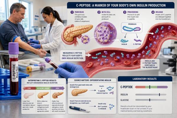
A C-peptide blood test shows how much insulin your pancreas is making. Beta cells release insulin and C-peptide in equal amounts when they split proinsulin into its active parts. Injected insulin does not contain C-peptide, so the test can separate insulin made inside the body from insulin taken as medicine.
C-peptide is useful when the type of diabetes is uncertain, when an insulin-treated person may still have meaningful beta-cell function, or when doctors investigate unexplained low blood sugar. A high result can reflect insulin resistance, kidney impairment, a medicine that stimulates insulin release, or rarely an insulin-producing tumor. A low result can reflect type 1 diabetes, advanced beta-cell failure, pancreatic damage, or appropriately reduced secretion during low glucose. The glucose level at the same moment is essential: the same C-peptide number can be reassuring with normal glucose and inadequate when glucose is very high. Laboratories also use different assays and ranges, so interpretation should start with the report’s own reference interval.
- C-peptide measures insulin made by the pancreas; injected insulin does not raise it.
- A fasting adult reference interval may be around 1.1–4.4 ng/mL in one major laboratory, but ranges vary by assay.
- Low C-peptide with high glucose suggests limited beta-cell function, while high C-peptide with high glucose often suggests insulin resistance.
- Kidney impairment can raise C-peptide because the kidneys help clear it.
- For hypoglycemia, C-peptide must be drawn during a documented low-glucose episode with insulin, proinsulin, and a medication screen.
- Fasting, random, and stimulated results are not interchangeable.
Table of Contents
- What C-Peptide Measures
- Why the Test Is Ordered
- Fasting, Random, and Stimulated Tests
- Normal Ranges and Units
- Causes of Low C-Peptide
- Causes of High C-Peptide
- Results in Diabetes and Hypoglycemia
- Preparation, Follow-Up, and Limitations
What C-Peptide Measures
Pancreatic beta cells first make proinsulin, a single chain that folds into a specific shape. Enzymes then cut proinsulin into insulin and connecting peptide, usually called C-peptide. Both products enter the bloodstream in equal molar amounts.
Insulin and C-peptide behave differently after release. The liver removes a large portion of insulin during its first pass through the portal circulation, and circulating insulin has a short half-life of only several minutes. C-peptide largely bypasses first-pass liver extraction and remains in the circulation longer, commonly around 20 to 30 minutes. Its concentration is therefore steadier and often easier to use as a marker of beta-cell secretion.
The kidneys clear and break down much of the circulating C-peptide. Reduced kidney function can raise the result even when pancreatic production has not increased. This is one of the most important interpretation limits.
A C-peptide test does not measure how well insulin works. A person with insulin resistance may have a high result because the pancreas is producing extra insulin to overcome poor tissue response. A person with insulin deficiency may have a low result because beta cells cannot produce enough. The test answers “How much endogenous insulin appears to be produced?” rather than “Is the body sensitive to insulin?”
Injected insulin lacks C-peptide. That feature makes the marker particularly useful in people already using insulin. Although insulin assays can be affected by the type of insulin analog and by insulin antibodies, C-peptide continues to reflect endogenous secretion.
C-peptide is measured in serum or plasma with an immunoassay. Results commonly appear in nanograms per milliliter (ng/mL), nanomoles per liter (nmol/L), or picomoles per liter (pmol/L). A frequently used approximate conversion is:
- 1 ng/mL C-peptide ≈ 0.331 nmol/L
- 1 ng/mL C-peptide ≈ 331 pmol/L
Conversions are useful for reading studies, but a result should still be compared with the performing laboratory’s own method and interval.
Why the Test Is Ordered
C-peptide is ordered when knowing endogenous insulin production could change diagnosis or treatment. It is not required for every person with diabetes.
Clarifying the type of diabetes
Type 1 diabetes usually causes low or absent C-peptide after beta-cell destruction progresses. Type 2 diabetes commonly begins with normal or high secretion because insulin resistance drives compensation, but C-peptide may decline after years of disease. Adults can also have autoimmune diabetes that initially resembles type 2 diabetes, monogenic diabetes, pancreatogenic diabetes, or mixed features.
C-peptide helps organize that uncertainty, especially when paired with diabetes autoantibodies, age at diagnosis, body habitus, history of ketoacidosis, family history, medication response, and disease duration. It does not classify diabetes perfectly by itself.
Estimating residual beta-cell function
An insulin-treated person may still make a meaningful amount of insulin. Preserved C-peptide is often associated with lower glucose variability and less severe hypoglycemia, although it does not remove the need for treatment. The result can help guide whether non-insulin medicines are likely to contribute, whether insulin requirements are plausible, and whether a person may qualify for certain technology, research, or specialist pathways.
Investigating hypoglycemia
During genuine hypoglycemia, normal beta cells should sharply reduce insulin and C-peptide secretion. If both remain inappropriately elevated while glucose is low, endogenous hyperinsulinism is possible. Causes include sulfonylurea or meglitinide exposure and insulinoma. If insulin is high but C-peptide is suppressed, injected insulin becomes more likely.
The sample must be collected during the low-glucose event. A normal C-peptide drawn hours later cannot explain what happened during symptoms. A complete hypoglycemia hormone panel often includes plasma glucose, insulin, C-peptide, proinsulin, beta-hydroxybutyrate, and a screen for insulin-secretagogue medicines.
Assessing pancreatic disease
Chronic pancreatitis, pancreatic surgery, cystic-fibrosis-related diabetes, hemochromatosis, and other pancreatic disorders can reduce beta-cell mass. C-peptide can help show the degree of remaining secretion but cannot identify the anatomical cause.
Checking unusual insulin results
When insulin and the clinical picture disagree, C-peptide may clarify whether a high insulin level reflects endogenous release, injected insulin, reduced clearance, or assay interference. Interpretation sometimes uses the molar insulin-to-C-peptide ratio, but the ratio has a narrow role and requires correct unit conversion.
Fasting, Random, and Stimulated Tests
C-peptide can be measured under several conditions. The best method depends on the clinical question.
Fasting C-peptide
A fasting sample is usually collected after 8 to 12 hours without calories. It is often drawn with fasting glucose. Fasting testing reduces the immediate effect of food and is useful for a baseline assessment, but a low-normal glucose can suppress secretion and make beta-cell reserve look smaller than it is.
Fasting values are commonly used in research calculations and in assessments of insulin resistance. For diabetes classification, a random or stimulated value may be more informative because it tests the pancreas under a stronger glucose stimulus.
Random or nonfasting C-peptide
A random sample is easier to obtain and may reflect everyday secretion. The meal timing, carbohydrate content, and matching glucose must be recorded. A random C-peptide that is clearly substantial despite glucose in a stimulating range can show preserved beta-cell function.
Random thresholds used in studies or guidance should not be applied without checking units, kidney function, and the population in which they were developed.
Stimulated C-peptide
Stimulation produces a more direct measure of beta-cell reserve. Common approaches include:
- A mixed-meal tolerance test
- Glucagon stimulation
- A standardized meal or oral nutrient challenge
A mixed-meal test is often favored in research because it resembles normal nutrient stimulation. Samples may be drawn at baseline and at one or more later time points. Glucagon stimulation is shorter but can cause temporary nausea.
Stimulated testing should follow a defined protocol. The dose, timing, glucose level, recent insulin, and specimen handling all influence the result. A value from one protocol should not be compared with a cutoff developed for another.
Urine C-peptide
C-peptide can also be measured in urine, often as a urine C-peptide-to-creatinine ratio. This avoids a blood draw and integrates secretion over the collection period. It depends on kidney function, urine concentration, and correct collection. Blood testing remains more familiar for acute hypoglycemia and many classification questions.
The following comparison helps show why the label matters:
| Test condition | Main advantage | Main limitation |
|---|---|---|
| Fasting | Standardized baseline | May underestimate reserve when glucose is low |
| Random | Convenient and clinically practical | Meal timing and glucose vary |
| Mixed-meal stimulated | Strong physiological assessment | Requires protocol and multiple timed steps |
| Glucagon stimulated | Fast and standardized | Can cause nausea and differs from meal physiology |
| Urine ratio | Needle-free and integrated over time | Influenced by renal function and collection quality |
Normal Ranges and Units
C-peptide reference intervals differ by laboratory, assay, fasting status, age, and specimen. One major U.S. laboratory lists a fasting serum reference interval of 1.1 to 4.4 ng/mL, equivalent to about 0.36 to 1.46 nmol/L. Other laboratories use narrower or wider intervals.
A reference interval describes the distribution in a selected population; it is not a universal threshold for adequate beta-cell function. Clinical interpretation must account for the simultaneous glucose:
- A C-peptide of 1.0 ng/mL may be a reasonable response when glucose is 75 mg/dL.
- The same 1.0 ng/mL may be inappropriately low when glucose is 300 mg/dL.
- A value above the fasting range may be expected shortly after a carbohydrate-containing meal.
- A value within the fasting range may be inappropriate during documented hypoglycemia, when secretion should be nearly suppressed.
Studies and professional discussions often describe very low stimulated or random values around 0.2 nmol/L, or fasting values around 0.08 nmol/L, as indicating severe insulin deficiency in certain settings. These are not universal diagnostic cutoffs. Different guidance uses different thresholds, and assay standardization remains incomplete.
Results should be interpreted cautiously when estimated glomerular filtration rate is reduced. C-peptide can accumulate because renal clearance falls. A high value in chronic kidney disease may overstate secretion, while urine-based results may become unreliable.
Other factors can influence the result:
- Recent food or intravenous glucose
- Sulfonylureas and meglitinides
- GLP-1 receptor agonists and other medicines that change glucose or beta-cell stimulation
- Pregnancy
- Acute illness and stress hormones
- Hemolysis, which can lower C-peptide in some assays
- Heterophile antibodies or other immunoassay interference
- High proinsulin cross-reactivity in certain methods
The number should never be labeled “good” or “bad” without the context of glucose, kidney function, and the question being asked.
Causes of Low C-Peptide
Low C-peptide means endogenous insulin secretion was low at the time of testing. It can be pathological or physiologically appropriate.
Type 1 diabetes and autoimmune beta-cell loss
C-peptide often falls as immune-mediated beta-cell destruction progresses. At diagnosis, children and adults may retain some secretion, especially during a temporary “honeymoon” period after insulin treatment begins. Years later, many people have very low or undetectable values, although sensitive assays show that some retain small amounts for decades.
A detectable result does not rule out type 1 diabetes. Disease duration, glucose at collection, stimulated testing, and autoantibodies matter. Likewise, a low result alone does not prove autoimmunity.
Advanced type 2 diabetes
Type 2 diabetes is progressive for many people. Early insulin resistance may cause high C-peptide, but beta cells can eventually lose capacity. A low result with high glucose can signal insulin deficiency and a greater need for insulin-based treatment.
This does not mean that lifestyle or other medicines have “failed.” Beta-cell decline reflects disease biology, genetics, glucose toxicity, lipotoxicity, and time. Treatment should match current physiology rather than carry blame.
Pancreatic damage or removal
Chronic pancreatitis, pancreatic cancer treatment, major pancreatic surgery, trauma, cystic fibrosis, and iron overload can reduce insulin production. These forms are sometimes grouped as pancreatogenic or type 3c diabetes. Digestive enzyme insufficiency and nutritional problems may coexist.
Low glucose or prolonged fasting
When glucose is low, low C-peptide is normally appropriate. A fasting value should not be called deficient without checking the matching glucose. Recent injected insulin can lower glucose and suppress the pancreas, indirectly lowering C-peptide.
Other considerations
Severe illness can alter secretion in either direction. Sample hemolysis may produce an artifactually low value in some immunoassays. If the result conflicts strongly with glucose patterns and insulin needs, repeating it with careful collection may be appropriate.
Causes of High C-Peptide
High C-peptide usually means the pancreas released more insulin, clearance was reduced, or a medicine stimulated secretion.
Insulin resistance
Insulin resistance is the most common explanation for a high fasting result when glucose is normal or elevated. The pancreas compensates by making more insulin. This pattern occurs with central obesity, metabolic syndrome, prediabetes, early type 2 diabetes, polycystic ovary syndrome, sleep apnea, glucocorticoid exposure, and several endocrine disorders.
A high C-peptide does not quantify insulin resistance by itself. A fasting insulin test, glucose, triglycerides, HDL cholesterol, blood pressure, and waist measurement provide the broader context. The pancreas can secrete a large amount and still fail to maintain normal glucose.
Kidney impairment
Because the kidneys clear C-peptide, chronic kidney disease can increase the level. Interpretation should include creatinine and estimated glomerular filtration rate. The effect can be large enough to obscure the degree of true beta-cell secretion.
Insulin-secretagogue medicines
Sulfonylureas and meglitinides stimulate endogenous insulin and C-peptide release. During unexplained hypoglycemia, a medication screen is necessary because the laboratory pattern can resemble insulinoma.
Insulinoma or endogenous hyperinsulinism
An insulinoma is a rare pancreatic neuroendocrine tumor that releases insulin. Diagnosis does not rest on a high C-peptide during normal glucose. The critical finding is failure to suppress insulin, C-peptide, and proinsulin during verified hypoglycemia, often observed during a supervised fast. Low beta-hydroxybutyrate and a rise in glucose after glucagon may support insulin-mediated hypoglycemia.
Other causes
Pregnancy can increase insulin secretion. Cushing syndrome, acromegaly, and other states that oppose insulin can raise C-peptide through secondary compensation. Severe obesity can shift fasting values above a laboratory’s general reference interval.
An isolated high value without a simultaneous glucose is difficult to interpret. Repeat testing is not always needed, but the clinical question should be clarified before extensive investigation.
Results in Diabetes and Hypoglycemia
Pattern recognition is more useful than a single cutoff.
| Glucose | C-peptide | Common interpretation possibilities |
|---|---|---|
| High | Low | Marked beta-cell deficiency, type 1 diabetes, advanced type 2 diabetes, or pancreatic damage |
| High | Normal-high | Preserved secretion with insulin resistance or early type 2 diabetes |
| Normal | High fasting | Compensatory hyperinsulinemia, medicine effect, or reduced renal clearance |
| Low | Low | Appropriate suppression, injected insulin effect, or non-insulin cause of hypoglycemia |
| Low | Inappropriately high | Endogenous hyperinsulinism, sulfonylurea/meglitinide exposure, or insulinoma |
| Any | Unexpectedly high with kidney failure | Reduced clearance may be contributing |
Diabetes classification
C-peptide is most helpful after the immediate instability of diagnosis has passed and when glucose is high enough to stimulate secretion. A person with obesity can still have type 1 diabetes, and a lean adult can have type 2 or monogenic diabetes. Autoantibodies such as GAD, IA-2, or ZnT8 help identify autoimmune disease, while genetic testing may be appropriate for selected monogenic patterns.
A low result supports insulin deficiency but does not identify its cause. A high result supports preserved secretion but does not exclude autoimmune diabetes early in its course.
Treatment implications
Preserved C-peptide may support the use of therapies that depend partly on endogenous beta-cell function, but treatment decisions also consider A1c, glucose patterns, cardiovascular and kidney disease, weight, hypoglycemia risk, and patient preferences. A low value can justify more emphasis on insulin education, ketone safety, and technology for glucose management.
Never stop insulin solely because C-peptide is detectable. A person with autoimmune diabetes can retain secretion and still need insulin to prevent ketoacidosis.
Hypoglycemia interpretation
The diagnostic sample should be a “critical sample” drawn while plasma glucose is low, generally before glucose treatment when this can be done safely. Insulin, C-peptide, and proinsulin should be suppressed in a healthy fasting response. Detectable numbers are not automatically abnormal; they must be judged against the severity of hypoglycemia and the assay’s decision limits.
The insulin/C-peptide ratio can support suspected injected-insulin exposure, but it can be distorted by kidney disease, insulin antibodies, assay recognition of insulin analogs, and unit errors. Direct interpretation of the complete critical sample is safer than relying on the ratio alone.
Preparation, Follow-Up, and Limitations
Follow the order’s instructions. For a fasting test, avoid calories for the requested period, commonly 8 to 12 hours, while drinking plain water unless told otherwise. Continue prescribed medicines unless the clinician gives a specific plan. Insulin may need carefully timed adjustment for a fasting or stimulation protocol; never omit it without medical instructions.
Tell the healthcare team about:
- All insulin types and the last dose time
- Sulfonylureas, meglitinides, GLP-1 medicines, and other diabetes drugs
- Kidney disease
- Pregnancy
- Recent severe hypoglycemia or ketoacidosis
- Pancreatic surgery or pancreatitis
- Biotin and supplements if the laboratory method may be affected
The blood draw has the usual minor risks of bruising, soreness, and lightheadedness. Stimulated tests may add nausea, especially after glucagon.
Follow-up depends on the pattern. A low result with high glucose may lead to diabetes autoantibodies, review of insulin treatment, or evaluation for pancreatic disease. A high result may lead to assessment of insulin resistance, kidney function, and medicines. A result drawn during hypoglycemia should be interpreted promptly as part of the full critical sample.
Repeat testing can help when the sample was hemolyzed, glucose was not measured, fasting status was unclear, kidney function changed, or the value conflicts with the clinical picture. For tracking over time, use the same laboratory and similar test conditions.
C-peptide should not be used as a stand-alone screening test for diabetes. Diagnosis still relies on glucose or A1c criteria. It also cannot show whether insulin is reaching tissues effectively, predict an exact insulin dose, or guarantee protection from future beta-cell decline.
Seek urgent care for symptoms of severe hypoglycemia such as confusion, seizure, fainting, or inability to swallow; for signs of diabetic ketoacidosis such as vomiting, abdominal pain, deep breathing, or marked drowsiness; or for persistently extreme glucose with illness. Immediate treatment should never be delayed to obtain a C-peptide result.
References
- C‐peptide determination in the diagnosis of type of diabetes and its management: A clinical perspective 2022 (Review)
- The evolution of C-peptide’s role in diabetes care 2025 (Review)
- Call for Standardization of C-Peptide Measurement 2025 (Position Statement)
- Exploring the potential role of C-peptide in type 2 diabetes management 2025 (Review)
- Persistent C-peptide secretion is associated with favourable outcomes in people with type 1 diabetes 2025 (Research Study)
- CPR – Overview: C-Peptide, Serum 2026 (Official Test Catalog)
Disclaimer
C-peptide results cannot diagnose or classify diabetes, explain hypoglycemia, or determine treatment on their own. A qualified clinician should interpret the value with the simultaneous glucose, test conditions, kidney function, medicines, diabetes history, and other relevant laboratory findings. Do not change or stop insulin based only on a C-peptide result.