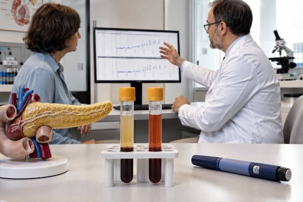
The insulin/C-peptide ratio is a calculated laboratory value used mainly during the investigation of documented hypoglycemia. Insulin and C-peptide are released together when the pancreas makes insulin, but injected insulin contains no C-peptide. Converting both results to the same molar units and comparing them can therefore help distinguish exogenous insulin exposure from continued pancreatic insulin production. The ratio is not a routine screening test for diabetes, insulin resistance, or metabolic health, and a result obtained while glucose is normal usually cannot answer the question for which the calculation was designed. Interpretation requires a properly timed critical sample that includes laboratory plasma glucose, insulin, C-peptide, proinsulin, beta-hydroxybutyrate, and a sulfonylurea or meglitinide screen. Kidney function, insulin antibodies, assay cross-reactivity, specimen handling, and treatment given before the blood draw can all alter the apparent pattern. The ratio should be treated as supporting evidence within a complete clinical evaluation, not as proof of intentional medication use or of a pancreatic tumor.
- The ratio must use molar units; dividing the numbers printed in different conventional units produces a meaningless result.
- A molar insulin/C-peptide ratio greater than 1 during true hypoglycemia may support exogenous insulin exposure, especially when C-peptide is suppressed.
- A ratio of 1 or less with inappropriately high insulin and C-peptide suggests endogenous insulin secretion but does not distinguish insulinoma from an insulin-releasing drug.
- Insulin antibodies, kidney dysfunction, poorly detected insulin analogs, and mistimed sampling can overturn a simple ratio-based interpretation.
- Severe confusion, seizure, unconsciousness, or inability to swallow during suspected hypoglycemia requires immediate treatment; diagnostic sampling must not delay rescue.
Table of Contents
- What the Insulin/C-Peptide Ratio Measures
- When the Ratio Is Used
- How to Calculate the Molar Ratio
- Interpreting a Ratio Greater Than 1
- Interpreting a Ratio of 1 or Less
- Critical-Sample Tests and Result Patterns
- Limitations and Common Pitfalls
- Follow-Up, Safety, and Next Steps
What the Insulin/C-Peptide Ratio Measures
The insulin/C-peptide ratio compares the circulating molar concentration of insulin with the molar concentration of C-peptide. It is derived from two laboratory measurements rather than being a separate hormone assay. Its value comes from the way beta cells manufacture and secrete insulin.
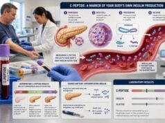
Insulin begins as preproinsulin, which is processed to proinsulin. Proinsulin folds so that the future A and B chains of insulin are connected by a segment called C-peptide. When a beta cell prepares a secretory granule, enzymes split proinsulin into mature insulin and C-peptide. The two products are released in equal numbers of molecules.
Equal secretion does not produce equal blood concentrations. Insulin first enters the portal circulation, and the liver removes a substantial proportion before the hormone reaches peripheral blood. Circulating insulin also has a relatively short half-life. C-peptide is not extracted to the same extent by the liver, remains in circulation longer, and is cleared mainly by the kidneys. Consequently, in ordinary endogenous secretion, the peripheral molar insulin/C-peptide ratio is usually 1 or less.
Injected insulin changes that relationship in two ways. Pharmaceutical insulin contains insulin or an insulin analog but no C-peptide. It also enters the peripheral circulation rather than passing first through the portal vein and liver. At the same time, falling glucose normally suppresses the person’s own beta-cell secretion, so endogenous C-peptide becomes low. The combination can reverse the usual ratio, producing more measured insulin than C-peptide on a molar basis.
This physiology explains why the ratio is most informative during hypoglycemia. At a normal or high glucose concentration, pancreatic insulin secretion may be expected, and the values may reflect a recent meal, insulin resistance, diabetes treatment, stress, or ordinary biological variation. A ratio calculated in that setting is not a validated shortcut for determining whether someone has insulin resistance or whether they use insulin.
When the Ratio Is Used
Clinicians use the insulin/C-peptide ratio primarily when investigating hypoglycemia that appears to be mediated by insulin. In an adult without diabetes, a formal evaluation generally begins after Whipple’s triad has been demonstrated:
- Symptoms or signs compatible with hypoglycemia
- A reliably measured low plasma glucose concentration
- Improvement after glucose is raised
Symptoms may include sweating, tremor, hunger, palpitations, tingling, anxiety, blurred vision, confusion, unusual behavior, weakness, difficulty speaking, seizure, or loss of consciousness. Home glucose meters and continuous glucose monitors can reveal patterns, but their accuracy is poorer at low concentrations. A laboratory plasma glucose obtained during symptoms is usually needed before an uncommon hypoglycemic disorder is diagnosed.
The ratio may be considered when the critical sample shows insulin that is not appropriately suppressed for the low glucose. It is especially relevant when clinicians need to distinguish insulin entering from outside the body from insulin released by pancreatic beta cells. Typical settings include:
- Unexplained fasting or overnight hypoglycemia
- Recurrent severe episodes in a person with access to insulin
- A mismatch between reported medication use and the biochemical findings
- Possible insulinoma or another endogenous hyperinsulinemic disorder
- Suspected insulin autoimmune hypoglycemia
- Unexpected hypoglycemia in a hospitalized person
The calculation is less useful when hypoglycemia is clearly explained by prescribed insulin therapy and the management question is simply how to prevent another dosing-related event. It is also inappropriate as a stand-alone test for ordinary post-meal fatigue, nonspecific dizziness, weight gain, or suspected metabolic syndrome.
A properly timed sample is crucial. The insulin and C-peptide values should be drawn at the same time as the low laboratory glucose, ideally before carbohydrate, intravenous dextrose, glucagon, or another treatment changes the physiology. If an episode cannot be sampled spontaneously, an endocrinologist may use a supervised fast for fasting symptoms or a supervised mixed-meal test for reproducible postprandial symptoms. Neither test should be attempted without medical supervision.
How to Calculate the Molar Ratio
The ratio must compare insulin and C-peptide in the same molar units. Laboratory reports commonly express insulin in micro-international units per milliliter and C-peptide in nanograms per milliliter. Dividing those printed values directly is incorrect because the units measure different quantities.
A commonly used conversion is:
Insulin in pmol/L = insulin in µIU/mL × 6.945
C-peptide in pmol/L = C-peptide in ng/mL × 331
Then calculate:
Molar insulin/C-peptide ratio = insulin in pmol/L ÷ C-peptide in pmol/L
Laboratories may use slightly different conversion factors or may report one or both results directly in SI units. The interpreting clinician should use the factors specified by the performing laboratory and should confirm that both values came from the same blood draw.
Worked example suggesting exogenous insulin
Suppose a critical sample collected during confirmed hypoglycemia shows:
- Insulin: 100 µIU/mL
- C-peptide: 0.5 ng/mL
Convert the values:
- Insulin: 100 × 6.945 = 694.5 pmol/L
- C-peptide: 0.5 × 331 = 165.5 pmol/L
The molar ratio is:
694.5 ÷ 165.5 = approximately 4.2
A ratio greater than 1, combined with a high insulin concentration and suppressed C-peptide during low glucose, supports exogenous insulin exposure. The conclusion still depends on whether the insulin assay detects the suspected preparation and whether antibodies or laboratory interference could explain the result.
Worked example suggesting endogenous secretion
Suppose another critical sample shows:
- Insulin: 20 µIU/mL
- C-peptide: 5 ng/mL
Convert the values:
- Insulin: 20 × 6.945 = 138.9 pmol/L
- C-peptide: 5 × 331 = 1,655 pmol/L
The molar ratio is:
138.9 ÷ 1,655 = approximately 0.084
If glucose was genuinely low and both hormones were inappropriately elevated, this pattern supports continued endogenous insulin secretion. It does not, by itself, determine whether the cause is an insulinoma, a sulfonylurea or meglitinide, post-bariatric hypoglycemia, or another beta-cell disorder.
When the denominator is nearly zero
A very low or undetectable C-peptide can produce a very large or mathematically undefined ratio. In practice, the qualitative pattern—high insulin with suppressed C-peptide during low glucose—is often more informative than reporting an extreme number. Results below the assay’s detection limit should not be replaced with zero for casual calculation. The laboratory or endocrinologist may use the assay’s lower reporting boundary and describe the ratio as greater than a minimum value.
Rounding should occur only after both conversions and the final division. Excessive decimal precision implies more certainty than the assays provide. Usually one or two significant decimal places are adequate for a clinical explanation.
Interpreting a Ratio Greater Than 1
A molar ratio greater than 1 is a reversal of the usual peripheral relationship and can support exogenous insulin administration. The strongest pattern includes all of the following during confirmed hypoglycemia:
- Insulin is elevated or inappropriately detectable
- C-peptide is low or undetectable
- Proinsulin is low or appropriately suppressed
- Beta-hydroxybutyrate is low, showing insulin-like suppression of ketone production
- A sulfonylurea and meglitinide screen is negative
Injected insulin lowers glucose, suppresses endogenous beta-cell secretion, and prevents normal fasting ketone production. C-peptide and proinsulin fall because the pancreas is responding appropriately to low glucose, while the circulating pharmaceutical insulin remains active.
The ratio should not be used to accuse a person of surreptitious use. In someone prescribed insulin, the same pattern may be expected after excess dosing. Clinicians should review every insulin formulation, dose, timing, injection device, pump setting, recent exercise, alcohol intake, meal pattern, kidney function, and possibility of a medication mix-up. In children, dependent adults, or people receiving care from others, accidental or imposed exposure must also be considered with appropriate safeguarding.
A ratio greater than 1 is not specific for exogenous insulin. Insulin autoantibodies can bind insulin and prolong its apparent circulation, producing very high measured insulin and a reversed ratio. Insulin autoimmune syndrome can cause alternating post-meal hyperglycemia and delayed hypoglycemia as insulin binds to and later dissociates from antibodies. People previously treated with insulin may also develop antibodies that complicate interpretation.
Assay mismatch is another major issue. Some immunoassays detect recombinant human insulin well but recognize particular analogs poorly or not at all. If the injected product is not detected, the reported insulin may be unexpectedly low, and the classic high-ratio pattern may disappear. Conversely, a different assay may detect the same analog strongly. When suspicion remains despite discordant results, the laboratory should be asked which insulin preparations its method recognizes. Testing with another immunoassay or a specialized method capable of identifying analogs may be necessary.
Timing can also alter the ratio. Insulin and C-peptide have different half-lives. A sample drawn long after the causative dose, after glucose treatment, or while glucose is recovering may not reproduce the peak pattern. The complete timeline should document symptoms, glucose measurements, blood collection, food, dextrose, glucagon, and medication administration.
Thus, “greater than 1” means that the result deserves an exogenous-insulin and antibody-focused interpretation. It does not establish the source, intent, or exact preparation without corroborating evidence.
Interpreting a Ratio of 1 or Less
A molar ratio of 1 or less is the usual relationship when insulin originates from the pancreas. During hypoglycemia, the key question is whether insulin, C-peptide, and proinsulin are appropriately suppressed. If all three are low, a ratio below 1 is simply consistent with normal beta-cell shutdown and does not suggest endogenous hyperinsulinism.
If insulin and C-peptide remain inappropriately elevated during low glucose, a ratio of 1 or less supports endogenous insulin secretion. Possible causes include:
- Insulinoma
- Sulfonylurea or meglitinide exposure
- Post-bariatric hypoglycemia
- Non-insulinoma pancreatogenous hypoglycemia syndrome
- Rare congenital or genetic hyperinsulinism
- Insulin autoimmune syndrome in some phases or assay settings
An insulinoma is a pancreatic neuroendocrine tumor that secretes insulin without normal suppression. A typical critical sample shows low glucose, measurable insulin, C-peptide and proinsulin that are not suppressed, low beta-hydroxybutyrate, and a negative insulin-secretagogue screen. The ratio supports pancreatic origin but cannot localize a tumor. Biochemical confirmation should come before pancreatic imaging because small incidental lesions can lead to false attribution.
Sulfonylureas and meglitinides stimulate the person’s own beta cells. Their biochemical pattern can be indistinguishable from insulinoma: insulin, C-peptide, and proinsulin are present, ketones are suppressed, and the ratio remains 1 or less. A properly timed drug screen is therefore essential. The panel must include the relevant medications and metabolites; a negative screen may be misleading if the drug is not covered, the sample was collected too late, or the assay lacks adequate sensitivity.
Post-bariatric hypoglycemia most often occurs one to several hours after eating in people who have undergone procedures that accelerate nutrient delivery to the small intestine. The diagnosis depends on the timing, documented low glucose, and exclusion of other causes. An insulin/C-peptide ratio alone cannot distinguish this condition from other forms of endogenous secretion.
Kidney dysfunction complicates a low-ratio interpretation because C-peptide is cleared mainly by the kidneys. Reduced filtration can raise C-peptide disproportionately and push the ratio downward. Insulin clearance may also change. Creatinine and estimated glomerular filtration rate should be reviewed before a low ratio is taken as evidence of unusually strong pancreatic secretion.
A ratio of 1 or less at normal glucose is not evidence of insulinoma. Most people producing their own insulin will have a ratio in that range because of normal hepatic extraction and C-peptide kinetics. The diagnostic meaning comes from failure of the beta-cell markers to suppress during hypoglycemia.
Critical-Sample Tests and Result Patterns
The ratio is one component of a broader hypoglycemia hormone test panel. A complete critical sample usually includes laboratory plasma glucose, insulin, C-peptide, proinsulin, beta-hydroxybutyrate, and a screen for insulin secretagogues. Clinicians may also obtain insulin antibodies, cortisol, free fatty acids, liver and kidney tests, or other studies based on the circumstances.
| Pattern during confirmed hypoglycemia | Molar ratio | Most likely interpretation |
|---|---|---|
| Insulin high; C-peptide low; proinsulin low; ketones low | Often greater than 1 | Exogenous insulin, subject to analog detection and antibody caveats |
| Insulin, C-peptide, and proinsulin inappropriately present; ketones low; drug screen negative | Usually 1 or less | Endogenous hyperinsulinism, including insulinoma |
| Insulin, C-peptide, and proinsulin inappropriately present; ketones low; drug screen positive | Usually 1 or less | Sulfonylurea or meglitinide effect |
| Very high insulin with variable C-peptide and positive insulin antibodies | May be greater than 1 | Insulin antibody interference or insulin autoimmune syndrome |
| Insulin, C-peptide, and proinsulin suppressed; ketones high | Not diagnostically useful | Non-insulin-mediated fasting hypoglycemia |
Beta-hydroxybutyrate tests the biological effect of insulin. During ordinary fasting, low insulin permits fat breakdown and ketone production, so beta-hydroxybutyrate should rise. A low ketone concentration during hypoglycemia supports insulin or insulin-like activity even when the insulin assay is difficult to interpret. Low ketones can also occur in IGF-2-mediated tumor hypoglycemia and selected metabolic disorders, so they do not identify the source alone.
Proinsulin is valuable because beta-cell tumors may release incompletely processed hormone. An inappropriately high proinsulin result during low glucose strengthens evidence for endogenous secretion. A normal fasting proinsulin measured on a different day cannot replace the critical-sample result.
After blood is collected, a clinician may administer intravenous glucagon and measure the glucose response. A substantial rise can support excessive insulin or insulin-like activity because insulin has preserved liver glycogen while preventing its release. The response depends on nutrition, fast duration, and liver function and should remain supportive rather than decisive.
When endogenous hyperinsulinism is confirmed and a secretagogue screen is negative, the next stage may include pancreatic imaging, endoscopic ultrasound, or selective arterial calcium stimulation in specialized centers. Imaging should not precede biochemical confirmation merely because the ratio is low; incidental pancreatic findings are possible, while many insulinomas are small.
Limitations and Common Pitfalls
The ratio appears simple but is vulnerable to clinical, mathematical, and laboratory errors.
Using mismatched units
The most common calculation error is dividing insulin in µIU/mL by C-peptide in ng/mL. That produces a numerical value with no valid physiological interpretation. Both concentrations must be converted to pmol/L or another identical molar unit first.
Sampling when glucose is not low
Insulin and C-peptide may be detectable after food or whenever glucose is normal. The ratio then describes ordinary physiology rather than inappropriate insulin action. A normal outpatient fasting draw cannot retrospectively diagnose the cause of an episode that occurred hours or days earlier.
Assay-specific insulin analog detection
Insulin immunoassays differ markedly in cross-reactivity with lispro, aspart, glulisine, glargine and its metabolites, detemir, degludec, and newer preparations. A report labeled “insulin” does not guarantee that every pharmaceutical analog was measured. The laboratory’s method documentation is part of the interpretation.
Kidney and liver dysfunction
C-peptide is cleared mainly by the kidneys, while insulin undergoes substantial hepatic extraction and both hormones have renal clearance components. Kidney failure can raise both values, often affecting C-peptide strongly. Liver disease can alter insulin clearance. A ratio threshold derived from people with normal organ function may be less reliable in these settings.
Insulin antibodies and immunoassay interference
Insulin antibodies can prolong measured insulin, release biologically active insulin unpredictably, or interfere with an assay. Heterophile antibodies and antibodies directed against assay components can also cause falsely high or low values. When the result does not fit the symptoms and companion markers, the laboratory may repeat testing with dilution, blocking reagents, polyethylene glycol precipitation, or a different platform.
Hemolysis and specimen processing
Hemolysis can lower measured insulin because red blood cells release insulin-degrading enzymes, and it can also reduce C-peptide results. Delayed glucose processing allows blood cells to consume glucose after collection, creating pseudohypoglycemia. Tube type, separation, freezing, and transport requirements differ by assay. A mathematically precise ratio cannot repair a compromised specimen.
Incomplete medication screens
A negative sulfonylurea or meglitinide screen is meaningful only if the test includes the relevant drug and the specimen was obtained while enough drug or metabolite remained. Clinicians should compare the assay panel with all household and workplace medications, including combination tablets.
Different samples or different times
Insulin and C-peptide must come from the same critical sample. Combining an insulin value drawn during symptoms with a C-peptide value obtained after dextrose can produce an artificial ratio. Their different half-lives make even modest timing differences important.
Overreliance on a cutoff
A ratio greater than 1 is supportive, not infallible, and a ratio of 1 or less does not exclude injected analogs that the assay misses. Interpretation should integrate absolute concentrations, glucose, ketones, proinsulin, drug testing, renal function, antibodies, and the clinical timeline.
Follow-Up, Safety, and Next Steps
A clinician reviewing an insulin/C-peptide ratio should first verify the context rather than starting with the numerical cutoff. Useful questions include:
- Was Whipple’s triad documented?
- What was the laboratory plasma glucose at the exact blood draw?
- Were insulin and C-peptide measured from the same specimen time?
- Were both values converted to identical molar units?
- What were proinsulin and beta-hydroxybutyrate?
- Was a sufficiently broad insulin-secretagogue screen obtained?
- Which insulin analogs does the assay detect?
- Are kidney dysfunction, liver dysfunction, antibodies, or specimen problems present?
- Was glucose, glucagon, food, or medication given before sampling?
If the biochemical pattern suggests prescribed insulin excess, the immediate plan may involve dose adjustment, device review, carbohydrate education, continuous glucose monitoring, or changes for kidney function and exercise. Medication changes should be made with the treating clinician because reducing insulin without an alternative plan can cause severe hyperglycemia or ketoacidosis.
If the pattern suggests endogenous hyperinsulinism, referral to endocrinology is appropriate. Confirmation may require review of the original specimens, repeat critical sampling, a supervised fast, or a mixed-meal test. Only after the biochemical diagnosis is secure should localization studies for insulinoma proceed.
When results raise the possibility of unreported medication exposure, communication should remain nonjudgmental. Factitious disorder is a psychiatric diagnosis, not a laboratory value. Accidental exposure, cognitive impairment, unsafe caregiving, prescribing errors, and malicious administration are distinct possibilities. A multidisciplinary approach may include endocrinology, laboratory medicine, pharmacy, psychiatry, social work, toxicology, or safeguarding teams.
Anyone with suspected severe hypoglycemia should receive rapid treatment. A conscious person able to swallow can generally take fast-acting carbohydrate and recheck glucose according to their care plan. A person who is unconscious, seizing, unable to swallow, or not improving requires emergency assistance, glucagon when available and appropriate, and urgent medical evaluation. Do not force food or drink into an unconscious person.
The ratio is most useful when it clarifies a physiological question: is the insulin effect coming from continued pancreatic secretion or from insulin introduced into the body? Used within a complete critical-sample evaluation, it can guide the next investigation and prevent unnecessary imaging. Used without low glucose, correct units, or assay context, it can be misleading.
References
- CPR – Overview: C-Peptide, Serum 2026 (Laboratory test information)
- INS – Overview: Insulin, Serum 2026 (Laboratory test information)
- Non-Diabetic Hypoglycemia: Evaluation and Management in Adults 2025 (Review)
- Investigation and Causes of Spontaneous (Non-Diabetic) Hypoglycaemia in Adults: Pitfalls to Avoid 2023 (Review)
- Evaluation and management of adult hypoglycemic disorders: an Endocrine Society Clinical Practice Guideline 2009 (Clinical practice guideline)
Disclaimer
This article is for general education and cannot diagnose the cause of hypoglycemia or establish medication exposure. Insulin/C-peptide ratios require clinician and laboratory interpretation using the glucose concentration, assay characteristics, companion tests, and medical context. Severe or persistent hypoglycemia is an emergency and should be treated immediately.