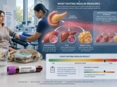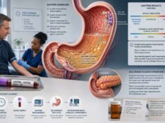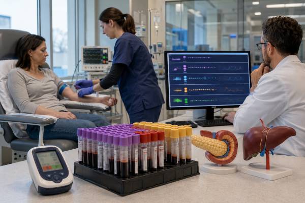
A hypoglycemia hormone panel is a group of blood tests collected while plasma glucose is genuinely low. The core measurements—glucose, insulin, C-peptide, proinsulin, and beta-hydroxybutyrate—show whether insulin activity is appropriately suppressed or is continuing when it should be nearly absent. A sulfonylurea and meglitinide screen is usually essential, and clinicians may also measure cortisol, insulin antibodies, free fatty acids, or the glucose response to glucagon. Timing is decisive. Normal hormone values obtained when glucose is normal cannot confirm or exclude an insulinoma, hidden insulin exposure, or another cause of hypoglycemia. In adults without diabetes, evaluation usually begins only after Whipple’s triad is documented: compatible symptoms, a low reliable plasma glucose, and symptom relief when glucose rises. Because severe hypoglycemia can cause seizure, coma, injury, or death, immediate treatment takes priority; the diagnostic sample should be drawn first only when doing so does not delay rescue.
- The panel must be collected during low laboratory plasma glucose; a routine fasting draw with normal glucose is not a valid “critical sample.”
- Inappropriately detectable insulin, C-peptide, and proinsulin with suppressed ketones suggests endogenous insulin or an insulin-releasing drug.
- High insulin with low C-peptide and low proinsulin suggests injected insulin, but insulin analog assays can complicate this pattern.
- Beta-hydroxybutyrate should rise during ordinary fasting; a low value during hypoglycemia indicates insulin-like suppression of ketone production.
- Severe confusion, seizure, unconsciousness, or inability to swallow is an emergency and should be treated immediately.
Table of Contents
- What the Panel Measures
- When a Critical Sample Is Needed
- How the Tests Are Collected
- Interpreting Insulin, C-Peptide, and Proinsulin
- Interpreting Beta-Hydroxybutyrate and Glucagon
- Common Result Patterns and Causes
- Limitations and Testing Pitfalls
- Follow-Up, Safety, and Next Steps
What the Panel Measures
The panel is designed around one physiological fact: when plasma glucose falls, normal beta cells nearly stop releasing insulin. Low insulin allows the liver to produce glucose and the body to release fatty acids and make ketones. If insulin action remains excessive, those rescue pathways stay suppressed.
The usual critical sample includes:
- Laboratory plasma glucose, which confirms the biochemical event
- Insulin, the hormone that lowers glucose and suppresses fat breakdown and ketone production
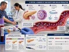
- C-peptide, released in equal molar amounts with endogenous insulin when proinsulin is split inside beta cells
- Proinsulin, the precursor molecule normally processed into insulin and C-peptide
- Beta-hydroxybutyrate, the main circulating ketone body during fasting
- Sulfonylurea and meglitinide screen, which looks for medicines that stimulate pancreatic insulin release
Additional tests depend on the suspected cause. They may include cortisol, growth hormone in selected pediatric cases, insulin antibodies, free fatty acids, liver and kidney tests, IGF-1 or IGF-2-related studies, and toxicology testing. A clinician may administer intravenous glucagon after the critical sample and measure how much glucose rises.
Each marker answers a different question:
| Marker | Main question during hypoglycemia |
|---|---|
| Insulin | Is measurable insulin present when it should be suppressed? |
| C-peptide | Is the insulin coming from the person’s pancreas? |
| Proinsulin | Is beta-cell peptide secretion inappropriately continuing? |
| Beta-hydroxybutyrate | Has fasting ketone production been appropriately activated? |
| Drug screen | Could a sulfonylurea or meglitinide be driving endogenous insulin release? |
C-peptide is especially useful because pharmaceutical insulin does not contain it. However, the pattern is not infallible. Kidney dysfunction can raise C-peptide, insulin antibodies can distort insulin measurements, and some laboratory assays detect certain insulin analogs poorly.
When a Critical Sample Is Needed
Adults without diabetes should generally undergo a formal hypoglycemia workup only when Whipple’s triad is present:
- Symptoms or signs compatible with hypoglycemia
- A low plasma glucose measured by a reliable laboratory method
- Improvement after glucose is raised
Symptoms may be autonomic—sweating, tremor, hunger, palpitations, anxiety, tingling—or neuroglycopenic, meaning the brain lacks glucose. Neuroglycopenic features include blurred vision, unusual behavior, confusion, weakness, impaired speech, seizure, and loss of consciousness.
A continuous glucose monitor or home meter can identify timing and trends, but neither should be the sole biochemical proof for an uncommon spontaneous hypoglycemia disorder. Sensors measure interstitial glucose and may lag behind blood glucose. Accuracy also declines in the low range. A suspicious device reading should be confirmed when safely possible.
The setting helps determine the test approach:
- Fasting or overnight episodes raise concern for excessive insulin, medication exposure, alcohol effects, critical illness, adrenal insufficiency, severe liver disease, kidney failure, malnutrition, or a non-islet-cell tumor.
- Post-meal episodes may occur after gastric surgery, with rare beta-cell disorders, or as symptoms without confirmed hypoglycemia.
- Episodes in a person using diabetes medication are commonly treatment-related and require medication, meal, exercise, alcohol, and kidney-function review.
- Ill hospitalized patients may have hypoglycemia from sepsis, organ failure, poor intake, or medications rather than a pancreatic tumor.
A common diagnostic error is ordering insulin and C-peptide the morning after an episode, when glucose has returned to normal. At normal glucose, measurable insulin and C-peptide may be entirely appropriate. The sample must capture the abnormal regulation during the low value.
The glucose threshold that triggers a diagnostic sample depends on the protocol and symptoms. In a supervised fast, testing is often completed when plasma glucose is below about 55 mg/dL and compatible symptoms or signs occur, although clinicians may use additional criteria. In a person with recurrent neuroglycopenia, the endocrinology team may obtain samples at a lower or protocol-defined threshold under close observation.
How the Tests Are Collected
The best sample is obtained during a spontaneous episode before carbohydrate, dextrose, or glucagon is given. Safety comes first. Blood collection should never delay treatment in a person who is seizing, unconscious, unable to swallow, or rapidly deteriorating.
If spontaneous sampling is not possible, an endocrinologist may reproduce the circumstances under supervision.
Supervised fast
A supervised fast is used mainly for suspected fasting hypoglycemia. It may continue for up to 72 hours, though many insulinomas declare themselves earlier. The test is performed in a controlled setting with scheduled and symptom-triggered glucose checks. Calorie-free fluids are permitted according to protocol, and nonessential medicines may be held under medical direction.
As glucose falls, blood is collected more frequently. At the endpoint, the full critical sample is drawn before the fast is ended. The clinician may then give intravenous glucagon and measure plasma glucose over the next several minutes. Food or intravenous dextrose follows as required.
A 72-hour fast is not a self-test. Prolonged fasting at home can cause dangerous neuroglycopenia and fails to ensure that the correct specimens are collected and processed.
Mixed-meal test
A supervised mixed-meal test may be used when symptoms occur several hours after eating. The person consumes a meal similar to one that triggers symptoms or a standardized liquid meal. Glucose and relevant hormones are measured for several hours, especially when symptoms occur.
An oral glucose tolerance test is generally a poor way to evaluate ordinary postprandial symptoms because a pure glucose load can provoke low values even in people who do not have a clinically important disorder. The meal should resemble the real-world trigger.
Specimen details
Glucose samples must be processed promptly because blood cells continue to consume glucose after collection. Delay can create pseudohypoglycemia. Insulin, C-peptide, and proinsulin have assay-specific tube and storage requirements. The drug screen may require a separate serum or plasma tube and must be ordered at the time of the event; it cannot be reliably added days later if no specimen remains.
Record the exact timing of symptoms, glucose, blood draw, medication, food, dextrose, and glucagon. A sample obtained after treatment may no longer represent the causal physiology.
Interpreting Insulin, C-Peptide, and Proinsulin
During true fasting hypoglycemia, insulin secretion should be nearly shut off. The important finding is not necessarily an insulin value above the ordinary fasting reference range. It is an insulin value that is inappropriately detectable for the low glucose concentration.
Classic adult guidance has used findings such as plasma glucose below 55 mg/dL with insulin at least 3 µIU/mL, C-peptide at least 0.6 ng/mL, and proinsulin at least 5 pmol/L as evidence supporting endogenous hyperinsulinemia. These are not universal stand-alone cutoffs. Modern insulin assays, insulin analog cross-reactivity, renal function, specimen timing, and the complete pattern must be considered.
Insulin
A high or inadequately suppressed insulin value supports excessive insulin action. A “normal-range” insulin can still be abnormal during hypoglycemia because normal physiology should reduce it to very low or undetectable levels.
Insulin assays differ in their detection of analogs such as lispro, aspart, glulisine, glargine metabolites, detemir, and degludec. A low reported insulin level does not always exclude injected analog exposure. Specialized mass spectrometry or an assay with known analog cross-reactivity may be needed when the clinical suspicion remains high.
C-peptide
C-peptide indicates endogenous secretion. When the pancreas releases insulin, it releases C-peptide in equal molar amounts. Injected insulin usually produces high insulin with suppressed C-peptide because the person’s own beta cells reduce secretion.
C-peptide remains in circulation longer than insulin and is cleared mainly by the kidneys. Chronic kidney disease can raise it and blur thresholds. Interpretation should always include creatinine or estimated glomerular filtration rate.
Proinsulin
Proinsulin is normally converted efficiently before secretion, but insulin-secreting tumors often release an increased proportion of incompletely processed hormone. An inappropriately elevated proinsulin during hypoglycemia strengthens evidence for endogenous beta-cell secretion, particularly when insulin is borderline.
A proinsulin blood test obtained when glucose is normal cannot substitute for a critical-sample result. The diagnostic value comes from failure to suppress during hypoglycemia.
Interpreting Beta-Hydroxybutyrate and Glucagon
Beta-hydroxybutyrate provides a functional readout of insulin action. During fasting, falling insulin allows fat cells to release fatty acids. The liver converts part of that fuel into ketones, including beta-hydroxybutyrate. Therefore, ketones should rise as fasting progresses.
Insulin strongly suppresses lipolysis and ketogenesis. A beta-hydroxybutyrate concentration of about 2.7 mmol/L or lower at the end of a hypoglycemic fast has traditionally supported insulin- or IGF-mediated hypoglycemia. A clearly elevated ketone level makes excessive insulin less likely, though it does not identify the cause by itself.
Low ketones can occur with:
- Endogenous hyperinsulinism, including insulinoma
- Injected insulin
- Sulfonylurea or meglitinide exposure
- Insulin autoimmune hypoglycemia
- IGF-2-mediated non-islet-cell tumor hypoglycemia
- Certain fatty-acid oxidation disorders
High ketones during hypoglycemia suggest that insulin is appropriately suppressed. Causes may include prolonged fasting, low glycogen stores, alcohol-related impaired gluconeogenesis, adrenal insufficiency, malnutrition, or other non-insulin mechanisms.
After the critical sample, intravenous glucagon may be given. Glucagon signals the liver to release stored glycogen. A glucose rise of about 25 mg/dL or more has traditionally supported excess insulin or insulin-like activity because hepatic glycogen was preserved while insulin blocked its release. A small response may occur when glycogen stores are depleted by prolonged fasting, liver failure, malnutrition, or alcohol.
The glucagon response is supportive, not decisive. It depends on liver glycogen, fast duration, liver function, and prior nutrition. It should be interpreted with beta-hydroxybutyrate and peptide results.
Common Result Patterns and Causes
The panel is most useful when the markers are interpreted together.
| Pattern during low plasma glucose | Likely interpretation |
|---|---|
| Insulin high or detectable; C-peptide high; proinsulin high; ketones low; drug screen negative | Endogenous hyperinsulinism, including insulinoma or a less common beta-cell disorder |
| Insulin high; C-peptide low; proinsulin low; ketones low | Exogenous insulin exposure, subject to analog assay limitations |
| Insulin high; C-peptide high; proinsulin high; ketones low; drug screen positive | Sulfonylurea or meglitinide effect |
| Insulin low; C-peptide low; proinsulin low; ketones high | Non-insulin-mediated fasting hypoglycemia |
| Insulin low; C-peptide low; ketones low | Consider IGF-mediated tumor hypoglycemia or a fatty-acid oxidation problem |
Endogenous hyperinsulinemia does not automatically equal insulinoma. Differential diagnoses include:
- Sulfonylurea or meglitinide exposure missed by an incomplete or poorly timed screen
- Insulin autoimmune syndrome
- Post-bariatric hypoglycemia
- Non-insulinoma pancreatogenous hypoglycemia syndrome
- Rare inherited or congenital hyperinsulinism presenting later
The medication screen must cover the drugs used in the local market and should be sensitive enough at the time of sampling. A negative result cannot exclude a drug that the panel does not detect or that has already fallen below the detection limit.
Non-insulin causes include critical illness, sepsis, liver failure, kidney failure, malnutrition, adrenal insufficiency, alcohol, medication effects, and non-islet-cell tumors. These conditions may be evident from history and routine laboratory findings before a prolonged fast is considered.
In a person with diabetes, the most common causes are excess insulin, sulfonylurea or meglitinide therapy, reduced food intake, increased exercise, alcohol, kidney dysfunction, or dosing errors. A tumor workup is not the first step unless the pattern remains unexplained after treatment factors are addressed.
Limitations and Testing Pitfalls
Several errors can make the panel misleading.
Sampling at normal glucose: Insulin and C-peptide cannot be judged as “too high” without simultaneous low plasma glucose.
Relying on a fingerstick alone: Poor circulation, contaminated fingers, strip limitations, and low-range inaccuracy can produce false readings. Confirm with laboratory plasma glucose when feasible.
Delayed glucose processing: Ongoing glycolysis in the tube can falsely lower glucose while hormone levels reflect a nonhypoglycemic state.
Treatment before sampling: Dextrose raises glucose and stimulates endogenous insulin. Glucagon changes glucose. Samples drawn afterward may obscure the original pattern.
Incomplete drug screen: Not every assay detects every sulfonylurea or meglitinide. Timing and metabolite detection matter.
Insulin analog mismatch: Some immunoassays barely detect certain injected analogs, potentially creating an apparently low insulin value.
Kidney dysfunction: Reduced clearance raises insulin, C-peptide, and proinsulin and increases susceptibility to medication-related hypoglycemia.
Insulin antibodies: Antibodies can bind insulin, create very high total immunoreactive levels, and release insulin unpredictably. Specialized testing may be required.
Imaging before biochemical proof: Small pancreatic lesions are common incidental findings, while insulinomas can be tiny and hard to see. Imaging should localize a source after endogenous hyperinsulinemic hypoglycemia is established, not replace biochemical diagnosis.
Mislabeling postprandial symptoms: Palpitations, fatigue, or shakiness after food can occur without low glucose. Whipple’s triad prevents unnecessary invasive investigation.
Thresholds should never be applied without considering the assay. The ratio between insulin and C-peptide also requires molar units and can be altered by kidney function and antibodies. A dedicated insulin/C-peptide ratio is supplementary rather than definitive.
Follow-Up, Safety, and Next Steps
If the critical sample supports endogenous hyperinsulinism and the drug screen is negative, the next phase is localization. A specialist may use pancreas-protocol CT, MRI, endoscopic ultrasound, or functional imaging. If noninvasive studies do not identify a lesion, selective arterial calcium stimulation with hepatic venous sampling may be considered at an experienced center.
If injected insulin or an insulin-releasing drug is detected or strongly suspected, clinicians must address accidental, prescribing, pharmacy, occupational, or intentional exposure with sensitivity and safety. The immediate priority remains prevention of recurrent severe hypoglycemia.
When the pattern is non-insulin-mediated, follow-up targets the likely mechanism: cortisol testing for adrenal insufficiency, liver and kidney evaluation, nutritional assessment, infection workup, medication review, or tumor assessment when IGF-2-mediated hypoglycemia is suspected.
Until the cause is controlled, safety planning may include:
- Avoiding driving, swimming alone, heights, and hazardous machinery when episodes are possible
- Carrying rapid-acting glucose if the person can safely self-treat
- Teaching family or close contacts how to recognize severe symptoms
- Using prescribed glucagon when appropriate
- Maintaining regular meals according to the clinician’s plan
- Recording symptoms, food, activity, medication, and glucose timing
- Using continuous glucose monitoring as a warning tool when recommended, while confirming diagnostic lows appropriately
A conscious person able to swallow can often take fast-acting carbohydrate according to an individualized plan. An unconscious or seizing person should not be given food or drink by mouth. Use glucagon if available and trained to do so, call emergency services, and place the person safely on their side.
The panel’s strength comes from its coordinated physiology. Insulin, C-peptide, and proinsulin show the source of insulin-like activity; beta-hydroxybutyrate shows whether fat metabolism is suppressed; the drug screen separates endogenous secretion from medication stimulation; and glucose anchors every conclusion. Without that simultaneous low glucose, the numbers lose their diagnostic meaning.
Documenting the Critical Sample
The value of the panel depends on a precise timeline. The record should state when symptoms began, the plasma glucose at the hormone draw, whether a bedside meter or continuous monitor prompted sampling, and exactly when food, dextrose, glucagon, insulin, or another medicine was given. Even a short delay after treatment can suppress ketones, raise glucose, alter insulin secretion, and blur the original pattern.
A complete critical sample often includes laboratory glucose, insulin, C-peptide, proinsulin, beta-hydroxybutyrate, and a sulfonylurea/meglitinide screen. Depending on age and context, clinicians may add cortisol, growth hormone, free fatty acids, lactate, ammonia, acylcarnitines, insulin antibodies, or other metabolic studies. The panel should be tailored rather than ordered as an indiscriminate battery.
Medication screening requires special attention. A negative result does not exclude every secretagogue because panels differ in the drugs and metabolites they detect, and concentrations fall with time. The laboratory list should be compared with all prescribed, over-the-counter, household, and workplace medicines. Sampling during the episode is preferable to testing much later.
When spontaneous episodes cannot be captured, the provoking test should match the history. A supervised fast evaluates fasting or overnight symptoms, whereas a mixed-meal study is more appropriate for reproducible post-meal episodes. An oral glucose tolerance test can produce low values in people without a true hypoglycemic disorder and is generally not the preferred test for this purpose.
Treatment always takes priority over perfect sampling when a person is severely symptomatic. Whenever it is safe and does not delay care, blood and urine specimens can be obtained immediately before rescue therapy. If that opportunity is missed, the episode should still be treated and a safer diagnostic plan arranged.
References
- Evaluation and management of adult hypoglycemic disorders: an Endocrine Society Clinical Practice Guideline 2009 (Guideline)
- Investigation and Causes of Spontaneous (Non-Diabetic) Hypoglycaemia in Adults: Pitfalls to Avoid 2023 (Review)
- Non-Diabetic Hypoglycemia: Evaluation and Management in Adults 2025 (Review)
- A Systematic Review of the Accuracy of Insulin and C-peptide Secretion Ratios During the Oral Glucose Tolerance Test to Diagnose Insulinoma 2024 (Systematic Review)
- High Risk for Hypoglycemia Guideline Resources 2022 (Guideline)
Disclaimer
This article is educational and cannot diagnose the cause of hypoglycemia. A critical sample requires clinician supervision and method-specific interpretation, and severe hypoglycemia is a medical emergency. Do not attempt a prolonged diagnostic fast at home or delay treatment to collect laboratory tests.