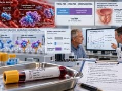
The Prostate Health Index, or PHI, is a blood test that combines three forms of prostate-specific antigen—total PSA, free PSA, and [-2]proPSA, usually written p2PSA—to estimate the likelihood of prostate cancer more accurately than total PSA alone. PHI is most useful when PSA is elevated but not clearly high enough to make the next step obvious, especially when a clinician is deciding whether prostate MRI, biopsy, or continued observation is appropriate. A higher PHI generally means a greater chance of finding prostate cancer and clinically significant disease on biopsy, but PHI is not a cancer diagnosis and there is no single cutoff that applies to every patient. Age, prostate size, digital rectal examination findings, family history, prior biopsy results, MRI, and personal preferences all influence how a PHI result is used. The test is best viewed as a risk-refinement tool that can reduce unnecessary biopsies without treating a low score as a permanent all-clear.
- PHI combines total PSA, free PSA, and p2PSA in one calculated score rather than measuring a completely separate tumor protein.
- Higher PHI values generally correspond to higher prostate cancer risk, including a higher probability of Grade Group 2 or greater disease.
- PHI is especially useful in the PSA gray zone, where total PSA alone cannot reliably separate benign enlargement from cancer.
- There is no universal “normal PHI” cutoff; laboratories and clinical pathways use risk bands or thresholds in context with MRI and other findings.
- No special fasting is usually required, but temporary PSA-altering factors such as infection, urinary retention, recent prostate procedures, and some medications still matter.
Table of Contents
- What the PHI Test Measures
- How PHI Is Calculated
- How to Interpret PHI Results
- PHI Versus PSA and Other Tests
- Preparation and Testing Process
- Limitations and Common Pitfalls
- What to Do After a PHI Result
What the PHI Test Measures
PHI is built from three related PSA measurements. Understanding them makes the score much easier to interpret.
Total PSA is the overall concentration of prostate-specific antigen circulating in blood, including PSA that is free and PSA that is bound to blood proteins. PSA is produced mainly by prostate tissue, so it is organ-associated but not cancer-specific. Benign prostatic hyperplasia, prostatitis, infection, urinary retention, and recent instrumentation can all raise PSA. The prostate-specific antigen test therefore remains the starting point for understanding PHI.
Free PSA is the portion of PSA circulating without being attached to proteins. In men with a borderline total PSA, a lower proportion of free PSA is generally associated with a higher probability of prostate cancer. The percent-free PSA test uses that relationship directly.
p2PSA, or [-2]proPSA, is a precursor isoform of PSA that is more strongly associated with prostate cancer tissue than total PSA alone. PHI gives p2PSA more weight by placing it in the numerator of the calculation and free PSA in the denominator.
The result is not simply an average of the three laboratory values. It is a calculated index designed to improve discrimination between benign conditions and cancer. Studies consistently show that PHI generally performs better than total PSA alone for predicting biopsy-detected cancer and clinically significant prostate cancer.
PHI does not measure tumor size, stage, or whether cancer has spread. It is primarily a pre-biopsy risk marker. A high PHI cannot tell whether a suspicious area is in the left or right side of the prostate, and it cannot replace prostate MRI or biopsy pathology. Its value lies in helping decide who is more or less likely to benefit from those next steps.
How PHI Is Calculated
The standard formula is:
PHI = (p2PSA ÷ free PSA) × √total PSA
The three component measurements are obtained from the same blood sample using a compatible laboratory assay system. Because PHI depends mathematically on all three, analytical differences in the components can affect the final score. This is one reason serial PHI measurements are easiest to compare when performed through the same laboratory platform.
The formula also explains the direction of the score. PHI tends to increase when p2PSA rises, when free PSA falls, or when total PSA rises. Those patterns are associated, in aggregate, with a higher probability of cancer.
For example, two men can both have a total PSA of 5 ng/mL but different PHI values. If one has a relatively high free PSA and low p2PSA, his PHI may be lower. If the other has lower free PSA and higher p2PSA, his PHI may be much higher even though total PSA is identical. That is the main advantage of the index: it extracts more information from PSA biology than a single total PSA number.
PHI is commonly used in men with a moderately elevated PSA, historically around 2–10 ng/mL or 4–10 ng/mL depending on the clinical pathway and regulatory setting. Current practice is broader and more individualized, especially when MRI is part of evaluation. The relevant question is whether PHI can meaningfully reclassify the patient’s risk before biopsy.
PHI can also be adjusted for prostate size as PHI density, calculated by dividing PHI by prostate volume. Research suggests PHI density may further refine risk in selected patients, particularly when MRI and prostate volume are already available. However, it is less standardized than PHI itself and should not be substituted casually for established clinical pathways.
How to Interpret PHI Results
A PHI result is best interpreted as a continuous risk score: the higher the value, the greater the probability of prostate cancer and clinically significant disease. There is no sharp biological point at which cancer suddenly appears.
Some laboratories and older manufacturer data divide PHI into approximate risk bands such as values below the high 20s, around the upper 20s to mid-30s, the mid-30s to mid-50s, and values above the mid-50s. Cancer probability rises across these bands. These ranges can help explain the result, but they should not be treated as universal modern biopsy thresholds because patient populations, assay calibration, MRI use, and definitions of clinically significant cancer differ.
A practical interpretation looks at four layers:
- The absolute PHI value. Is it relatively low, intermediate, or high for the laboratory and population used?
- The baseline PSA situation. Was PSA only mildly elevated, repeatedly rising, or clearly abnormal after repeat testing?
- Other risk factors. Age, family history, ancestry, digital rectal examination, previous biopsy, prostate volume, and medications all matter.
- Imaging information. A PI-RADS 4 or 5 lesion on MRI may outweigh a modestly reassuring PHI, while a negative MRI plus low PHI and low PSA density can support observation in some patients.
The main endpoint in contemporary decision-making is often Grade Group 2 or higher prostate cancer, not just any cancer. This is important because a test can appear “accurate” by detecting many low-grade tumors without necessarily improving clinically useful risk stratification. Meta-analyses suggest PHI has good sensitivity for clinically significant prostate cancer, but it still misses some cases and generates false-positive results.
A PHI result should therefore be discussed in terms of what it changes. If the pretest probability of significant cancer was 15% and PHI meaningfully lowers it, the patient may reasonably choose MRI or surveillance rather than immediate biopsy. If PHI raises an intermediate risk into a range the patient considers unacceptable, biopsy may become more attractive.
PHI Versus PSA and Other Tests
PHI is not intended to replace PSA screening. It is generally a second-line refinement test after PSA creates uncertainty.
Compared with total PSA, PHI improves specificity because it incorporates PSA isoforms more associated with cancer biology. Compared with percent-free PSA alone, PHI adds p2PSA and total PSA into a formal calculation. This can provide better discrimination in the borderline PSA range.
PHI is often discussed alongside the 4Kscore test. Both are blood tests used to estimate the chance of clinically significant prostate cancer, but they are built differently. PHI combines three PSA-related measurements. 4Kscore uses four kallikrein proteins—total PSA, free PSA, intact PSA, and hK2—plus clinical variables in a proprietary risk model. Neither test has proven universally superior for every patient. Availability, insurance coverage, clinician familiarity, MRI findings, and the exact decision being made can determine which is more practical.
Urine tests such as PCA3, SelectMDx, ExoDx, and MyProstateScore approach risk from gene-expression or molecular signals rather than serum PSA isoforms. The SelectMDx urine test, for example, evaluates prostate cancer-associated gene expression. Choosing among these tests is generally more sensible than ordering all of them simultaneously.
PHI can also complement MRI. MRI provides anatomic and functional information about suspicious lesions; PHI provides a blood-based molecular risk estimate. A patient with an equivocal PI-RADS 3 lesion may benefit from PHI or PSA density to decide whether biopsy is warranted. Conversely, a highly suspicious MRI may make additional biomarker testing unnecessary.
PSA density is particularly relevant because it corrects PSA for prostate volume. A large benign prostate can raise total PSA without implying the same cancer risk as an identical PSA from a small prostate. The PSA density test is widely used in MRI-based biopsy decisions and may be combined with PHI in selected cases.
Preparation and Testing Process
PHI requires a standard venous blood draw. Fasting is usually not needed. The most important preparation issue is not food; it is avoiding or recognizing conditions that temporarily alter PSA biology.
Before the blood draw, tell the clinician if you have symptoms of a urinary tract infection or prostatitis, such as fever, burning urination, pelvic discomfort, or urinary frequency. Also report urinary retention, recent catheterization, cystoscopy, prostate biopsy, prostate surgery, or other manipulation. These can raise total and free PSA and may distort PHI interpretation.
Ejaculation can cause a small transient PSA increase in some men. When a borderline result could change management, clinicians may advise avoiding ejaculation for about 24–48 hours before sampling. Some also recommend avoiding vigorous cycling shortly before a repeat PSA-based test, although evidence is less consistent.
Medication history matters. Finasteride and dutasteride can lower PSA substantially after months of use. A clinician should interpret PHI components with awareness of these drugs rather than applying unadjusted assumptions. Testosterone therapy and treatments for prostate cancer can also change PSA dynamics.
The process itself is straightforward:
- A clinician decides that PHI is likely to help with a biopsy or MRI decision.
- Blood is drawn and the laboratory measures total PSA, free PSA, and p2PSA.
- The laboratory calculates PHI using the standard formula.
- The result is interpreted with other risk information rather than as a stand-alone positive or negative test.
If PSA is newly elevated and there is no urgent concern, repeating PSA before PHI may be reasonable because temporary PSA elevations are common enough to affect downstream testing. The exact interval depends on the likely cause, severity of elevation, and clinical context.
Limitations and Common Pitfalls
PHI is useful because it improves risk estimation, not because it eliminates uncertainty. A low result can miss clinically significant cancer, and a high result can occur in someone whose biopsy is benign or shows only low-grade disease.
Common interpretation errors include:
- Calling PHI a cancer test. It estimates risk; biopsy establishes diagnosis.
- Using one cutoff for every person. Thresholds depend on the acceptable tradeoff between avoiding biopsy and missing significant cancer.
- Ignoring MRI. In modern practice, MRI often provides important independent information about lesion risk and biopsy targeting.
- Ignoring prostate size. PSA and PHI may be interpreted differently in a very large benign prostate than in a small prostate.
- Reacting to a single borderline measurement without checking for reversible PSA causes. Infection, retention, and recent procedures can create misleading elevations.
- Comparing results from different assay systems as if they were identical. Small analytical differences can matter in a calculated index.
- Assuming a low PHI permanently rules out cancer. Prostate cancer risk changes over time, so follow-up still matters.
PHI performance can also differ across populations. Studies use different biopsy strategies, MRI pathways, PSA ranges, and definitions of clinically significant disease. Published sensitivity and specificity therefore cannot be applied mechanically to one individual.
Measurement uncertainty deserves attention because PHI is calculated from three separate laboratory values. A small change in PHI may reflect analytical and biological variation rather than a meaningful biological shift. Trends should be interpreted cautiously, particularly when values are close together.
What to Do After a PHI Result
A PHI result should end with a decision plan. The most useful discussion is: What was my estimated risk before PHI, what is it now, and what action fits that risk?
If PHI is relatively low and other features are reassuring, options may include repeat PSA at an agreed interval, MRI before biopsy, or continued observation. If PHI is high, particularly alongside high PSA density, an abnormal digital rectal exam, concerning family history, or suspicious MRI, biopsy becomes more likely to provide useful information.
When MRI is performed, a biopsy may target MRI-visible lesions and also include systematic sampling depending on the clinical setting. A negative MRI does not reduce significant cancer risk to zero, so PHI and PSA density may remain useful when suspicion persists.
If biopsy is deferred, follow-up should be explicit. The plan may include repeat PSA in months rather than years, reassessment of PHI, MRI, or urology review depending on the patient’s risk. The purpose is to gain the benefit of avoiding an unnecessary procedure without losing the opportunity to detect a clinically important cancer later.
PHI is especially valuable when the decision is genuinely uncertain. If clinical evidence already makes biopsy clearly appropriate, another biomarker may only delay care. If overall risk is already very low and testing would not change management, PHI may add cost without benefit.
The test works best as part of a structured prostate cancer biomarker assessment that combines biochemical results, imaging, and patient-specific risk rather than asking any one number to carry the entire decision.
One additional point is that PHI should be interpreted against the clinical question rather than compared only with a previous PHI value. A patient being evaluated for a first biopsy has a different baseline risk from someone with a previous negative biopsy or someone already on active surveillance. The same numerical score may therefore lead to different actions. When serial PHI values are available, a consistent upward pattern can justify reassessment, but there is no validated “PHI velocity” rule comparable to a formal recurrence criterion. Changes should be confirmed and reviewed alongside total PSA, MRI, and prostate volume.
References
- Prostate health index (PHI) as a reliable biomarker for prostate cancer: a systematic review and meta-analysis 2022 (Systematic Review)
- Diagnostic Accuracy of Liquid Biomarkers for Clinically Significant Prostate Cancer Detection: A Systematic Review and Diagnostic Meta-analysis of Multiple Thresholds 2023 (Systematic Review)
- Early Detection of Prostate Cancer: AUA/SUO Guideline Part II: Considerations for a Prostate Biopsy 2023 (Guideline)
- Optimizing Prostate Cancer Care: Clinical Utility of the Prostate Health Index 2025 (Review)
- Measurement Uncertainty and the Prostate Health Index-A Review 2025 (Review)
- Blood- and urine-based biomarkers for the detection of clinically significant prostate cancer: a contemporary review 2025 (Review)
Disclaimer
This article provides general educational information and is not a substitute for medical advice, prostate cancer screening counseling, or individualized biopsy decisions. PHI thresholds and interpretation vary with assay, age, PSA level, prostate size, MRI findings, prior biopsy history, and other risk factors. Discuss your result with the clinician who ordered the test before changing follow-up or treatment.