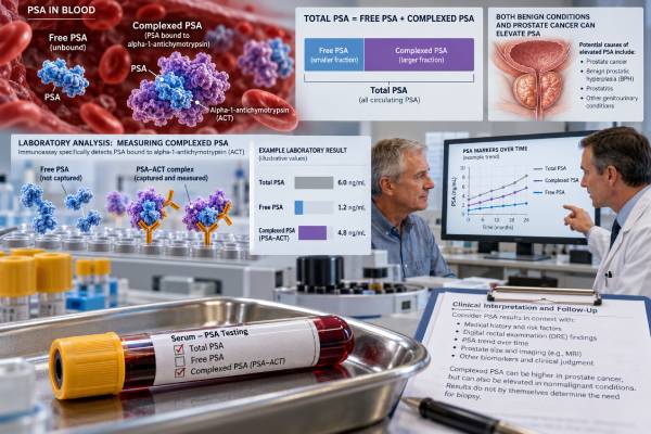
The complexed PSA test measures the portion of prostate-specific antigen that is bound to blood proteins, mainly alpha-1-antichymotrypsin. In men with prostate cancer, a larger share of measurable PSA tends to circulate in complexed rather than free form, which led to the development of complexed PSA, or cPSA, assays as a way to improve cancer-risk assessment beyond total PSA alone. The test was studied most heavily in men with mildly to moderately elevated PSA, especially in the traditional diagnostic “gray zone.” Higher cPSA generally raises concern for prostate cancer, but there is no single modern universal cutoff that should be used independently of the assay, age, prostate size, MRI findings, and other risk factors. Today, complexed PSA is much less commonly used than total PSA, percent free PSA, PSA density, MRI, PHI, 4Kscore, and other contemporary tools. A cPSA result should therefore be interpreted as one piece of prostate-risk information rather than as a stand-alone screening or biopsy decision.
- Complexed PSA is the protein-bound portion of measurable PSA, especially PSA bound to alpha-1-antichymotrypsin.
- Prostate cancer tends to be associated with a higher proportion of complexed PSA and a lower proportion of free PSA.
- Higher cPSA can increase cancer suspicion, but assay-specific cutoffs vary and no single value confirms prostate cancer.
- Complexed PSA is now a less commonly used risk-refinement test than percent free PSA, PSA density, MRI, PHI, or 4Kscore.
- Abnormal cPSA should be followed by overall prostate-risk assessment rather than immediate treatment; biopsy is required to diagnose cancer.
Table of Contents
- What Complexed PSA Measures
- Why Complexed PSA Was Developed
- How to Interpret a Complexed PSA Result
- Complexed PSA Versus Total and Free PSA
- What Can Raise Complexed PSA
- How Complexed PSA Fits Into Modern Prostate Testing
- Follow-Up After an Abnormal Result
What Complexed PSA Measures
PSA is a protein made primarily by prostate gland cells. Most people are familiar with the total PSA test, but total PSA is actually a combination of different molecular forms circulating in blood.
The two clinically important broad forms are:
- Free PSA, which is not bound to a major serum protein.
- Complexed PSA, which is bound to protease-inhibitor proteins in blood.
The major measurable complex is PSA bound to alpha-1-antichymotrypsin, often written PSA-ACT. Smaller amounts of PSA can be associated with other proteins. Some PSA bound to alpha-2-macroglobulin is not readily measured by standard PSA immunoassays because the PSA molecule becomes hidden within the complex.
When laboratories report total PSA, they generally measure immunoreactive free PSA plus the major measurable complexed forms. A dedicated cPSA assay attempts to measure the bound fraction directly.
The biological reason cPSA attracted interest is that prostate cancer tends to shift the balance of PSA forms. Men with cancer often have a lower percentage of free PSA and a higher percentage of complexed PSA than men whose PSA elevation is caused by benign prostatic hyperplasia. This relationship is why a percent free PSA test and cPSA provide related information.
Complexed PSA is usually reported in ng/mL, like total PSA. However, the same numeric value should not be interpreted using total-PSA cutoffs because cPSA measures a different fraction. The laboratory method and reference information therefore matter.
Importantly, cPSA is a risk marker, not a tumor-specific substance. Benign prostate tissue also produces PSA, and noncancerous prostate conditions can raise both total and complexed PSA.
Why Complexed PSA Was Developed
Total PSA is sensitive to prostate abnormalities but lacks cancer specificity. Benign prostate enlargement, inflammation, infection, urinary retention, recent instrumentation, and normal age-related changes can all increase PSA. That means many men with an elevated total PSA do not have prostate cancer on biopsy.
Researchers looked for ways to improve specificity without losing too much sensitivity. PSA density, PSA velocity, age-specific ranges, percent free PSA, and complexed PSA were all developed as refinements.
The appeal of cPSA was straightforward: if prostate cancer produces a greater proportion of protein-bound PSA, directly measuring that fraction might distinguish cancer from benign enlargement better than total PSA alone. Early multicenter studies found that cPSA could provide modestly better specificity than total PSA at comparable sensitivity in men undergoing biopsy.
For example, historical prospective work evaluated men with cPSA values corresponding to total PSA ranges of roughly 2–10 ng/mL. Some studies reported that a cPSA-based threshold could reduce false-positive results compared with a total-PSA threshold while maintaining similar cancer detection sensitivity.
The test also offered a practical laboratory advantage. Percent free PSA requires measurement of both free and total PSA and then calculation of a ratio. cPSA was proposed as a single-analyte alternative that might provide similar risk refinement.
Over time, however, prostate-cancer diagnostics moved beyond this question. Percent free PSA became widely established, and newer approaches added more information: the Prostate Health Index incorporates p2PSA, while 4Kscore combines multiple kallikrein markers with clinical variables. MRI and PSA density now play major roles in biopsy decision-making.
As a result, complexed PSA remains biologically valid but has a smaller role in contemporary practice than it once appeared likely to have.
How to Interpret a Complexed PSA Result
There is no single universal cPSA cutoff that can be applied to every laboratory, patient, and clinical setting. Interpretation depends on the assay, the population in which the cutoff was validated, and the broader prostate-cancer risk profile.
Historical studies used several cPSA ranges and decision thresholds. In one large prospective multicenter trial, cPSA values of 1.5–8.3 ng/mL were evaluated across ranges corresponding approximately to total PSA values of 2–10 ng/mL. The investigators found improved specificity for cPSA over total PSA at matched sensitivity. Other studies used cutoffs near 2.1–2.2 ng/mL in men with total PSA around 2–4 ng/mL, or higher cutoffs in populations with higher PSA levels.
These numbers should not be treated as modern universal reference ranges. They were generated with particular assays and study populations. Laboratory calibration, patient selection, biopsy practice, and MRI use have changed substantially since many cPSA studies were performed.
A practical interpretation looks like this:
| Pattern | General meaning | What usually matters next |
|---|---|---|
| Lower cPSA relative to clinical risk | May be more compatible with benign causes, but cancer is not excluded | Review total PSA, prior values, prostate size, and other risk factors |
| Higher cPSA | Raises suspicion for prostate cancer | Consider repeat testing, MRI, PSA density, or biopsy depending on overall risk |
| Discordant cPSA and other markers | No single marker should dominate | Use multivariable risk assessment and clinical judgment |
The result should never be read as “cPSA above X means cancer.” Only prostate tissue can confirm the diagnosis. Conversely, a lower value does not guarantee that clinically significant cancer is absent.
If a laboratory still offers cPSA, use the assay-specific reference information supplied with the result rather than a cutoff copied from an older publication or another laboratory’s method.
Complexed PSA Versus Total and Free PSA
Total PSA, free PSA, and complexed PSA are related measurements, but they answer slightly different questions.
Total PSA measures the main immunoreactive forms of PSA together. It remains the standard blood marker for prostate cancer screening and follow-up. Its main weakness is limited specificity: benign conditions can raise it.
Free PSA measures unbound PSA. The clinically common calculation is percent free PSA, or free PSA divided by total PSA. A lower percentage of free PSA is associated with a higher probability of prostate cancer, especially when total PSA is in an intermediate range.
Complexed PSA measures primarily the protein-bound fraction. Because total PSA is roughly composed of free plus measurable complexed PSA, a higher complexed proportion generally corresponds to a lower free proportion.
This relationship explains why cPSA and percent free PSA often show similar diagnostic behavior. Early research did not establish a decisive, durable advantage for cPSA that displaced other PSA derivatives.
Modern practice also uses measures that incorporate different information. PSA density divides PSA by prostate volume, helping distinguish PSA production from a large benign gland versus a smaller gland with disproportionately high PSA. MRI can identify suspicious lesions and estimate prostate volume. PHI and 4Kscore use additional molecular forms or kallikreins.
The choice of test therefore depends on the clinical question. If the goal is to refine biopsy risk after an elevated PSA, a clinician may prefer a tool that has stronger contemporary validation, local availability, insurance coverage, and integration with MRI-based pathways.
Complexed PSA can still be informative when it is already available, but it should not be assumed to be superior simply because it is a more specific molecular form of PSA.
What Can Raise Complexed PSA
Because cPSA comes from prostate tissue, many of the same conditions that raise total PSA can increase complexed PSA.
Potential causes include:
- Prostate cancer. Cancer tends to increase the proportion of PSA present in complexed form.
- Benign prostatic hyperplasia. A larger prostate can release more PSA overall, including complexed PSA.
- Prostatitis or urinary infection. Inflammation can disrupt tissue barriers and increase PSA leakage into blood.
- Acute urinary retention. Retention can temporarily raise PSA.
- Recent prostate manipulation or instrumentation. Biopsy, catheterization, cystoscopy, and prostate procedures can change PSA measurements.
- Recent prostate treatment. Surgery, radiation, or other interventions alter PSA production and interpretation.
Medications can also change the context. Finasteride and dutasteride lower PSA over time by shrinking androgen-sensitive prostate tissue. A result from a patient taking a 5-alpha-reductase inhibitor should be interpreted with that treatment in mind.
Short-term biological variation can occur even without a clear cause. When the result is only mildly abnormal and there is no urgent clinical concern, clinicians often confirm an unexpected PSA-related result before escalating to biopsy.
The timing of repeat testing depends on why the value may be distorted. A urinary infection should be treated and allowed to resolve; a recent biopsy or prostate procedure requires more time. The clinician should choose the interval based on the event rather than applying one arbitrary waiting period to every situation.
Symptoms such as fever, painful urination, acute inability to urinate, or severe pelvic pain call for clinical evaluation because they may indicate infection or retention. The priority in that setting is treating the acute condition, not interpreting a tumor marker in isolation.
How Complexed PSA Fits Into Modern Prostate Testing
Complexed PSA is best understood as a historically important PSA derivative with a limited modern role. Current major prostate-cancer diagnostic pathways emphasize total PSA, repeat PSA confirmation, risk calculators, PSA density, MRI, and selected secondary biomarkers.
The 2026 AUA/SUO early-detection update focuses on PSA-based screening, MRI, risk assessment, and available biomarker strategies. Contemporary EAU guidance similarly emphasizes PSA, PSA density, MRI, and modern blood or urine biomarkers. cPSA is not a central component of these current diagnostic algorithms.
This does not make an existing cPSA result meaningless. It means the result should be translated into today’s decision framework rather than used with an outdated stand-alone biopsy rule.
A clinician evaluating an elevated PSA may consider:
- Age and life expectancy.
- Repeat total PSA and PSA trend.
- Prostate volume and PSA density.
- Digital rectal examination findings when clinically appropriate.
- Family history and inherited cancer risk.
- Prior biopsy history.
- Multiparametric MRI findings.
- A secondary biomarker such as PHI, 4Kscore, or another validated test when it could change the biopsy decision.
One advantage of modern multivariable tools is that they answer a more clinically useful question than whether one PSA fraction is elevated. They often estimate the risk of clinically significant prostate cancer, such as Grade Group 2 or higher disease, which is closer to the decision clinicians need to make.
If cPSA is the only specialized test available, it can still add context. But if the choice is between ordering cPSA and using a better-validated contemporary pathway, most clinicians will prioritize the latter.
The historical interest in complexed PSA came from a practical observation: men with prostate cancer tended, on average, to have a larger fraction of circulating PSA bound to protease inhibitors, while men with benign prostate enlargement often had a larger free-PSA fraction. Measuring the complexed portion directly was therefore proposed as a way to improve specificity compared with total PSA alone, particularly in men whose total PSA fell into an intermediate “gray zone.”
Older multicenter studies found that complexed PSA could maintain similar cancer-detection sensitivity while reducing some unnecessary biopsies. Those findings were clinically meaningful at the time, but they predated widespread multiparametric MRI, modern risk calculators, PSA density, PHI, 4Kscore, and contemporary biopsy pathways. As a result, a test can be analytically valid and historically useful without remaining central to current decision making.
If a laboratory still reports complexed PSA, the number should be interpreted using that laboratory’s assay, reference information, and the patient’s total PSA context. Values from different assay platforms should not be assumed to be directly interchangeable, and a historical cutoff from a study should not be applied mechanically to a modern patient. The key question is whether the result adds information beyond a repeated PSA, percent free PSA, prostate volume, MRI, and other current risk measures.
Modern follow-up should therefore focus less on the isolated cPSA number and more on the patient’s overall probability of clinically significant cancer. A persistently concerning pattern may justify MRI, a validated secondary biomarker, or biopsy even when cPSA itself is only modestly abnormal.
Follow-Up After an Abnormal Result
An abnormal cPSA should lead to risk clarification, not immediate treatment. The first step is to confirm what was measured and why the test was ordered.
Ask the ordering clinician or laboratory:
- Was this a direct complexed PSA assay or a calculated ratio?
- What assay-specific reference range or decision threshold applies?
- What was the total PSA at the same time?
- Was free PSA also measured?
- Could infection, urinary retention, recent instrumentation, or medication have affected the result?
- Has PSA been rising, stable, or fluctuating over time?
If the result remains concerning, the next step may be repeat PSA testing, prostate MRI, PSA density calculation, a contemporary secondary biomarker, or prostate biopsy. The sequence depends on baseline risk and what testing has already been completed.
A suspicious MRI lesion, high PSA density, strong hereditary risk, or persistently rising PSA can outweigh a reassuring cPSA result. Likewise, a mildly high cPSA in a man with a large benign prostate, low PSA density, stable PSA history, and reassuring MRI may not automatically require biopsy.
Biopsy is the test that confirms prostate cancer and assigns a Grade Group. Even then, a positive biopsy does not automatically mean immediate treatment; some low-risk cancers can be monitored with active surveillance.
The most useful interpretation of complexed PSA is therefore comparative rather than absolute. It helps answer whether the pattern of PSA forms makes cancer more or less likely, but modern follow-up should integrate stronger current risk tools. If a cPSA result appears on an older record, it can be useful historical context, but current management should be based on the patient’s present PSA, prostate imaging, clinical risk, and contemporary diagnostic standards.
References
- Updates to Early Detection of Prostate Cancer: AUA/SUO Guideline (2026) 2026 (Guideline)
- Diagnostic Evaluation – EAU Guidelines on Prostate Cancer 2026 (Guideline)
- Complexed prostate specific antigen improves specificity for prostate cancer detection: results of a prospective multicenter clinical trial. 2003
- Complexed prostate-specific antigen for early detection of prostate cancer in men with serum prostate-specific antigen levels of 2 to 4 nanograms per milliliter 2002
- Assays for Complexed Prostate-Specific Antigen and Other Advances in the Diagnosis of Prostate Cancer 2003 (Review)
Disclaimer
This article is for general education and does not replace evaluation by a urologist or other qualified clinician. Complexed PSA cutoffs are assay dependent, and older published thresholds should not be applied automatically to current testing. Prostate cancer diagnosis requires appropriate clinical assessment and, when indicated, tissue biopsy.