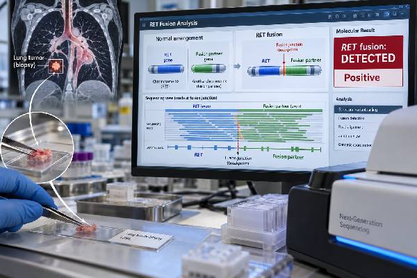
A RET fusion test looks for a rearrangement that joins the RET gene to another gene and creates an abnormal RET kinase that can drive non-small cell lung cancer. A confirmed RET fusion is an actionable molecular result because selective RET inhibitors can target this signaling pathway. RET fusions are uncommon, occurring in roughly 1%–2% of NSCLC, most often in lung adenocarcinoma. KIF5B is the most common fusion partner, with CCDC6 and other partners found less often. Broad next-generation sequencing—especially RNA-based fusion testing—is a strong approach because it can identify an expressed, functional fusion while checking many other lung cancer genes at the same time. FISH and RT-PCR can also detect some RET rearrangements, but both can miss or misclassify certain events. A positive result should represent a functional RET fusion, not simply RET protein expression, a point mutation, or a structural change of uncertain significance.
- What a positive RET result means: The tumor contains a RET fusion that is likely acting as a driver and may make the cancer eligible for RET-targeted therapy.
- How common it is: RET fusions occur in about 1%–2% of non-small cell lung cancers and are most often found in adenocarcinoma.
- Common fusion partners: KIF5B is the most frequent partner in lung cancer, followed by CCDC6 and several rarer genes.
- Preferred testing approach: Broad NGS, particularly RNA-based NGS, can identify functional RET fusions and avoid some limitations of single-gene FISH or RT-PCR.
- What a negative result means: No RET fusion was detected by that assay; an inadequate or failed specimen should not be treated as a true negative.
Table of Contents
- What a RET Fusion Is
- Who Should Be Tested for RET Fusions
- How RET Fusions Are Detected
- Understanding a Positive RET Result
- Negative, Equivocal, and Uncertain Results
- RET and Other Lung Cancer Biomarkers
- Practical Next Steps After RET Testing
What a RET Fusion Is
RET stands for rearranged during transfection. The RET gene encodes a receptor tyrosine kinase, a signaling protein involved in normal cell development and growth. In some lung cancers, a chromosome rearrangement breaks RET and joins its kinase-containing portion to a partner gene.
The new fusion can produce a continuously active RET protein. Instead of switching on only when appropriate, the kinase sends persistent growth and survival signals. That makes the RET fusion an oncogenic driver: a molecular alteration that helps maintain the cancer and can be directly targeted with drugs designed to inhibit RET.
In lung adenocarcinoma, KIF5B::RET is the dominant fusion, accounting for a large majority of RET-positive cases in many series. CCDC6::RET is the next most common, while partners such as NCOA4, TRIM33, KIAA1217 and others are less frequent. The exact fusion partner can be important for laboratory confirmation and research, but the main clinical question is whether the event creates a functional RET kinase fusion.
RET fusions occur in about 1%–2% of NSCLC. They are enriched in adenocarcinoma, younger patients, and never-smokers or light smokers. Those associations are not strong enough to replace testing. A person can have a RET-positive tumor without fitting the typical demographic pattern.
A RET fusion is not the same as a RET point mutation. Activating RET point mutations are well known in medullary thyroid carcinoma and some inherited syndromes, while lung cancer is more characteristically driven by RET fusions. A molecular report that lists a RET missense variant therefore needs a different interpretation from a report that identifies KIF5B::RET or another established fusion.
This distinction also matters when a sequencing report contains several RET-related lines. A laboratory may separately report a fusion, a copy-number change, and one or more small sequence variants. Only the alteration with established oncogenic and therapeutic evidence should drive the RET-specific treatment interpretation. When the report is complex, a molecular tumor board can review whether the detected event is known to activate the kinase, whether it has been validated at the RNA level, and whether the evidence matches the disease setting. That extra review can prevent both false reassurance from an incomplete negative test and overtreatment based on an uncertain structural finding.
Who Should Be Tested for RET Fusions
RET fusion testing is part of broad molecular profiling for appropriate patients with non-small cell lung cancer, especially advanced non-squamous NSCLC and lung adenocarcinoma. The low prevalence makes systematic panel testing more effective than trying to identify likely cases from clinical characteristics.
A lung cancer NGS panel can search for RET at the same time as EGFR, ALK, ROS1, BRAF, KRAS, MET, HER2, NTRK, and other clinically relevant genes. This reduces tissue use and helps deliver a complete molecular profile before systemic treatment decisions are finalized.
Testing should not be withheld because a patient smoked. Although RET fusions are more common in people with little or no smoking history, smokers can still have targetable kinase fusions. Similarly, age and sex are probability clues, not testing rules.
The strongest need for a complete molecular profile is in metastatic disease, where a driver result can directly change the preferred first-line treatment. In earlier-stage disease, testing strategy can depend on current guidelines, planned therapy, specimen availability, and local practice.
Repeat testing may become useful at progression. A tumor treated with a selective RET inhibitor can acquire on-target resistance mutations in RET or activate bypass pathways such as MET. A new tissue biopsy or liquid biopsy can sometimes identify these changes and help explain why treatment stopped working.
If an older tumor sample was tested before RET became routinely included on lung cancer panels, the original report should be reviewed. “Molecular testing negative” from an older limited panel may simply mean that RET was never examined.
How RET Fusions Are Detected
RET fusion testing can be performed by RNA-based NGS, DNA-based NGS, fluorescence in situ hybridization, RT-PCR, and some rapid multiplex RNA assays. Current evidence supports broad NGS as a preferred approach when it is available and the sample is adequate.
RNA-based NGS
RNA sequencing is especially useful because it detects the actual fusion transcript. It can identify the partner, fusion junction, and whether a transcript consistent with a functional kinase fusion is expressed. The 2024 RETING study used RNA-based NGS as the criterion-standard method and showed that some RET fusions were missed by FISH or RT-PCR.
The main limitation is RNA quality. Formalin-fixed tissue can be degraded, and tiny biopsies may not yield enough RNA. A failed RNA assay is technically indeterminate, not biologically negative.
DNA-based NGS
DNA panels can detect multiple alteration types in one test, including mutations and structural variants. Their ability to identify RET fusions depends on whether the relevant introns and breakpoints are covered. When DNA detects an unusual rearrangement, RNA confirmation can demonstrate whether it produces an expressed functional fusion.
Fluorescence in situ hybridization
RET FISH uses break-apart probes positioned around the RET locus. Separated signals indicate a rearrangement. The method does not require prior knowledge of the partner gene, which is an advantage.
Its limitation is specificity for functional fusions. A structural break can produce a FISH-positive pattern without generating a biologically active RET transcript. Studies have documented variable concordance between RET FISH and RNA sequencing, so borderline or unexpected FISH results often need molecular confirmation.
RT-PCR and rapid RNA assays
RT-PCR can be very sensitive for known fusion junctions but can miss rare partners or breakpoints that are not included in the assay design. Newer multiplex RNA assays can test several lung cancer fusions quickly, but the report should still state which fusion classes are covered and how unexpected events are handled.
Unlike ALK, there is no broadly accepted RET immunohistochemistry assay that reliably serves as a stand-alone clinical fusion test in NSCLC. RET protein staining is therefore not considered equivalent to demonstrating a RET gene fusion.
| Method | Useful feature | Important limitation |
|---|---|---|
| RNA NGS | Detects expressed fusions and identifies partners | Can fail when RNA is degraded or scarce |
| DNA NGS | Profiles many genomic alteration types together | Fusion sensitivity depends on intronic coverage |
| FISH | Shows RET locus rearrangement without knowing partner | Can detect nonfunctional rearrangements and miss complex events |
| RT-PCR | Sensitive for predefined fusion junctions | May miss rare or novel partners |
Understanding a Positive RET Result
A confirmed positive RET fusion means that the tumor has a rearrangement expected to create an active RET kinase. This is a high-value molecular finding because it can directly guide targeted treatment.
A clear report may say KIF5B::RET fusion detected, identify the relevant exons, and classify the event as pathogenic or oncogenic. The report may also state the assay used and whether the fusion was detected in RNA, DNA, or both.
The result has several layers of meaning:
- Diagnostic molecular subtype: The tumor belongs to the RET fusion-positive NSCLC subgroup.
- Predictive meaning: Selective RET inhibitors can produce substantial responses in RET fusion-positive advanced NSCLC.
- Resistance relevance: If the disease later progresses, repeat molecular testing may identify an on-target RET resistance mutation or an alternative pathway that has become dominant.
- No direct staging meaning: The fusion does not show whether disease is stage I or stage IV.
A positive result also does not mean the cancer was inherited. Most RET fusions in lung cancer are somatic, acquired within the tumor. Germline RET testing is a different question, usually considered in contexts such as medullary thyroid cancer or a suggestive family history, not because a lung tumor carries a fusion.
Treatment choice depends on current approvals and guidelines as well as brain metastases, prior therapy, organ function, interactions, and patient goals. Selective RET inhibitors such as selpercatinib and pralsetinib established RET as a therapeutically actionable driver. The oncology team chooses among available options rather than treating every RET-positive patient identically.
Negative, Equivocal, and Uncertain Results
A negative result means no reportable RET fusion was detected by the assay. It does not always prove that no fusion exists.
The reliability of a negative result depends on sample quality and method. A successful RNA-based NGS assay with adequate tumor content provides strong evidence. A failed RNA test, a small specimen with few tumor cells, or a targeted RT-PCR assay covering only common partners provides less reassurance.
Common reasons for a false-negative or noninformative result include:
- low tumor percentage;
- severe RNA degradation;
- uncommon fusion partners not included in a targeted assay;
- breakpoints outside DNA panel coverage;
- complex rearrangements that do not create a classic FISH pattern;
- low circulating tumor DNA in a plasma sample;
- technical failure during library preparation or sequencing.
The RETING study is a useful example of why method matters: RNA NGS identified RET fusions that were not detected by some FISH or RT-PCR approaches. This does not mean those methods have no value; it means discordant or low-confidence results should be resolved with an orthogonal method when clinically important.
A report may also identify a RET rearrangement of uncertain significance. The key questions are whether the RET kinase domain is retained, whether an in-frame transcript is produced, and whether the event has evidence of oncogenic function. A structural variant should not automatically be treated as an actionable fusion.
If RET is truly negative, the search shifts to other drivers. A ROS1 fusion test, ALK fusion testing, EGFR sequencing, MET exon 14 assessment, NTRK fusion testing, or other components of broad profiling may identify the relevant alteration.
RET and Other Lung Cancer Biomarkers
RET is one of several kinase drivers in NSCLC. It is molecularly distinct from ALK, ROS1, NTRK, EGFR, MET, BRAF, KRAS, and HER2, even though these genes can all influence treatment.
Driver alterations are often mutually exclusive because one dominant signaling event can be sufficient to sustain the tumor. However, co-alterations can occur, and the complete sequencing report matters for understanding both the primary driver and potential resistance biology.
PD-L1 testing answers a different question. The PD-L1 TPS test measures immune-checkpoint protein expression rather than a kinase fusion. A RET-positive tumor can also have high PD-L1, but the presence of a targetable oncogenic driver strongly affects treatment sequencing in advanced NSCLC.
Tumor mutational burden also differs from RET. TMB estimates the number of somatic mutations per megabase, while a RET test searches for one specific driver class. Neither result can substitute for the other.
Pathology markers such as TTF-1 and p40 are used to classify tumor type. RET fusion-positive cancers are commonly adenocarcinomas and may be TTF-1 positive, but immunophenotype cannot reliably predict whether the fusion is present. Molecular testing is required.
The overall biomarker strategy therefore has several layers: histology identifies what the tumor is, molecular profiling identifies what may be driving it, and immune biomarkers help evaluate checkpoint-based treatment options. RET belongs in the driver layer.
Practical Next Steps After RET Testing
RET testing does not require fasting or special patient preparation. The critical practical issue is obtaining enough viable tumor and preserving tissue for all required biomarkers.
After the report arrives, review these questions:
- Is the finding a true RET fusion or another RET alteration?
- Was the fusion partner identified? KIF5B and CCDC6 are common, but rarer partners can still be valid.
- Which method detected it? RNA NGS usually provides the clearest evidence of an expressed fusion.
- Did the assay pass quality controls? A failed or inadequate test is not a negative result.
- Has broad molecular profiling been completed? The full report may contain other clinically important changes.
- Does the result alter treatment now? The answer depends on stage, prior therapy, current approvals, and oncology guidance.
- Would repeat testing help at progression? Tissue or plasma profiling can reveal resistance mechanisms in some cases.
If the only result is “RET FISH positive,” it can be reasonable to ask whether an RNA-based method confirmed the event, particularly if the pattern is borderline or the clinical profile is unusual. If RT-PCR is negative but the assay covered only common fusion partners, broad NGS can provide a more complete answer.
For a positive, well-characterized fusion, the molecular meaning is straightforward: the cancer has activated RET signaling that can be therapeutically targeted. For a negative result, the key question is whether the test truly had the technical ability to exclude RET. That distinction—biologically negative versus technically uninformative—is essential in rare fusion testing and can directly affect access to precision treatment.
References
- RET Fusion Testing in Patients With NSCLC: The RETING Study 2024
- Rearranged During Transfection Rearrangement Detection by Fluorescence In Situ Hybridization Compared With Other Techniques in NSCLC 2024 (Review)
- Targeting RET alterations in non-small cell lung cancer 2024 (Review)
- Current status of molecular diagnostics for lung cancer 2024 (Review)
- Multicenter evaluation of an automated, multiplex, RNA-based molecular assay for detection of ALK, ROS1, RET fusions and MET exon 14 skipping in NSCLC 2024
- Gene Fusion Detection in NSCLC Routine Clinical Practice: Targeted-NGS or FISH? 2023
Disclaimer
RET fusion results should be interpreted by qualified molecular pathology and oncology professionals using the complete diagnosis and assay-quality information. A confirmed fusion can have major treatment implications, while a point mutation, uncertain rearrangement, or technically limited negative result may mean something different. Do not start, stop, or change cancer treatment based on a RET report without specialist review.