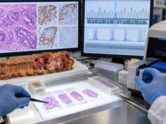
A colorectal cancer biomarker panel combines tumor and blood tests that answer different clinical questions: Which treatments are likely to work, could the cancer be related to Lynch syndrome, what is the tumor’s prognosis, and how can disease be monitored over time? In metastatic colorectal cancer, core molecular testing commonly includes KRAS and NRAS, BRAF V600E, and mismatch repair or microsatellite instability status. Depending on the tumor and treatment setting, testing may expand to HER2 amplification, NTRK fusions, and broader next-generation sequencing. CEA is different from these genomic markers; it is a serum protein used mainly for monitoring and recurrence surveillance. Results should be available early enough to guide treatment, because a RAS mutation can rule out standard anti-EGFR therapy, MSI-high/dMMR can support immunotherapy, and BRAF V600E can identify a targeted treatment pathway. No single biomarker describes the whole cancer, so the panel should be interpreted as an integrated molecular profile.
- KRAS and NRAS mutations predict resistance to standard anti-EGFR antibodies in metastatic colorectal cancer.
- BRAF V600E provides prognostic information and can identify patients for BRAF-plus-EGFR targeted therapy.
- MSI-high or dMMR status predicts strong sensitivity to immune checkpoint inhibitors and can raise concern for Lynch syndrome.
- CEA is a blood monitoring marker, not a treatment-selection mutation test.
- Broader profiling can add HER2, NTRK, and other rare actionable alterations when clinically appropriate.
Table of Contents
- What the Biomarker Panel Is For
- KRAS and NRAS Testing
- BRAF V600E Testing
- MSI and Mismatch Repair Testing
- CEA and Disease Monitoring
- Expanded Molecular Testing
- How to Read a Complete Result
What the Biomarker Panel Is For
A biomarker panel is not one laboratory test. It is a coordinated group of assays selected to answer treatment, hereditary, prognostic, and monitoring questions.
The most important distinction is between tumor biomarkers and blood tumor markers. Tumor biomarkers such as KRAS, NRAS, BRAF, and MSI/MMR describe the cancer’s molecular makeup. They are usually tested on formalin-fixed tumor tissue or, in advanced disease, sometimes through circulating tumor DNA. CEA is measured in blood and changes over time with disease activity in some patients.
In metastatic colorectal cancer, biomarker testing should be performed early because first-line treatment can depend on the results. A left-sided, RAS-wild-type tumor may be considered for an anti-EGFR antibody, while a RAS-mutant tumor should not receive that strategy. An MSI-high/dMMR tumor may be treated with immunotherapy. A BRAF V600E tumor has a distinct targeted-treatment pathway.
The panel also supports hereditary assessment. MMR immunohistochemistry or MSI testing is recommended broadly in colorectal cancer because mismatch repair deficiency can be the first clue to Lynch syndrome.
Testing should use adequate tumor material. A pathologist confirms tumor content and may mark areas for macrodissection. If the tissue is old, scant, or exhausted, a new biopsy or plasma ctDNA assay may be considered depending on the clinical need.
KRAS and NRAS Testing
KRAS and NRAS are members of the RAS family, which transmits growth signals downstream of the epidermal growth factor receptor (EGFR). Activating RAS mutations keep the pathway turned on even when EGFR is blocked.
That biology makes RAS status a negative predictive biomarker for anti-EGFR therapy. Standard cetuximab or panitumumab strategies do not provide the expected benefit when a qualifying KRAS or NRAS mutation is present.
Modern extended RAS testing evaluates key hotspots in KRAS and NRAS exons 2, 3, and 4, not just the classic KRAS codon 12 and 13 variants. A report that says “KRAS wild type” but does not mention NRAS or extended exons may be incomplete for metastatic treatment planning.
A KRAS test for colon cancer is therefore only one component of full RAS assessment. NRAS testing completes the common anti-EGFR resistance evaluation.
Tumor sidedness adds another layer. Even among RAS-wild-type cancers, first-line anti-EGFR therapy generally has the strongest role in left-sided metastatic primary tumors. Right-sided tumors have different biology and are often treated with other first-line strategies.
BRAF V600E Testing
BRAF lies downstream of RAS in the same MAPK signaling pathway. The V600E mutation activates BRAF independently and defines a clinically important colorectal cancer subtype.
In metastatic disease, BRAF V600E is associated with poorer average prognosis compared with BRAF-wild-type disease. It also predicts benefit from a combination strategy that blocks both BRAF and EGFR. As of February 2026, U.S. FDA approval includes encorafenib plus cetuximab with fluorouracil-based chemotherapy for adults with BRAF V600E metastatic colorectal cancer, including first-line treatment. Later-line BRAF-plus-EGFR strategies can also remain relevant depending on prior treatment and the current guideline or regulatory setting. This is why BRAF testing is performed alongside RAS rather than left for later.
BRAF has an additional role in Lynch syndrome triage. If MMR immunohistochemistry shows loss of MLH1 and PMS2, a positive BRAF V600E result strongly supports sporadic MLH1 promoter methylation. A negative result does not establish Lynch syndrome, so direct MLH1 methylation testing or germline evaluation may still be needed.
The dedicated BRAF V600E test therefore has two clinical meanings: treatment/prognosis in advanced cancer and hereditary triage in MLH1-deficient tumors.
Non-V600 BRAF mutations are less common and should not automatically be managed like V600E. Broad sequencing helps distinguish the exact variant class.
MSI and Mismatch Repair Testing
Mismatch repair proteins correct errors that occur when DNA is copied. The core proteins are MLH1, PMS2, MSH2, and MSH6. When the pathway fails, repetitive DNA sequences called microsatellites become unstable.
Two common testing approaches are:
- MMR immunohistochemistry (IHC), which looks for loss of MLH1, PMS2, MSH2, or MSH6 protein expression; and
- MSI testing, performed by PCR or NGS, which detects instability at microsatellite loci.
Tumors with absent repair protein function are called dMMR, and those with a high level of microsatellite instability are called MSI-H. The categories overlap strongly, although discordant cases occasionally require review.
The result has two major uses. First, MSI-H/dMMR metastatic colorectal cancer is highly responsive to immune checkpoint blockade compared with conventional chemotherapy in many patients. Second, dMMR can indicate Lynch syndrome.
The specific IHC pattern guides hereditary workup. Loss of MLH1/PMS2 often leads to BRAF V600E and/or MLH1 promoter methylation testing. Loss of MSH2/MSH6, isolated MSH6, or isolated PMS2 more directly raises concern for a germline mismatch repair gene defect.
A detailed MMR IHC test interpretation can help explain why each protein-loss pattern points to a different next step.
CEA and Disease Monitoring
CEA is fundamentally different from RAS, BRAF, and MSI. It is a circulating glycoprotein, not a tumor DNA alteration.
CEA is commonly measured before treatment to establish a baseline. If it is elevated and then falls after surgery or chemotherapy, future serial values can act as a personalized disease marker. A sustained rise after curative treatment can be an early clue to recurrence and may prompt CT or other imaging.
Many laboratories use an upper limit around 3 ng/mL for nonsmokers and 5 ng/mL for smokers, although ranges vary. Smoking, liver disease, inflammatory bowel disease, lung inflammation, pancreatitis, and other cancers can also raise CEA.
A normal CEA does not rule out active colorectal cancer, because not all tumors secrete it. The CEA test for colon cancer is most useful when the original cancer clearly produced CEA.
CEA should not be used to decide whether anti-EGFR therapy or immunotherapy will work. It also does not replace surveillance colonoscopy or cross-sectional imaging.
Expanded Molecular Testing
Broad molecular profiling can identify less common but actionable alterations that are not captured by the core panel.
HER2 amplification or overexpression occurs in a small subset of colorectal cancers, enriched among RAS/BRAF-wild-type tumors. HER2-targeted combinations can be effective in selected metastatic patients, so testing becomes important after standard options or as part of comprehensive profiling.
NTRK fusions are rare in colorectal cancer, but their importance is disproportionate to their frequency because TRK inhibitors can produce substantial responses across tumor types. A NTRK fusion test is especially relevant when broad NGS finds an unusual rearrangement or when the tumor is RAS/BRAF wild type with atypical molecular features.
Other assays may report tumor mutational burden, POLE/POLD1 mutations, RET or ALK fusions, and additional rare targets. Not every detected alteration has an approved treatment in colorectal cancer, so “mutation found” does not automatically mean “actionable.”
RNA-based sequencing can improve fusion detection, while DNA-based panels are strong for point mutations, small insertions/deletions, and copy-number changes. Some laboratories combine DNA and RNA for more complete coverage.
Liquid biopsy can be valuable when tissue is unavailable or turnaround is urgent. Its main limitation is false negativity when the tumor sheds little circulating DNA. Tissue testing is still needed when a negative plasma result would leave an important treatment question unresolved.
How to Read a Complete Result
A useful colorectal biomarker report should answer several questions in sequence.
| Marker | Main result categories | Clinical meaning |
|---|---|---|
| KRAS/NRAS | Mutant or wild type | Predicts anti-EGFR resistance when activating RAS mutation is present |
| BRAF | V600E, other variant, or wild type | Prognosis, targeted therapy, and MLH1-deficient Lynch workup |
| MSI/MMR | MSI-H/dMMR or MSS/pMMR | Immunotherapy selection and Lynch syndrome screening |
| CEA | Serum level and trend | Monitoring and recurrence surveillance |
| HER2 | Amplified/overexpressed or negative | Potential HER2-targeted therapy in selected metastatic disease |
| NTRK | Fusion present or absent | Potential tumor-agnostic TRK therapy |
The next step depends on the entire pattern. For example, an MSI-H, BRAF V600E tumor may be eligible for immunotherapy and may also have a BRAF-targeted option later. A left-sided RAS/BRAF-wild-type, MSS tumor may be a strong anti-EGFR candidate. A RAS-mutant tumor generally is not.
Ask whether the report is complete for the current stage of disease. A localized stage II cancer may need MSI/MMR primarily for prognosis and hereditary assessment, while metastatic disease requires a wider treatment-selection profile.
Also distinguish not detected from not tested. A report can look reassuring while actually lacking an assay for a clinically relevant biomarker.
Finally, biomarker results are not static instructions. New therapies and indications continue to emerge. Molecular findings should be revisited at each major treatment transition to determine whether a previously “nonactionable” alteration has become clinically relevant.
When biomarker testing should be repeated
Most foundational colorectal cancer driver alterations are stable enough that they do not need to be retested repeatedly without a reason. A high-quality tissue result for KRAS, NRAS, BRAF, and MSI/MMR usually remains valid for initial treatment planning. Repeat testing becomes more useful when the original assay was incomplete, tissue quality was poor, the disease has evolved after multiple targeted therapies, or a new technology can answer a question the first panel could not.
Circulating tumor DNA can be especially helpful after anti-EGFR therapy. Resistant RAS or EGFR-pathway clones can emerge under treatment pressure and later decline when that pressure is removed. In selected settings, ctDNA may help determine whether a rechallenge strategy is biologically reasonable. This is a specialized use and should not be confused with routine baseline RAS testing.
For hereditary assessment, germline testing is separate from tumor retesting. If tumor MSI/MMR raises concern for Lynch syndrome, a blood or saliva germline panel may be needed even though the tumor panel is already complete.
Why specimen quality can change the apparent result
Molecular tests require enough viable tumor. If a specimen contains mostly fibrosis, necrosis, mucin, or normal tissue, a true mutation can fall below the assay’s detection threshold. Pathologists often macrodissect a block to enrich tumor before DNA extraction.
Different metastases can also contain different resistant subclones, although major early drivers such as KRAS or BRAF are often shared. A liver biopsy obtained years after diagnosis may therefore reveal additional alterations that were absent or below detection in the original primary tumor.
The report’s technical section matters. Tumor percentage, sequencing depth, limit of detection, genes covered, and whether RNA was included can explain why a result is confidently negative or only weakly informative.
Predictive, prognostic, and monitoring biomarkers are not interchangeable
A common source of confusion is using one biomarker category to answer another category’s question. Predictive biomarkers estimate whether a treatment is likely to work; RAS status for anti-EGFR therapy and MSI-H/dMMR for checkpoint inhibition are examples. Prognostic biomarkers describe average disease behavior independent of a specific treatment; BRAF V600E has an important prognostic role. Monitoring biomarkers track disease burden over time; CEA is the classic example.
One test can serve more than one role. BRAF V600E is both prognostic and predictive. MSI/MMR is predictive for immunotherapy and can trigger hereditary evaluation. The key is to know which clinical question is being asked at that moment.
This framework also prevents overreaction to CEA. A high CEA does not make a patient eligible for a targeted drug. Likewise, a KRAS mutation does not tell whether the total tumor burden is rising this month.
How to prepare for a treatment-planning visit
A complete biomarker packet should include the pathology diagnosis, stage, primary tumor location, KRAS/NRAS result, BRAF result, MSI/MMR result, HER2 or NTRK findings when tested, and the baseline CEA. If NGS was performed, bringing the full report is more useful than a summary that says only “no actionable mutations.”
Ask whether every required biomarker was actually tested, whether the panel was done on tissue or plasma, and whether any result is indeterminate. If the cancer is left-sided and RAS/BRAF wild type, ask how that affects anti-EGFR options. If it is MSI-H/dMMR, ask how that changes first-line therapy and whether Lynch syndrome evaluation is needed.
A panel becomes clinically valuable only when each result is connected to a decision. The goal is not to collect molecular data for its own sake, but to make sure the right treatment and hereditary questions are answered early enough to matter.
Why tumor sidedness belongs next to the biomarker panel
Primary tumor location is not a molecular test, but it changes how molecular results are used. A left-sided RAS-wild-type metastatic cancer is generally a stronger candidate for first-line anti-EGFR therapy than a right-sided RAS-wild-type cancer. This is a reminder that precision treatment combines anatomy with genomics rather than using mutations in isolation.
The original operative and colonoscopy reports should therefore identify the primary site accurately. “Colon cancer” alone may be insufficient when treatment decisions depend on whether the tumor arose to the right or left of the splenic flexure.
Bottom-line interpretation
A complete colorectal panel should be actionable, not merely comprehensive. The clinician should be able to point to each major result and explain what it changes: RAS for anti-EGFR eligibility, BRAF for prognosis and targeted therapy, MSI/MMR for immunotherapy and Lynch evaluation, and CEA for longitudinal monitoring. If a result has no clear implication, its evidence level and trial relevance should be stated rather than presented as an established target. This approach makes a long sequencing report easier to use and protects patients from overinterpreting rare variants.
The most useful panel is one whose results arrive before the treatment choice, are technically reliable, and are explained in language the patient and care team can act on.
References
- FDA grants traditional approval to encorafenib for metastatic colorectal cancer with a BRAF V600E mutation 2026 (Official Page)
- Treatment of Metastatic Colorectal Cancer: ASCO Guideline 2023 (Guideline)
- Pan-Asian adapted ESMO Clinical Practice Guidelines for the diagnosis, treatment and follow-up of patients with metastatic colorectal cancer 2023 (Guideline)
- Tumor Biomarker Testing for Metastatic Colorectal Cancer: a Canadian Consensus Practice Guideline 2022 (Guideline)
- Canadian consensus for the assessment and testing of Lynch syndrome 2025 (Position Statement)
- Japanese society for cancer of the colon and rectum (JSCCR) guidelines 2024 for the clinical practice of hereditary colorectal cancer 2025 (Guideline)
Disclaimer
Colorectal biomarker results guide treatment and hereditary evaluation but do not replace pathology, staging, imaging, or individualized oncology care. The clinical meaning depends on disease stage, tumor sidedness, exact variant, assay method, and current treatment guidelines. Genetic counseling is appropriate when MSI/MMR or family history raises concern for Lynch syndrome.