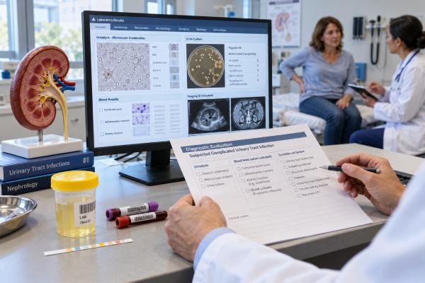
A complicated UTI test panel is not one standardized laboratory package. It is a clinical workup used when a urinary infection may extend beyond simple bladder cystitis, involve systemic illness, or occur with factors that can change treatment or increase the risk of treatment failure. The core tests are usually urinalysis and urine culture with antibiotic susceptibility. If fever, flank pain, sepsis, kidney dysfunction, obstruction, or another complication is suspected, clinicians may add a complete blood count, creatinine and electrolytes, blood cultures, pregnancy testing when relevant, and imaging such as ultrasound or CT. C-reactive protein, procalcitonin, and lactate can provide information about inflammation or illness severity in selected patients, but none of them can diagnose a complicated UTI by itself. The term “complicated UTI” is also evolving: newer guidelines emphasize infection extending beyond the bladder and systemic features, while older definitions often focused on anatomy, catheters, male sex, or comorbid conditions. Testing should therefore match the patient’s actual syndrome and risk factors.
- The core tests for suspected complicated UTI are usually urinalysis plus urine culture and antibiotic susceptibility testing.
- Fever, flank pain, low blood pressure, vomiting, or systemic illness may justify blood tests, blood cultures, and assessment for kidney infection or sepsis.
- Creatinine and eGFR help show whether kidney function is impaired and can affect antibiotic dosing and imaging decisions.
- Ultrasound or CT is used when obstruction, stones, abscess, urinary retention, or failure to improve is a concern; imaging is not required for every UTI.
- CRP and procalcitonin may support severity assessment, but symptoms, urine testing, cultures, kidney function, and imaging are more important for diagnosis and source control.
Table of Contents
- What Counts as a Complicated UTI
- Urinalysis and Urine Culture: Core Tests
- Blood Tests, Kidney Function, and Infection Markers
- When Blood Cultures and Sepsis Testing Are Used
- When Ultrasound or CT Imaging Is Needed
- Risk Factors That Change the Workup
- How Results Guide Treatment and Follow-Up
What Counts as a Complicated UTI
“Complicated UTI” has never had one universal definition. Older frameworks often classified a UTI as complicated when the patient was male, had a catheter, urinary obstruction, stones, abnormal anatomy, kidney disease, diabetes, immunosuppression, or another condition thought to increase treatment failure.
The 2025 Infectious Diseases Society of America (IDSA) guideline moved toward a more clinically immediate classification for treatment decisions. It emphasizes whether infection appears confined to the bladder or has progressed beyond it, especially when fever or other systemic features are present. The guideline also considers factors apparent at the point of care, such as catheterization. Importantly, IDSA states that this classification was designed to guide treatment, not to replace diagnostic judgment.
In practice, clinicians still use the term in more than one way. A person with fever and flank pain, a patient with an obstructed infected kidney, and a catheterized patient with systemic infection may all be described as having complicated UTI. The useful question is therefore not the label alone, but what makes the case higher risk.
Features that may prompt a broader workup include:
- fever, chills, flank pain, or costovertebral-angle tenderness;
- vomiting or inability to maintain hydration;
- low blood pressure, rapid breathing, confusion, or other sepsis features;
- pregnancy;
- indwelling urinary catheter or recent urinary instrumentation;
- kidney stones, urinary retention, hydronephrosis, or known structural disease;
- impaired kidney function or a kidney transplant;
- recurrent infection or recent treatment failure;
- prior resistant organisms or substantial recent antibiotic exposure;
- immunocompromise or serious comorbid illness.
A patient with classic lower-bladder symptoms and no systemic illness may need a much smaller workup. Someone with suspected kidney infection, obstruction, or sepsis may need a full pyelonephritis evaluation, blood tests, and imaging in addition to urine testing.
Urinalysis and Urine Culture: Core Tests
Urinalysis gives rapid clues about inflammation and bacterial activity. It may include leukocyte esterase, nitrite, white blood cells, red blood cells, bacteria, protein, pH, and specific gravity. In a patient with compatible symptoms, pyuria supports urinary inflammation, and nitrite can increase the likelihood of infection when positive.
No urinalysis finding proves a complicated UTI. Nitrite can be negative when the infecting organism does not produce nitrite, when urine has not stayed in the bladder long enough, or when the bacterial burden is low. Pyuria can occur with stones, sexually transmitted infections, catheter irritation, and other inflammatory conditions. A UTI urinalysis is therefore a screening and supporting test, not a substitute for symptoms and culture.
Urine culture is especially important when pyelonephritis or another complicated infection is suspected. The culture can identify the organism, quantify growth, and provide susceptibility testing that helps clinicians move from empiric antibiotics to a narrower effective drug.
Collection should occur before antibiotics whenever this can be done without delaying urgent treatment. A midstream clean-catch culture is common. If a urinary catheter is present, the sample should be obtained using an appropriate catheter-sampling technique rather than from the drainage bag.
The report must be interpreted in context. A traditional threshold of 10⁵ CFU/mL is not a universal cutoff for every symptomatic patient. Lower colony counts can be significant when a typical uropathogen grows from a properly collected sample and symptoms fit. Conversely, bacteriuria can represent colonization rather than infection in catheterized or asymptomatic patients.
Susceptibility testing becomes particularly valuable in complicated cases because resistant organisms are more likely after prior healthcare exposure, recurrent UTIs, or recent antibiotics. If the laboratory reports ESBL production, carbapenem resistance, or another major resistance pattern, an antibiotic-resistant UTI result can substantially change therapy.
Blood Tests, Kidney Function, and Infection Markers
Blood tests are not needed for every lower UTI, but they become more useful when a patient is systemically ill, dehydrated, older or medically vulnerable, or has possible kidney involvement.
A complete blood count (CBC) can show leukocytosis, leukopenia, anemia, and platelet abnormalities. A high white blood cell count can support infection or inflammation, but it is nonspecific. A low white blood cell count in severe infection can be more concerning than a modest elevation because it may occur in sepsis or bone-marrow suppression.
Serum creatinine is one of the most practical kidney tests in this setting. It is used to estimate glomerular filtration rate (eGFR), assess for acute kidney injury, and help determine medication dosing. Dehydration, sepsis, obstruction, pre-existing chronic kidney disease, and nephrotoxic medications can all raise creatinine.
A basic metabolic panel commonly provides creatinine, blood urea nitrogen, sodium, potassium, bicarbonate, and glucose. Abnormal electrolytes can reflect dehydration, vomiting, kidney dysfunction, or severe systemic illness. A rising creatinine may prompt a more urgent search for obstruction or an acute kidney injury caused or worsened by infection.
Inflammatory markers can add context. C-reactive protein (CRP) rises with many infectious and noninfectious inflammatory states. Procalcitonin may increase with significant bacterial infection and has been associated with more severe pyelonephritis in some studies. A 2025 meta-analysis of outpatient UTI risk factors found that elevated procalcitonin and CRP were associated with hospitalization, along with hypotension, fever, elevated creatinine, older age, and diabetes.
These markers should not be treated as yes-or-no UTI tests. There is no single CRP or procalcitonin cutoff that can reliably distinguish complicated UTI from every other infection. They are most useful as part of severity assessment, particularly when the clinical picture is uncertain or the patient is very ill.
Pregnancy testing is also relevant in people who could be pregnant when pregnancy cannot be excluded from history. Pregnancy changes antibiotic choices, imaging decisions, and the significance of bacteriuria.
When Blood Cultures and Sepsis Testing Are Used
Blood cultures are not routinely necessary for uncomplicated cystitis. They become appropriate when there is sepsis, severe systemic illness, suspected bloodstream infection, or another reason to think bacteria may have spread beyond the urinary tract.
The 2026 European Association of Urology guidance recommends considering routine blood tests in systemic UTI and obtaining blood cultures in patients with sepsis or severe illness. It also highlights Staphylococcus aureus in urine as a special finding that can be associated with bloodstream infection and deserves careful assessment.
Blood cultures are ideally drawn before antibiotics when this does not meaningfully delay treatment. If the same organism grows in urine and blood, that supports a urinary source of bacteremia. However, a negative blood culture does not rule out pyelonephritis or complicated UTI.
Lactate is often measured when sepsis or shock is suspected. Elevated lactate can signal impaired tissue perfusion or severe physiologic stress, but it is not a kidney-infection-specific marker. A normal lactate also does not exclude serious infection. It should be interpreted with blood pressure, mental status, urine output, oxygenation, and organ-function tests.
Other tests may be added depending on severity. Liver enzymes, coagulation studies, arterial or venous blood gas measurements, and repeated kidney-function tests can help assess organ dysfunction in sepsis. These are not part of a fixed “UTI panel”; they are part of a systemic-illness workup.
Urine output itself is also clinically important. A sharp fall in urine volume can reflect dehydration, low kidney perfusion, acute kidney injury, or urinary obstruction. In a sick patient, clinicians may track fluid intake and urine output over time rather than relying on a single creatinine value. A creatinine that is still “within range” can lag behind an evolving acute injury, so trends, urine output, blood pressure, and the patient’s baseline all matter.
Warning signs that justify urgent sepsis assessment include low blood pressure, new confusion, very rapid breathing, mottled or cool skin, markedly reduced urine output, severe weakness, or rapidly worsening illness. In that setting, collecting cultures matters, but treatment and source control should not be delayed while waiting for results.
When Ultrasound or CT Imaging Is Needed
Imaging does not diagnose most simple UTIs, and routine CT scanning for every case would expose patients to unnecessary radiation and cost. Imaging becomes valuable when clinicians need to identify a structural problem or complication that laboratory tests cannot show.
Important reasons include suspected urinary obstruction, hydronephrosis, kidney or ureteral stones, urinary retention, renal or perinephric abscess, emphysematous infection, an infected cyst, or poor response to appropriate therapy.
Ultrasound is often the first imaging test because it does not use ionizing radiation and can show hydronephrosis, bladder retention, and some stones or abscesses. It is especially useful when radiation should be minimized, such as during pregnancy, though the exact imaging plan depends on the clinical problem.
CT provides greater anatomic detail and is often preferred when obstruction, abscess, complicated pyelonephritis, or another abdominal diagnosis is strongly suspected. European guidance recommends additional cross-sectional imaging when hydronephrosis or upper-tract obstruction is suspected, and when symptoms persist despite 48–72 hours of appropriate therapy. Immediate advanced imaging may be warranted when the patient deteriorates.
A 2026 systematic review of acute and obstructive pyelonephritis found CT sensitivities reported between 71% and 100% for obstructive complications across included studies, reinforcing its value when a dangerous obstruction is suspected. The key issue is not simply proving inflammation in the kidney; it is finding a problem that requires drainage, stenting, surgery, or another change in management.
MRI can be used in selected patients when CT is undesirable or when specific soft-tissue detail is needed. Imaging choice should account for pregnancy, kidney function, contrast risks, prior imaging, local availability, and the suspected complication.
Risk Factors That Change the Workup
Risk factors influence both testing and initial treatment because they change the likelihood of resistance, treatment failure, obstruction, or severe disease. The most useful risk factors are those tied to the current patient rather than broad labels.
Prior urine cultures are particularly valuable. The 2025 IDSA cUTI guideline notes that recent prior susceptibility results can help predict current susceptibility and recommends avoiding antibiotics to which a previous uropathogen was resistant. More recent cultures are generally more informative than remote ones.
Recent antibiotic exposure also matters. IDSA specifically advises caution with fluoroquinolones when that class was used during the previous 12 months. More broadly, repeated antibiotics can select resistant Enterobacterales and other organisms.
Other factors that may broaden the workup include:
- an indwelling catheter or recent instrumentation;
- a history of ESBL-producing or carbapenem-resistant bacteria;
- recurrent UTI or a recent relapse;
- urinary stones or known obstruction;
- neurogenic bladder or incomplete bladder emptying;
- kidney transplant or significant chronic kidney disease;
- diabetes with severe illness or poor control;
- immunosuppressive therapy;
- pregnancy;
- recent hospitalization or residence in a healthcare facility.
A catheterized patient requires special interpretation because pyuria and bacteriuria are common even without symptomatic infection. A catheter urine culture should be obtained for a clinical reason and from the correct sampling site, not simply because urine looks cloudy.
Recurrent infections also change the value of culture. A recurrent UTI workup often depends on comparing current and prior organisms, susceptibility patterns, treatment response, and whether the same organism returns soon after therapy.
How Results Guide Treatment and Follow-Up
Complicated UTI testing is useful only if the results change management. At the start, clinicians combine illness severity, prior microbiology, recent antibiotics, allergies, kidney function, local resistance data, and the suspected infection site to choose empiric therapy.
When culture and susceptibility results become available, treatment should usually be narrowed to an effective targeted antibiotic. The 2025 IDSA guideline explicitly supports moving away from unnecessarily broad empiric therapy once the pathogen and susceptibility pattern are known.
Kidney-function results may require dose adjustment or a different drug. Imaging may reveal that antibiotics alone are not enough. An infected obstructed kidney, for example, can require urgent urinary drainage because source control is critical; changing antibiotics repeatedly without relieving the obstruction may fail.
Response over the first 48–72 hours also provides diagnostic information. Fever and pain should generally begin improving with effective therapy, although exact timing varies. Persistent or worsening fever, new hypotension, rising creatinine, ongoing vomiting, or increasing pain should prompt reassessment for resistance, an incorrect diagnosis, obstruction, abscess, or another infection source.
Routine post-treatment urine cultures are usually unnecessary when symptoms have fully resolved, even after pyelonephritis. A post-treatment culture becomes more useful when symptoms persist, recur soon after therapy, pregnancy creates a specific test-of-cure indication, or a clinician needs to document clearance for another reason.
A complicated UTI panel is therefore best understood as a decision-based set of tests rather than a fixed checklist. Urinalysis and urine culture identify urinary inflammation and the likely organism. Blood tests show systemic impact and kidney function. Blood cultures look for bacteremia in selected patients. Imaging finds obstruction or complications. Together, those results help clinicians answer the most important questions: Is this truly a UTI, how sick is the patient, is the kidney or urinary tract obstructed, what organism is responsible, and what treatment is most likely to work safely?
References
– IDSA 2025 Guideline Update on Complicated Urinary Tract Infections 2025 (Guideline) – EAU Guidelines on Urological Infections – THE GUIDELINE 2026 (Guideline) – Guidelines for the Prevention, Diagnosis, and Management of Urinary Tract Infections in Pediatrics and Adults: A WikiGuidelines Group Consensus Statement 2024 (Guideline) – Clinical Characteristics, Microbiological Spectrum, Biomarkers, and Imaging Insights in Acute Pyelonephritis and Its Complicated Forms-A Systematic Review 2026 (Systematic Review) – Risk factors for poor prognosis in adult outpatient urinary tract infection: a meta-analysis 2025 (Meta-Analysis) – The EFLM European Urinalysis Guideline 2023 2024 (Guideline)
Disclaimer
This article provides general information about testing for complicated urinary tract infection and does not replace medical evaluation. The appropriate tests depend on symptoms, pregnancy status, kidney function, urinary anatomy, illness severity, and local guidance. Seek urgent care for signs of sepsis, severe flank pain, persistent vomiting, inability to urinate, pregnancy with systemic symptoms, or rapidly worsening illness.