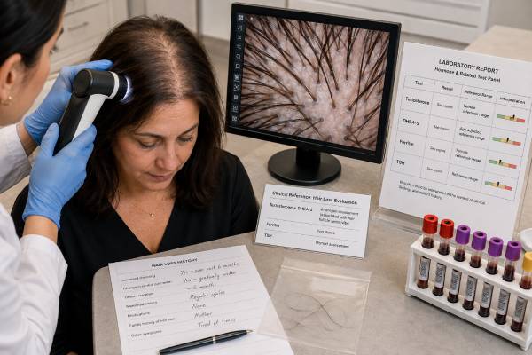
A female hair loss blood panel can identify medical conditions that increase shedding or contribute to patterned thinning, but no single panel diagnoses every cause. Testosterone and DHEA-S help look for androgen excess when hair loss occurs with acne, excess facial or body hair, irregular periods, or virilization. Ferritin estimates iron stores, while thyroid-stimulating hormone, or TSH, screens for thyroid dysfunction that can cause diffuse shedding. Many women with female pattern hair loss have normal hormone results, so scalp examination, hair-loss pattern, timing, medications, nutrition, illness, pregnancy history, and family history remain essential. Tests should be selected from the clinical picture rather than ordered as a fixed package for everyone. Abnormal results also need confirmation and context: low ferritin may reflect iron deficiency, a high testosterone result may depend on assay quality, and an unusual TSH usually requires free T4. Sudden patchy loss, scalp scarring, inflammation, or rapid virilization needs prompt specialist assessment rather than blood testing alone.
- The panel checks common contributors, not every hair disorder: examination usually determines which tests are appropriate.
- Testosterone and DHEA-S are most useful when signs of androgen excess are present.
- Ferritin reflects stored iron: low values support iron deficiency, but the hair-growth threshold is not universally agreed.
- TSH screens thyroid function: an abnormal value is usually interpreted with free T4 and symptoms.
- Normal blood tests do not rule out female pattern hair loss, telogen effluvium, alopecia areata, or scarring alopecia.
Table of Contents
- What a Female Hair Loss Test Panel Can Find
- Hair Loss Patterns That Guide Testing
- Testosterone and DHEA-S Results
- Ferritin and Iron Status
- TSH and Thyroid Testing
- Preparation and Result Accuracy
- Follow-Up, Treatment, and Red Flags
What a Female Hair Loss Test Panel Can Find
A hair loss panel is a group of blood tests chosen to look for treatable factors that can trigger shedding or worsen an existing scalp condition. The common combination of total or free testosterone, DHEA-S, ferritin, and TSH addresses three broad areas: androgen excess, iron deficiency, and thyroid disease.
These tests do not examine the hair follicle directly. They cannot show miniaturization, inflammation, scarring, breakage, or the pattern of follicle loss. A clinician still needs to inspect the scalp and ask how the problem began.
The panel may help detect:
- hyperandrogenism from polycystic ovary syndrome, an adrenal disorder, ovarian hyperthecosis, or rarely a hormone-producing tumor;
- depleted iron stores from heavy periods, pregnancy, low iron intake, gastrointestinal blood loss, or poor absorption;
- hypothyroidism or hyperthyroidism;
- a clue that additional testing is needed, such as free T4, complete blood count, sex hormone-binding globulin, prolactin, zinc, vitamin D, or metabolic tests.
A broad panel is not automatically better. For gradual central scalp thinning with no irregular periods, acne, hirsutism, or virilization, androgen testing may be normal and may not change treatment. For sudden diffuse shedding after childbirth, fever, surgery, rapid weight loss, or severe stress, the history may strongly suggest telogen effluvium, while testing focuses on unresolved triggers such as iron or thyroid disease.
What the panel cannot diagnose by itself
Normal testosterone and DHEA-S do not rule out female pattern hair loss. Hair follicles can be sensitive to normal androgen concentrations because of genetics, local enzyme activity, receptor sensitivity, and age-related changes. Likewise, a low ferritin value does not prove that iron deficiency is the only cause of thinning, and a mildly abnormal TSH does not explain every scalp symptom.
A dermatologist may use dermoscopy, also called trichoscopy, to look for variation in hair shaft diameter, miniaturized hairs, yellow dots, broken hairs, inflammation, or loss of follicular openings. A scalp biopsy is sometimes needed when the diagnosis remains uncertain or when a scarring alopecia is possible.
The most useful panel is therefore a targeted one. Blood tests should answer questions raised by the history and examination, not replace them.
Hair Loss Patterns That Guide Testing
The pattern and speed of hair loss help determine whether hormones, iron, or thyroid testing is likely to help.
Female pattern hair loss
Female pattern hair loss usually develops slowly. The central part widens, density decreases over the crown, and the frontal hairline is often preserved. Some women notice more scalp visibility before they notice increased shedding. The condition can begin after puberty, become more apparent around menopause, and run in families.
Most affected women do not have markedly high circulating androgens. Androgen testing becomes more important when patterned thinning occurs with irregular or absent periods, infertility, hirsutism, severe or persistent acne, acanthosis nigricans, or rapid progression. These features can suggest PCOS or another source of androgen excess.
Telogen effluvium
Telogen effluvium causes increased shedding across the scalp, often two to three months after a trigger. Common triggers include childbirth, high fever, major surgery, a serious infection, rapid weight loss, very low calorie intake, emotional stress, medication changes, thyroid dysfunction, and iron deficiency. The part may look wider because overall density falls, which can make telogen effluvium resemble or uncover female pattern hair loss.
Acute telogen effluvium often improves within several months after the trigger resolves, although visible density takes longer to recover because hair grows slowly. Persistent shedding beyond six months deserves reassessment for ongoing triggers or another diagnosis.
Alopecia areata and scarring alopecia
Alopecia areata commonly produces smooth, sharply defined patches, though it can also cause diffuse loss. It is immune-mediated and may be associated with autoimmune thyroid disease. TSH testing can be reasonable when symptoms, personal history, or family history suggest thyroid disease, but blood tests do not establish alopecia areata.
Scarring alopecias damage follicles permanently. Warning signs include burning, pain, marked itching, redness, scale around follicles, pustules, shiny skin, or disappearing follicular openings. Frontal fibrosing alopecia may cause recession at the front and temples along with eyebrow loss. Central centrifugal cicatricial alopecia often begins at the crown. These patterns require early dermatology review because delay can allow irreversible loss.
Hair breakage and traction
Broken hairs from chemical processing, heat, tight hairstyles, extensions, or repetitive pulling can mimic shedding. Traction alopecia often affects the hairline and areas under tension. Laboratory testing is less useful when the pattern and styling history explain the damage, although another condition can coexist.
A careful examination prevents the common mistake of treating every form of hair loss as a hormone imbalance.
Testosterone and DHEA-S Results
Testosterone and DHEA-S help assess androgen production. The ovaries and adrenal glands both contribute to testosterone, while DHEA-S is produced mainly by the adrenal glands. The pattern can help a clinician decide whether an abnormality appears more ovarian, adrenal, or mixed.
Which testosterone test is most useful
Total testosterone is often the first androgen measured. In women, concentrations are low enough that assay quality matters. Liquid chromatography–tandem mass spectrometry is generally preferred when available. Free testosterone represents the small fraction not tightly bound to proteins, but direct free-testosterone immunoassays can be unreliable. Clinicians may calculate free testosterone from total testosterone, sex hormone-binding globulin, and albumin or use a validated equilibrium-dialysis method.
A free testosterone test can be helpful when total testosterone is normal but sex hormone-binding globulin is low, as can occur with insulin resistance, obesity, hypothyroidism, or some medications. Low SHBG increases the biologically available fraction.
High testosterone
Mild to moderate elevation commonly occurs with PCOS, especially when hair loss accompanies irregular ovulation, hirsutism, or acne. Other causes include ovarian hyperthecosis, congenital adrenal hyperplasia, medications or supplements containing androgens, and laboratory interference.
Marked elevation, rapid symptom progression, deepening voice, increased muscle mass, clitoral enlargement, or loss of the usual frontal hairline raises concern for severe androgen excess. An ovarian or adrenal tumor is uncommon but must be considered. The absolute threshold depends on the assay and laboratory, so clinicians interpret both the number and the pace of symptoms.
High DHEA-S
A DHEA-S test points mainly toward adrenal androgen production. Mild elevations can occur with PCOS. Larger increases may prompt evaluation for an adrenal tumor, nonclassic congenital adrenal hyperplasia, Cushing syndrome, or exogenous DHEA use. DHEA-S normally declines with age, so an age-adjusted range is important.
A high DHEA-S does not prove that adrenal hormones caused the hair loss. It must be considered with testosterone, menstrual history, hirsutism, acne, cortisol-related symptoms, and sometimes 17-hydroxyprogesterone.
Normal and low androgen results
Normal androgen values are common in female pattern hair loss and do not make the condition less real. Low testosterone or DHEA-S is usually not a recognized cause of common female scalp thinning. Symptoms such as fatigue or low libido should not be used to justify over-the-counter DHEA or testosterone treatment without a separate medical indication. Androgen supplements can worsen acne, facial hair growth, scalp thinning, cholesterol levels, and pregnancy risk.
Hormonal contraception, glucocorticoids, antiandrogens, menopause, ovarian suppression, and age can change androgen values. These factors must be recorded before interpretation.
Ferritin and Iron Status
Ferritin is a protein that stores iron. Serum ferritin is the most common blood marker used to estimate iron reserves. Low ferritin strongly supports iron deficiency, which can contribute to diffuse shedding and may coexist with female pattern hair loss.
Iron is needed for rapidly dividing cells, including cells in the hair follicle. Deficiency can also cause fatigue, reduced exercise tolerance, headaches, restless legs, shortness of breath, paleness, and brittle nails. Hair loss may occur before severe anemia develops, so a normal hemoglobin does not always mean iron stores are adequate.
Interpreting ferritin
The laboratory’s lower limit is important, but there is no universally accepted ferritin target guaranteed to restore hair growth. Some hair clinics use higher thresholds than general medical laboratories, yet studies have not established one optimal number for every woman. A result below about 15 to 30 ng/mL often supports depleted stores in otherwise healthy adults, but inflammation, liver disease, infection, obesity, and recent illness can raise ferritin and hide deficiency.
For that reason, clinicians may interpret ferritin with:
- a complete blood count;
- serum iron;
- total iron-binding capacity or transferrin;
- transferrin saturation;
- C-reactive protein when inflammation is suspected;
- symptoms and likely causes of iron loss.
A ferritin result should not be treated as a hair-only value. The cause of iron deficiency matters. Heavy menstrual bleeding is common, but gastrointestinal bleeding, celiac disease, inflammatory bowel disease, bariatric surgery, frequent blood donation, pregnancy, and a restrictive diet can also deplete stores.
Iron treatment and hair recovery
Iron supplements are appropriate when deficiency is confirmed or strongly suspected, but more is not better. Excess iron can cause gastrointestinal side effects and, in severe cases, organ damage. The dose, formulation, schedule, and duration should be matched to the degree of deficiency and the cause. Some patients absorb alternate-day oral iron well; others need treatment of bleeding or malabsorption, and a minority require intravenous iron.
Ferritin may improve before visible hair density does. Shedding can take weeks to slow, and regrowth generally becomes noticeable over several months because follicles need time to re-enter and progress through the growth phase. If ferritin normalizes but loss continues, the diagnosis should be revisited rather than escalating iron indefinitely.
TSH and Thyroid Testing
TSH is produced by the pituitary gland and regulates thyroid hormone production. It is the usual first-line blood test for primary hypothyroidism or hyperthyroidism. Both can disrupt the hair cycle and cause diffuse shedding, though hair loss is rarely the only symptom.
High TSH
A high TSH often indicates that the thyroid is underactive. Free T4 helps determine whether hypothyroidism is overt or subclinical. Other possible symptoms include fatigue, cold intolerance, constipation, dry skin, weight change, slowed thinking, muscle aches, heavier periods, and reduced heart rate.
Hashimoto thyroiditis is a common autoimmune cause. Thyroid peroxidase antibodies may be checked when the diagnosis or future risk needs clarification, but antibody positivity alone does not prove that thyroid disease is causing the hair loss.
Low TSH
A low TSH may reflect an overactive thyroid, excessive thyroid medication, pregnancy-related changes, or a pituitary issue. Free T4 and sometimes free or total T3 clarify the pattern. Hyperthyroidism can cause diffuse fine hair, increased shedding, heat intolerance, tremor, palpitations, anxiety, sweating, and weight loss.
The TSH test must be interpreted using the laboratory range and the patient’s situation. Pregnancy has trimester-specific considerations. Biotin supplements can interfere with some thyroid assays, often creating an apparently low TSH and high thyroid hormone pattern. The laboratory or clinician can advise whether biotin should be held before collection.
Normal TSH
A normal TSH makes common primary thyroid dysfunction less likely but does not explain the type of alopecia. It also does not rule out every pituitary or thyroid condition. Further thyroid testing is based on symptoms, medication use, pregnancy, examination, and the degree of clinical suspicion.
Correcting true thyroid dysfunction may improve related shedding, but regrowth is delayed. Starting thyroid hormone when TSH and free T4 are normal does not treat female pattern hair loss and can cause harmful overtreatment.
Preparation and Result Accuracy
Most hair loss blood tests do not require fasting, although a clinician may request a morning fasting sample when combining them with glucose, insulin, lipid, or other metabolic tests. Androgens are often measured in the morning because daily variation can affect results, especially in younger women.
For menstruating women, early follicular-phase testing may reduce hormonal variation, but irregular cycles can make precise timing impossible. DHEA-S is relatively stable across the day and cycle compared with testosterone. Hormonal contraception can suppress ovarian androgens and raise SHBG, making androgen results difficult to interpret. When safe and clinically necessary, a specialist may plan testing after an appropriate medication-free interval rather than guessing from values obtained during treatment.
Before collection, report:
- birth control, menopausal hormone therapy, fertility medicines, spironolactone, finasteride, glucocorticoids, thyroid medicine, and antiseizure drugs;
- DHEA, testosterone, “hair growth,” thyroid, iron, or high-dose biotin supplements;
- pregnancy, recent childbirth, miscarriage, or breastfeeding;
- recent infection, surgery, major weight change, or intense training;
- heavy periods, dietary restrictions, gastrointestinal symptoms, or blood donation;
- the timing, pattern, and speed of hair loss.
Do not stop prescribed treatment without instructions. Stopping thyroid medicine or antiandrogen therapy abruptly can create more confusion and may be unsafe.
Useful additional tests
The initial panel may be expanded according to the history. A complete blood count can identify anemia. Prolactin may be useful with irregular periods or nipple discharge. 17-hydroxyprogesterone can screen for nonclassic congenital adrenal hyperplasia. Zinc, vitamin D, vitamin B12, folate, or protein status may be considered when diet, malabsorption, or deficiency risk supports testing, but routine testing of every nutrient often produces incidental abnormalities without proving causation.
Pregnancy testing is important before some medications used for hair loss because antiandrogens and 5-alpha-reductase inhibitors can harm a developing fetus. Baseline potassium or kidney tests may be needed before spironolactone in selected patients.
Follow-Up, Treatment, and Red Flags
Follow-up should address both the blood-test abnormality and the scalp diagnosis. Treating low ferritin or thyroid dysfunction helps when those conditions are present, but a woman may still need direct treatment for female pattern hair loss or another alopecia.
A typical plan may include:
- Confirm the hair-loss type. History, pull test, dermoscopy, and sometimes biopsy distinguish pattern loss, telogen effluvium, alopecia areata, traction, and scarring disease.
- Repeat surprising laboratory results. Markedly high testosterone, discordant thyroid values, or ferritin affected by inflammation may need confirmation with a better method or additional markers.
- Treat a documented medical cause. Restore iron stores, correct thyroid dysfunction, stop an offending supplement, or evaluate androgen excess.
- Use hair-specific treatment when appropriate. Topical minoxidil is a common first-line treatment for female pattern hair loss. Low-dose oral minoxidil, spironolactone, finasteride, or other options may be considered by a clinician after reviewing blood pressure, pregnancy potential, medications, and risks.
- Track progress realistically. Standardized photographs and part-width measurements every three to six months are more useful than daily inspection. Most effective treatments require at least six months to judge.
Minoxidil can cause temporary early shedding as follicles shift cycles. Topical products may irritate the scalp or cause unwanted facial hair if they spread. Oral minoxidil can affect blood pressure, heart rate, fluid retention, and body hair. Antiandrogens require pregnancy prevention when conception is possible and are not appropriate for everyone.
Prompt dermatology assessment is important for scalp pain, burning, pustules, heavy scale, shiny scarred areas, eyebrow loss, or rapid recession. Urgent endocrine evaluation is appropriate for rapidly increasing hirsutism, a deepening voice, clitoral enlargement, severe muscle changes, or a markedly elevated androgen result. Seek medical care for severe anemia symptoms, chest pain, fainting, or significant shortness of breath.
Hair growth is slow, and improvement often lags behind correction of the trigger. A normal panel should not end the evaluation when visible loss continues. It redirects attention to the scalp pattern, hair care, medications, genetics, recent stressors, and dermatologic causes.
How to tell whether the plan is working
Hair count changes cannot be judged on the same timeline as a blood test. After a trigger is corrected, shedding may continue for several weeks because follicles that already entered the resting phase still have to release their hairs. New growth then emerges slowly, often first as short tapered hairs along the part or hairline. A ferritin or TSH value may improve months before density looks different, and androgen treatment can reduce future miniaturization without restoring every follicle.
Use consistent evidence rather than mirror checks. Take photographs in the same room, lighting, hairstyle, camera distance, and part position every three months. Record wash-day shedding only if it can be done without increasing anxiety. A dermatologist may use dermoscopy, standardized global photography, or a hair-pull test. If shedding remains heavy after six months, the part continues to widen, or scalp symptoms develop, revisit the diagnosis rather than simply adding supplements. Iron, thyroid hormone, and antiandrogens can all cause harm when used without a documented indication.
References
- Diagnosis and treatment of female alopecia 2023 (Review)
- Recent Advances in Understanding of the Etiopathogenesis, Diagnosis, and Management of Hair Loss Diseases 2023 (Review)
- Female pattern hair loss and polycystic ovarian syndrome 2022 (Review)
- An Update on Alopecia and its Association With Thyroid Autoimmune Diseases 2023 (Review)
- Iron Deficiency and Nonscarring Alopecia in Women: Systematic Review and Meta-Analysis 2022 (Systematic Review)
- The Diagnostic Value of Serum Ferritin for Telogen Effluvium: A Cross-Sectional Comparative Study 2021 (Research Article)
Disclaimer
This article is for general education and does not replace examination by a clinician or dermatologist. Blood results must be interpreted with symptoms, medications, the laboratory method, and the observed hair-loss pattern. Rapid hair loss, scarring signs, severe anemia symptoms, or virilization requires prompt medical assessment.