
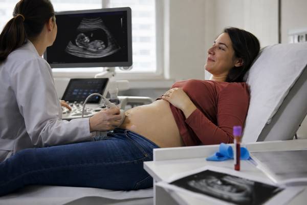
First-trimester screening combines an ultrasound measurement with pregnancy-related blood markers to estimate the chance of Down syndrome and, in many programs, trisomy 18 and trisomy 13. The ultrasound measures nuchal translucency—the fluid-filled space behind the fetal neck—while the blood test measures pregnancy-associated plasma protein A, or PAPP-A, and free beta or total human chorionic gonadotropin, or hCG. A calculation then incorporates gestational age, maternal age, weight, and other information. The result is a probability, such as 1 in 100 or 1 in 5,000; it is not a chromosome diagnosis. A higher-chance result may be followed by cell-free DNA screening or by diagnostic testing with chorionic villus sampling or amniocentesis. Low PAPP-A or an increased nuchal translucency may also matter beyond chromosome risk, so follow-up should address the individual marker pattern and ultrasound findings, not only whether the report crossed a screening cutoff.
- Main components: PAPP-A, hCG, nuchal translucency, age, and gestational data
- Timing: Blood and ultrasound must fall within validated first-trimester windows
- Purpose: Estimates chance of common trisomies; it does not diagnose them
- Result format: A ratio, percentage, or lower-chance/higher-chance category
- Important limitation: A normal calculation cannot exclude all fetal conditions
- Follow-up: cfDNA, detailed ultrasound, CVS, or amniocentesis depending on findings
Table of Contents
- What the Combined Test Measures
- Timing and Preparation
- How PAPP-A and hCG Affect the Calculation
- Nuchal Translucency and Ultrasound Findings
- Reading a 1-in-N Result
- Follow-Up After a Higher-Chance Result
- Special Situations and Common Problems
- Using the Result in Prenatal Care
What the Combined Test Measures
The first-trimester combined screen is a mathematical risk assessment built from several independent observations. It does not look directly at fetal chromosomes. Its major inputs are:
- Maternal age at the expected date of delivery
- Exact gestational age or crown-rump length
- Nuchal translucency measurement
- PAPP-A level
- Free beta-hCG or total hCG level, depending on the program
- Maternal weight and sometimes ethnicity, smoking, diabetes, conception method, and twin status
The laboratory converts the blood-marker concentrations into multiples of the median, abbreviated MoM. A value of 1.0 MoM is close to the median for an unaffected pregnancy at that gestational age after relevant adjustments. A value of 0.5 MoM is about half the median, while 2.0 MoM is about twice the median. MoM allows a result taken at one gestational age to be compared with a result taken at another.
The screening model compares the pattern with patterns observed in pregnancies affected and unaffected by specific trisomies. In trisomy 21, PAPP-A tends to be lower and free beta-hCG tends to be higher than expected. In trisomy 18 and trisomy 13, both markers often trend lower. These are statistical tendencies, not diagnostic rules. Many unaffected pregnancies have one low or high marker, and some affected pregnancies have marker values near the median.
The nuchal translucency measurement provides a separate ultrasound input. Combining age, serum markers, and ultrasound is more informative than any one component alone. The result may be reported separately for trisomy 21 and for trisomies 18 and 13, depending on the local program.
Names vary. “Combined screening,” “first-trimester screen,” “double test plus NT,” and “first-trimester aneuploidy screening” may refer to similar processes. A blood test that measures PAPP-A and hCG without a valid nuchal translucency is not the full combined test. Likewise, an NT scan alone does not provide the same screening performance.
This test differs from cell-free DNA prenatal screening. cfDNA analyzes placental DNA fragments and is more accurate for the common trisomies. Combined screening still provides early ultrasound information and may be part of regional programs, insurance pathways, or a broader assessment of placental and fetal development.
Timing and Preparation
Timing is essential because marker levels and fetal measurements change quickly during the first trimester. Programs define eligibility using gestational age and, more precisely, crown-rump length.
In the English screening program, the combined test is performed when the fetal crown-rump length is 45.0 to 84.0 millimeters, corresponding approximately to 11 weeks 2 days through 14 weeks 1 day. The blood sample can often be taken from 10 weeks. Other countries and laboratories may use slightly different validated windows.
The ultrasound appointment usually includes:
- Confirmation that the pregnancy is intrauterine
- Assessment of fetal number and chorionicity in twins
- Measurement of crown-rump length for dating
- Nuchal translucency measurement in a standardized fetal position
- A limited early review of visible anatomy
The pregnant person does not need to fast for PAPP-A or hCG testing. Some ultrasound units request a moderately full bladder early in pregnancy, but instructions vary. Bring the exact dates and details of fertility treatment, donor eggs, medications, smoking, diabetes, and previous pregnancies because these can affect the calculation.
If the fetus is too small, the sonographer may schedule another scan. If the fetus is too large for the validated crown-rump-length range, the combined calculation may not be possible. A second-trimester option or cfDNA screening may then be discussed. The quadruple screen is not simply a late combined test; it uses different markers and, in many programs, screens mainly for Down syndrome.
An accurate gestational age matters because the expected median concentrations change by week and day. A dating error can shift the calculated MoM and therefore the risk. Laboratories also need the correct maternal weight because blood volume affects measured marker concentration.
Twins require a specialized calculation. Nuchal translucency is measured for each fetus. Serum markers reflect the pregnancy as a whole and cannot be assigned cleanly to one twin. Chorionicity, whether the twins share a placenta, also affects interpretation. Higher-order multiples may have limited validated screening options.
If the blood sample and NT scan are done at separate sites, confirm that both results reach the same screening service. A calculation cannot be completed if one component is missing or if identifying details do not match.
How PAPP-A and hCG Affect the Calculation
PAPP-A is a protein produced largely by the placenta. hCG is a pregnancy hormone produced by placental tissue; it supports the corpus luteum early in pregnancy and is the hormone detected by pregnancy tests. First-trimester screening uses these substances as markers of placental biology.
For trisomy 21, the typical statistical pattern is:
- Lower PAPP-A
- Higher free beta-hCG
- Increased nuchal translucency in some pregnancies
For trisomy 18 or 13, the typical pattern more often includes:
- Lower PAPP-A
- Lower free beta-hCG
- Increased nuchal translucency or structural differences in some pregnancies
A laboratory algorithm weighs how far each value is from the expected median. It then combines the marker likelihoods with the age-related starting chance and the ultrasound input. The result is not derived by simply adding the values.
A low PAPP-A result can have significance even when the final chromosome screen is lower chance. Low PAPP-A is associated at a population level with placental complications such as fetal growth restriction, preeclampsia, preterm birth, and stillbirth. Most pregnancies with low PAPP-A do not experience all or any of these outcomes. The degree of reduction, other health factors, uterine-artery assessment, blood pressure, and later fetal growth all influence management.
Depending on local guidance, follow-up for markedly low PAPP-A may include:
- Review of aspirin eligibility for preeclampsia prevention
- Blood-pressure and urine monitoring
- Additional fetal growth ultrasounds later in pregnancy
- Doppler studies or antenatal surveillance when clinically indicated
Do not start aspirin solely from an online interpretation. Dose, timing, and eligibility depend on the complete risk profile and clinician guidance.
An isolated high hCG result is also nonspecific. It can be influenced by gestational dating, placental function, multiple pregnancy, and laboratory factors. It should not be treated as evidence that the fetus has Down syndrome.
Marker values can differ after IVF, use of donor eggs, smoking, diabetes, and in twin pregnancy. Validated algorithms apply correction factors. Incorrect background information can therefore distort the result. Patients should verify the demographic and pregnancy details printed on the report.
PAPP-A and hCG are not diagnostic biomarkers. A pattern may raise or lower the calculated chance, but only chromosome analysis of placental or fetal cells can establish whether trisomy is present.
Nuchal Translucency and Ultrasound Findings
Nuchal translucency, or NT, is the sonographic appearance of fluid beneath the skin at the back of the fetal neck. Every fetus has some fluid in this region during the first trimester. The measurement is interpreted according to crown-rump length rather than a single universal normal value.
A valid NT measurement requires a specific image. The fetus should be in a neutral position, the image should be magnified, and the calipers should be placed at the inner borders of the fluid space. Flexion can make the measurement look smaller; extension can make it look larger. Sonographer training and quality assurance are therefore important.
An increased NT is associated with a higher chance of:
- Trisomy 21, trisomy 18, trisomy 13, and other aneuploidies
- Congenital heart disease
- Copy-number variants detectable by chromosomal microarray
- Certain single-gene conditions, including some RASopathies
- Structural anomalies and genetic syndromes not detectable by routine aneuploidy screening
- Pregnancy loss
The degree of increase matters, but it does not determine outcome. Some fetuses with a markedly increased NT have a chromosome condition or major anomaly. Others have normal diagnostic testing and healthy outcomes. A normal NT also cannot exclude Down syndrome or every heart defect.
If NT is increased, relying only on a lower-chance cfDNA result may leave important questions unanswered. cfDNA mainly screens selected aneuploidies. Diagnostic testing with microarray may be offered because it examines a wider range of chromosome gains and losses. A detailed early anatomy assessment, later anatomy scan, and fetal echocardiogram may also be recommended.
An NT measurement should not be confused with a second-trimester nuchal fold. They are measured at different gestational ages and have different reference standards.
Other first-trimester ultrasound observations may influence counseling. A missing nasal bone, abnormal ductus venosus flow, tricuspid regurgitation, major structural anomaly, or severe growth discrepancy can alter the chance of aneuploidy. Some centers use these additional markers; others do not include them in routine calculation.
When the fetus cannot be positioned for a valid NT measurement, repeated attempts may be made within the appointment or another scan may be arranged. A guessed or technically poor measurement should not be entered into the risk algorithm.
Reading a 1-in-N Result
The report usually gives a chance ratio. A result of 1 in 100 means that among pregnancies with the same calculated pattern, about one would be expected to have the condition and 99 would not. It is equivalent to a 1% estimated chance.
Examples:
| Reported chance | Approximate percentage | Plain-language interpretation |
|---|---|---|
| 1 in 10 | 10% | Higher chance, but most pregnancies with this estimate are unaffected |
| 1 in 100 | 1% | Increased chance; not a diagnosis |
| 1 in 1,000 | 0.1% | Lower chance in many programs, but not zero |
| 1 in 10,000 | 0.01% | Very low estimated chance within the test’s scope |
Programs set a cutoff to determine which results are labeled “higher chance” or “screen positive.” The cutoff may be 1 in 150, 1 in 250, 1 in 300, or another value. A result just above the cutoff is not biologically different from one just below it. The cutoff is a service decision about when to offer further testing.
The final calculation begins with a prior chance, often strongly influenced by age, and then modifies it using the marker and NT pattern. Two people with identical PAPP-A, hCG, and NT values may receive different final ratios because their age-related starting chances differ.
A higher-chance result means further information should be offered. It does not mean the fetus has Down syndrome. Traditional combined screening has more false positives than cfDNA. Many patients with a higher-chance combined result receive a lower-chance cfDNA result or normal diagnostic testing.
A lower-chance result means the calculated chance falls below the program cutoff. It cannot guarantee typical chromosomes. The test may miss an affected pregnancy, and it does not screen for every chromosome or gene condition.
The report may also state that a result could not be calculated. Reasons include missing blood or ultrasound data, an invalid gestational age, sample timing outside the window, incorrect identification, or a pregnancy type not supported by the algorithm.
Do not interpret PAPP-A and hCG reference ranges from a standard laboratory portal without the screening calculation. Raw concentrations are expected to change with gestation, and the important values are corrected MoMs within the validated model.
Follow-Up After a Higher-Chance Result
Three broad options may follow a higher-chance combined screen: no further genetic testing, more accurate screening with cfDNA, or diagnostic testing.
Cell-free DNA screening can refine the probability without an invasive procedure. It is more accurate for trisomy 21, 18, and 13 than combined screening. A lower-chance cfDNA result can provide substantial reassurance, but it does not explain a markedly increased NT or a structural anomaly. A higher-chance cfDNA result still requires diagnostic confirmation before irreversible decisions.
Chorionic villus sampling obtains placental tissue and can often be performed during the first trimester. It provides earlier diagnostic information. The CVS genetic test may include rapid aneuploidy testing, karyotype, and/or microarray. Rare placental mosaicism can complicate results.
Amniocentesis samples amniotic fluid, usually from 15 weeks. The amniocentesis genetic test provides fetal cells for diagnostic analysis. Some patients prefer to wait for it because it is less affected by confined placental mosaicism.
The choice depends on:
- How high the calculated chance is
- NT measurement and any structural findings
- Gestational age
- Desire for certainty versus desire to avoid an invasive procedure
- Which conditions the patient wants assessed
- Procedure availability and local expertise
- How the information would affect pregnancy care or decisions
When NT is substantially increased or an anomaly is present, diagnostic testing with chromosomal microarray may be more informative than cfDNA alone. A normal cfDNA result would not rule out many copy-number variants or single-gene syndromes associated with increased NT.
A detailed ultrasound is often recommended regardless of the genetic-testing choice. Fetal echocardiography may be appropriate later when NT is increased. If all diagnostic and structural evaluations are normal, clinicians can provide a more reassuring—but never absolute—outlook.
A second serum screen should not be used as definitive confirmation. It may create another probability without resolving whether fetal chromosomes are affected.
Special Situations and Common Problems
First-trimester screening is more complex when the pregnancy or medical history differs from the assumptions of a standard singleton model.
Twins. The scan measures NT for each fetus, but maternal PAPP-A and hCG come from both placentas. In fraternal twins, each fetus may have a different chromosome status. The calculation and detection performance differ from singleton pregnancy. If one twin screens concerning, diagnostic sampling must identify each sac correctly.
Vanishing twin. A demised embryo can alter serum markers and cfDNA. The timing of demise matters. The screening laboratory and maternal-fetal medicine team should know about every gestational sac seen earlier.
IVF and donor eggs. Conception method can affect marker distributions. The age of the egg source is relevant to the baseline chance of aneuploidy, while the pregnant person’s weight and health affect marker correction. Provide accurate donor and embryo-transfer information.
Incorrect dating. A discrepancy between menstrual dates and crown-rump length can change expected medians. The ultrasound dating used by the program should be checked before interpreting unexpected marker values.
Unable to obtain NT. Fetal position, maternal anatomy, fibroids, and scan quality can prevent a valid measurement. Options depend on remaining time in the window and local pathways. They may include a repeat scan, serum-only method, cfDNA, or later screening.
Low PAPP-A but lower-chance aneuploidy result. The chromosome screen may be reassuring while the placental marker still leads to a growth or preeclampsia plan. Ask whether the report triggers separate obstetric follow-up.
High NT with lower-chance result. The ultrasound finding should not be dismissed. Genetic counseling, diagnostic testing, detailed anatomy scans, and fetal heart evaluation may still be indicated.
Previous affected pregnancy or parental chromosome rearrangement. A standard combined screen may not address the family-specific recurrence risk adequately. Direct diagnostic testing or targeted counseling may be more appropriate.
Medication and medical conditions. Diabetes, smoking, and other factors can influence marker values. Laboratories apply adjustments only when the information is supplied correctly.
The most common practical error is focusing on a single value. The combined result, marker MoMs, NT measurement, ultrasound anatomy, and clinical history should be reviewed together.
Using the Result in Prenatal Care
First-trimester screening is a decision tool, not a pass-or-fail judgment about the pregnancy. Its value is that it provides an early, individualized estimate and may reveal ultrasound or placental signals that deserve attention.
After receiving the report, verify:
- The gestational age and crown-rump length
- The NT measurement and whether it was technically valid
- PAPP-A and hCG MoM values
- Maternal age, weight, smoking, diabetes, and conception details
- Which conditions were screened
- The program cutoff
- Whether any separate recommendation was made for low PAPP-A or increased NT
Ask the clinician to translate the ratio into a percentage and to explain the residual chance after any follow-up test. A lower-chance cfDNA result following a higher-chance combined screen does not erase an abnormal ultrasound finding. A normal diagnostic chromosome result does not eliminate the need for anatomy assessment.
Routine prenatal care still includes a second-trimester anatomy ultrasound. Screening for open neural tube defects may require maternal serum AFP or ultrasound because first-trimester combined screening does not adequately address them. The maternal serum AFP test answers a different question.
A higher-chance result can cause intense anxiety even though it is not diagnostic. Request timely genetic counseling rather than trying to infer certainty from internet charts. A counselor can compare cfDNA, CVS, and amniocentesis and explain how each option fits the ultrasound findings and personal goals.
Some patients want the earliest definitive answer. Others want to avoid procedure risk and use additional screening. Some do not want information about fetal chromosomes. All are legitimate positions when informed consent is clear.
A sound interpretation keeps three ideas separate: the screen’s probability, any independent ultrasound or placental concern, and the diagnostic options available. This prevents a single cutoff label from overshadowing the full clinical picture.
References
- Screening for Down’s syndrome, Edwards’ syndrome and Patau’s syndrome 2026 (Program Handbook)
- Combined first-trimester screening 2025 (Clinical Resource)
- Presentation: Pregnant woman with a higher-chance first-trimester combined screening result 2025 (Clinical Guidance)
- Screening for Down’s syndrome, Edwards’ syndrome and Patau’s syndrome 2025 (Patient Guidance)
- Society for Maternal-Fetal Medicine Consult Series #74:Cell-free DNA screening for aneuploidies: Updated guidance 2025 (Guideline)
- Prenatal testing 2023 (Professional Guidance)
Disclaimer
This article provides general information about first-trimester combined screening and cannot interpret an individual report. Marker correction, screening cutoffs, test timing, and follow-up pathways vary by laboratory and country. Review PAPP-A, hCG, nuchal translucency, and any recommended next steps with an obstetric clinician, maternal-fetal medicine specialist, or genetic counselor.





