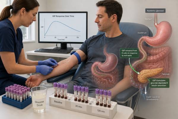
A glucose-dependent insulinotropic peptide test measures GIP, an incretin hormone released mainly from K cells in the upper small intestine after eating. GIP helps pancreatic beta cells release insulin when glucose is elevated and also participates in nutrient storage, fat metabolism, bone biology, and communication between the gut and brain. The test is specialized: it is used mostly in metabolic research, mixed-meal studies, and selected endocrine evaluations rather than routine diabetes care. GIP can be measured as an active hormone or as a total concentration that includes inactive breakdown products. Those results are not interchangeable. Food timing, meal composition, DPP-4 activity, kidney function, medication use, sample additives, processing speed, and the assay platform can all change the number. No universal fasting or post-meal GIP range diagnoses diabetes, obesity, insulin resistance, or “incretin dysfunction.” A meaningful interpretation compares the GIP pattern with glucose, insulin, C-peptide, GLP-1, symptoms, and the exact collection protocol.
- GIP is a meal-responsive incretin hormone; one fasting value usually gives less information than a standardized stimulated series.
- Active GIP is rapidly inactivated by DPP-4, so delayed processing can produce a falsely low result.
- Total GIP reflects secretion plus inactive forms, while active GIP reflects the intact receptor-stimulating fraction.
- High or low GIP alone does not diagnose diabetes, obesity, insulin resistance, or a gastrointestinal condition.
- Tirzepatide and other receptor-targeting medicines are not routinely monitored with an endogenous GIP blood test.
Table of Contents
- What GIP Is and What the Test Measures
- When GIP Testing Is Used
- Preparation, Collection, and Sample Handling
- Normal Ranges and Dynamic Results
- High GIP Levels
- Low GIP Levels
- GIP in Diabetes, Obesity, and Treatment
- How to Interpret a GIP Result
What GIP Is and What the Test Measures
GIP was first called gastric inhibitory polypeptide because early experiments suggested that it reduced stomach acid secretion. Its major physiological role was later recognized as stimulation of insulin secretion after oral nutrients, so the name glucose-dependent insulinotropic polypeptide became standard. Both names may appear in older records, test catalogs, and research articles.
K cells located mainly in the duodenum and jejunum release GIP when nutrients pass through the upper small intestine. Carbohydrate and fat are strong stimuli, and protein can contribute. GIP binds to the GIP receptor on pancreatic beta cells and amplifies insulin release when glucose is high enough. This is one part of the incretin effect: oral glucose provokes more insulin secretion than an intravenous glucose infusion that produces a similar glucose concentration because the gut releases signaling hormones during oral intake.
GIP does more than support insulin secretion. GIP receptors are expressed in several tissues, and research links the pathway to adipose tissue nutrient handling, glucagon regulation, bone remodeling, cardiovascular physiology, and central nervous system signaling. Some effects differ by glucose level, nutritional state, species, and whether the receptor receives a brief natural signal or prolonged pharmacologic stimulation.
A laboratory may offer several related measurements:
- Active or intact GIP, generally GIP(1-42), measures the form capable of strong classic receptor activation.
- Total GIP measures intact GIP plus the major DPP-4-generated metabolite, commonly GIP(3-42).
- Fasting GIP is measured before a nutrient challenge.
- Stimulated GIP is measured at timed intervals after oral glucose, a liquid meal, or a mixed meal.
- Peak, incremental area under the curve, or total area under the curve summarizes the response over time.
Dipeptidyl peptidase-4, or DPP-4, cleaves active GIP rapidly. Total GIP therefore tends to be higher and more stable than active GIP. A result labeled simply “GIP” is incomplete unless the report identifies which molecular pool the assay recognizes.
GIP is distinct from GLP-1. Both are incretins, but they arise from different intestinal cell populations, have different receptors, and show different physiological patterns. GIP often contributes more to the normal post-meal incretin effect, while GLP-1 has more consistent glucagon-suppressing and gastric-emptying effects at pharmacologic doses.
When GIP Testing Is Used
GIP measurement is not a standard screening or diagnostic test. Most hospital and outpatient laboratories do not include it in routine endocrine panels, and major diabetes diagnostic criteria do not use a GIP threshold.
Specialists and investigators may measure GIP to:
- Characterize incretin secretion during an oral glucose tolerance test or mixed-meal tolerance test
- Compare active and total incretin responses before and after a DPP-4 inhibitor
- Study beta-cell function in normal glucose tolerance, prediabetes, or type 2 diabetes
- Examine post-meal physiology after gastric bypass, sleeve gastrectomy, or other gastrointestinal surgery
- Investigate how meal composition, gastric emptying, or intestinal nutrient delivery affects hormone release
- Study obesity, weight change, appetite regulation, lipid handling, or metabolic adaptation
- Assess experimental therapies that activate or block the GIP receptor
- Explore unusual glucose and insulin patterns together with GLP-1, glucagon, insulin, and C-peptide
GIP testing may appear in a research metabolic profile, but that does not mean the result has an established individual treatment target. A study can identify average group differences without defining a clinically useful cutoff for one patient.
A clinician generally should not order GIP simply because someone has:
- Elevated fasting glucose or A1C
- Suspected insulin resistance
- Difficulty losing weight
- Strong hunger or food cravings
- A family history of diabetes
- A desire to predict response to tirzepatide
Those concerns are evaluated with established clinical history, physical examination, glucose-based testing, lipid measurements, blood pressure, liver assessment, medication review, sleep and nutrition factors, and other targeted tests. A fasting insulin test or HOMA-IR estimate may sometimes add context, but neither GIP nor fasting insulin independently defines metabolic health.
GIP measurement is also not the primary test for a neuroendocrine tumor. Rare tumors may secrete multiple peptides, but an unexplained GIP level without an appropriate syndrome, imaging question, or specialist plan is difficult to interpret. Broad “gut hormone panels” can generate incidental abnormalities because peptide assays have different reference populations and preanalytical requirements.
Preparation, Collection, and Sample Handling
Reliable GIP testing begins before the needle is inserted. The ordering team must specify whether the assay measures active or total GIP and whether the purpose is a fasting baseline or a dynamic response.
Before the test
Protocols commonly require an 8- to 12-hour overnight fast. Water is usually permitted. The laboratory may ask the person to avoid alcohol, strenuous exercise, smoking, and unusually large or high-fat meals before testing because these factors can alter glucose, gastrointestinal motility, and hormone release.
Medication instructions depend on the clinical question. Important examples include:
- DPP-4 inhibitors, which increase intact GIP by slowing conversion to GIP(3-42)
- Dual GIP/GLP-1 receptor agonists, which change appetite, gastric emptying, glucose, insulin, and glucagon but are not the same as endogenous GIP
- GLP-1 receptor agonists, which can alter gastric emptying and post-meal hormone patterns
- Insulin and sulfonylureas, which change glucose and insulin dynamics
- Metformin and other glucose-lowering drugs, which may influence the metabolic response indirectly
- Drugs affecting stomach emptying, intestinal absorption, or autonomic function
Prescription medicines should never be held solely to obtain a “clean” GIP number unless the prescribing clinician creates a safe plan. In many studies, the intent is to measure physiology on a stable regimen; in others, a supervised washout is part of the protocol.
Dynamic testing
A mixed-meal tolerance test is often more informative than a single fasting sample because GIP rises quickly when nutrients reach K cells. The test meal should be standardized. Differences in calories, fat, carbohydrate type, protein, volume, temperature, and consumption time can change the response.
A typical research sequence may collect samples at baseline and at several points during the next two to four hours. Early draws such as 10, 15, or 30 minutes help identify the rapid rise. Later samples show persistence and return toward baseline. Some protocols use a 75-g oral glucose drink instead of a mixed meal. Glucose is easier to standardize but does not reproduce the full fat- and protein-driven GIP response of ordinary food.
Timing errors matter. A specimen labeled “30 minutes” but drawn at 42 minutes cannot be treated as an exact 30-minute peak. The actual collection time should be recorded for every tube.
Protecting the sample
Active GIP is vulnerable to ex vivo degradation. Depending on the assay, collection may require chilled EDTA plasma, a DPP-4 inhibitor, and additional protease inhibitors. Tubes may need immediate mixing, placement on ice, refrigerated centrifugation, prompt separation, aliquoting, and freezing.
A tube processed as routine chemistry can show a falsely low active value. Total GIP is less affected by DPP-4 conversion because the assay intentionally recognizes both intact and cleaved forms, but uniform handling remains essential for comparing time points.
Modern immunoassays and mass-spectrometry methods may detect different molecular forms and have different lower limits of quantification. Cross-reactivity, matrix effects, calibration, and antibody epitope choice can produce substantial inter-assay differences. For longitudinal comparison, use the same laboratory, specimen type, assay version, and challenge protocol whenever possible.
Normal Ranges and Dynamic Results
There is no single accepted normal range for GIP. A value can be reported in picomoles per liter, picograms per milliliter, or another laboratory-specific format. Active and total assays yield different magnitudes, and fasting results cannot be compared directly with post-meal values.
The report should be interpreted using five layers of context:
| Layer | Questions to ask |
|---|---|
| Molecular form | Was intact GIP(1-42), total GIP, or a broader immunoreactive signal measured? |
| Stimulus | Was the person fasting, drinking glucose, or consuming a mixed meal? |
| Time course | Is this a baseline, early peak, later value, or calculated AUC? |
| Method | Which assay, calibration, detection limit, and specimen additives were used? |
| Metabolic response | What happened to glucose, insulin, C-peptide, GLP-1, and glucagon at the same time? |
In a typical physiological response, fasting GIP is relatively low, rises rapidly after nutrient intake, and then declines as absorption progresses. The exact peak and duration vary widely. Fat can generate a strong GIP response even when the glucose rise is modest, so a high post-meal GIP concentration cannot be judged without knowing the meal.
Researchers may calculate:
- Peak GIP, the highest measured concentration
- Incremental AUC, the area above the fasting baseline
- Total AUC, the entire concentration-time area
- Active-to-total ratio, an estimate influenced by secretion and DPP-4-mediated inactivation
- GIP-to-GLP-1 ratio, an exploratory measure of relative incretin secretion
These calculations are useful for comparing standardized groups or repeated interventions. They are not universally validated diagnostic indices. Different sampling schedules can change the apparent peak and AUC, even if biology is identical.
In type 2 diabetes, GIP secretion may be normal or elevated, while the insulin response to GIP is markedly reduced. This is sometimes described as GIP resistance or impaired beta-cell responsiveness. Therefore, a “normal” GIP concentration does not establish a normal incretin effect, and low insulin after a meal does not automatically mean low GIP secretion.
A laboratory reference interval, when provided, applies only to that assay and specimen protocol. If the report lacks a clear interval, an endocrinologist may compare the pattern with a matched research protocol, but such comparison remains descriptive rather than diagnostic.
High GIP Levels
High GIP most commonly reflects recent nutrient exposure, the type of meal, medication-related preservation of intact hormone, or altered post-meal physiology. It rarely represents a disease that can be diagnosed from GIP alone.
Possible explanations include:
- A recent meal, especially one rich in fat or rapidly absorbed carbohydrate
- Collection near the post-meal peak rather than at a true fasting baseline
- DPP-4 inhibitor use, which increases active GIP while total secretion may change little
- Obesity or insulin resistance, which can be associated with altered fasting or postprandial GIP in some studies but not uniformly
- Reduced beta-cell responsiveness, allowing high GIP to coexist with an inadequate insulin effect
- Gastric or intestinal surgery, which changes nutrient transit and gut hormone timing
- Kidney dysfunction, which may alter clearance of GIP forms and complicate comparisons
- Assay differences or interference, especially when the number conflicts with the clinical setting
A high total GIP result does not show how much active GIP remains. Conversely, a high active result during DPP-4 inhibitor treatment may indicate expected pharmacology rather than excess secretion.
GIP can stimulate glucagon under some low- or normal-glucose conditions and support insulin secretion at higher glucose. This glucose-dependent behavior means the same GIP concentration may have different downstream effects depending on the concurrent glucose level. Interpreting GIP without glucose is therefore incomplete.
High GIP has been studied in obesity because the hormone supports nutrient handling in adipose tissue, yet the relationship is not simple. Chronic pharmacologic activation of both GIP and GLP-1 receptors can produce substantial weight loss, while some experimental strategies investigate GIP receptor antagonism. These apparently opposite approaches reflect differences between native short-lived signaling, prolonged drug exposure, receptor interactions, central effects, and metabolic state. A high endogenous GIP result does not indicate that a person should avoid or receive a particular incretin medication.
When a fasting result is unexpectedly high, first verify that the person truly fasted and that the sample was not mislabeled. Review DPP-4 inhibitors, kidney function, recent surgery, and assay type. Repeat testing should use the same validated method and should be ordered only if the result will answer a defined question.
Low GIP Levels
Low GIP may reflect a valid low fasting concentration, weak nutrient stimulation, altered gastrointestinal delivery, or specimen degradation. Active GIP is especially sensitive to delayed inhibition of DPP-4.
Potential causes include:
- A true fasting state, when K-cell secretion is limited
- A low-calorie or low-fat challenge that provides little stimulus
- Delayed gastric emptying, which postpones nutrient arrival in the upper intestine
- Malabsorption or altered intestinal anatomy affecting K-cell exposure
- A sample drawn before the rise or after the response has subsided
- Missing DPP-4 inhibitor or delayed processing for an active assay
- Low assay sensitivity near the detection limit
- Biological variation or a reduced K-cell response
A low GIP result does not diagnose “incretin deficiency.” There is no accepted clinical syndrome defined solely by low circulating GIP. In people with diabetes, impaired insulin secretion after oral nutrients often reflects beta-cell dysfunction and reduced responsiveness to GIP rather than absent hormone release.
For an active assay, the laboratory should examine sample integrity before a biological explanation is accepted. If every stimulated active GIP point is unexpectedly low but total GIP rises normally, excessive ex vivo or in vivo inactivation may be relevant. If both active and total values remain flat, timing, meal delivery, assay performance, or secretion may need review.
Low post-meal GIP can occur after some gastrointestinal interventions or with altered nutrient routing, but GLP-1 may rise at the same time. This reciprocal pattern is one reason both hormones are sometimes measured together. The clinical consequence depends on glucose, insulin, gastric symptoms, and surgery type rather than on the GIP value by itself.
A single low result generally should not lead to dietary supplements, off-label hormone use, or medication changes. There is no standard treatment designed to “raise a low GIP blood level” in routine practice.
GIP in Diabetes, Obesity, and Treatment
GIP is deeply involved in metabolic physiology, but the blood test has a limited role in ordinary diabetes management. Diabetes and prediabetes are diagnosed using established glucose and A1C criteria. Treatment is monitored with glucose patterns, A1C, symptoms, weight, kidney function, cardiovascular risk, and medication-specific safety measures.
In healthy physiology, GIP and GLP-1 together amplify insulin secretion after oral nutrients. In type 2 diabetes, the incretin effect is reduced. Pharmacologic amounts of GIP historically produced a weak insulin response in people with poorly controlled type 2 diabetes, even when circulating GIP secretion was preserved. Better glucose control can partly restore responsiveness, showing that the defect is influenced by metabolic state rather than being permanently absent in every person.
GIP also affects alpha cells. Depending on glucose concentration, it may support glucagon release, helping protect against low glucose. This differs from GLP-1, which more reliably suppresses inappropriate glucagon during hyperglycemia. The combined effects of the two hormones vary across fasting, meal, and hypoglycemic states.
The success of dual GIP/GLP-1 receptor agonists has increased public interest in GIP testing. However, receptor agonist therapy does not require measurement of native GIP. A medicine such as tirzepatide is engineered for prolonged receptor activation and has pharmacology that cannot be inferred from a brief endogenous peptide concentration.
An endogenous GIP assay generally cannot answer:
- Whether a person will lose a specific amount of weight on a dual agonist
- Whether a medication dose should be increased
- Whether nausea or appetite reduction is “too much GIP”
- Whether the drug is present at a therapeutic concentration
- Whether future diabetes will develop
For suspected diabetes, clinicians use glucose-based testing. For questions about pancreatic insulin production, a C-peptide test is more established. For suspected endogenous hyperinsulinemic hypoglycemia, paired glucose, insulin, C-peptide, proinsulin, beta-hydroxybutyrate, and a drug screen during low glucose are far more informative than GIP.
Research on GIP remains active because receptor signaling may affect body weight, adipose biology, bone, cardiovascular function, and the brain. Those findings support drug development and mechanistic studies, but they should not be converted into unvalidated personal “optimal GIP” targets.
How to Interpret a GIP Result
Start by reconstructing exactly what happened around the blood draw. A useful review includes:
- The full assay name and whether it measured active or total GIP
- The unit and laboratory method
- Fasting duration and exact test meal or glucose dose
- Every actual collection time
- Specimen tube, DPP-4 inhibitor use, chilling, centrifugation, and freezing details
- Diabetes, gastrointestinal, kidney, and surgical history
- All medicines, especially DPP-4 inhibitors and incretin therapies
- Concurrent glucose, insulin, C-peptide, GLP-1, and glucagon results
- Symptoms during the test
- The clinical or research question the test was designed to answer
Consider a person whose GIP rises strongly after a mixed meal while glucose remains high and insulin increases only modestly. That pattern may suggest impaired beta-cell response in context, but it does not establish “GIP resistance” from a single patient-level cutoff. In contrast, an apparently absent active GIP response with a normal total response may point first to degradation, medication context, or assay-specific issues.
Post-bariatric symptoms require careful timing. Early rapid glucose and hormone peaks can be followed by a late glucose fall. GIP may rise, fall, or remain relatively unchanged depending on the operation and nutrient route, while GLP-1 may increase sharply. Diagnosis of postprandial hypoglycemia rests on documented low glucose associated with symptoms and appropriate recovery, not on a GIP threshold.
A repeat study is reasonable only when the result is likely to change management or clarify a defined physiological question. Repetition should match the original fasting period, meal, collection schedule, medication plan, and assay. Comparing a fasting active result from one laboratory with a total post-meal result from another is not meaningful.
Urgent care is based on symptoms and glucose, not GIP. Confusion, seizure, loss of consciousness, severe weakness, or inability to take oral carbohydrate during suspected hypoglycemia needs immediate treatment. Persistent vomiting, dehydration, or severe abdominal symptoms during incretin-based therapy also warrants prompt clinical assessment.
A GIP test can provide valuable detail in a controlled metabolic investigation. Its interpretation remains specialized because secretion is dynamic, active hormone is short-lived, assays are not harmonized, and receptor responsiveness cannot be read directly from the concentration. The strongest conclusions come from a complete time course paired with the hormones and metabolites that show what the pancreas and glucose system actually did.
References
- Glucose-dependent insulinotropic polypeptide (GIP) 2025 (Review)
- Beyond the pancreas: contrasting cardiometabolic actions of GIP and GLP1 2023 (Review)
- GIPR/GLP-1R dual agonist therapies for diabetes and weight loss—chemistry, physiology, and clinical applications 2023 (Review)
- A UHPLC/MS/MS method for the analysis of active and inactive forms of GLP-1 and GIP incretins in human plasma 2022
- Altered GIP/GLP-1 Secretion Ratio is Associated With Impaired β Cell Function in Humans 2025
Disclaimer
This article provides general information about endogenous GIP testing and is not a substitute for individual medical care. GIP values are assay- and protocol-specific and should be interpreted by a clinician familiar with incretin testing. Do not stop diabetes, gastrointestinal, or weight-management medicine for a test without direct instructions from the prescribing clinician.