A glucagon-like peptide-1 test measures GLP-1, a short-lived gut hormone released after food reaches the intestine. GLP-1 helps the pancreas release insulin when glucose is elevated, restrains glucagon, slows stomach emptying, and contributes to fullness. Despite its central role in modern diabetes and weight-loss treatment, an endogenous GLP-1 blood test is not a routine test for diagnosing diabetes, insulin resistance, or obesity. It is mainly used in specialized endocrine evaluation and research, often during an oral glucose tolerance test or mixed-meal test rather than as a single fasting measurement. Results depend heavily on whether the laboratory measures active or total GLP-1, how quickly the sample is chilled and processed, the meal or glucose stimulus, medications, and the assay method. A result therefore needs to be interpreted with the laboratory protocol, glucose, insulin, C-peptide, symptoms, and clinical question—not against a universal target number.
- A GLP-1 test measures the body’s own incretin hormone, not the blood concentration or dose effect of a GLP-1 receptor agonist medication.
- Active GLP-1 is rapidly broken down by DPP-4, so strict collection, inhibitor, chilling, separation, and freezing procedures are essential.
- There is no universal fasting or post-meal GLP-1 reference range; values are assay- and protocol-specific.
- A meal-stimulated series usually provides more information than one isolated fasting value.
- Low or high GLP-1 alone does not diagnose diabetes, insulin resistance, obesity, or a gastrointestinal disorder.
Table of Contents
- What the GLP-1 Test Measures
- Why the Test Is Ordered
- How GLP-1 Testing Is Performed
- Normal Ranges and Result Patterns
- High GLP-1 Results
- Low GLP-1 Results
- GLP-1, Diabetes, and Medications
- Interpreting Results and Next Steps
What the GLP-1 Test Measures
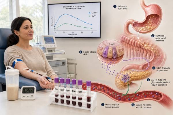
GLP-1 is an incretin hormone made mainly by intestinal enteroendocrine L cells. Nutrients in a meal—especially carbohydrate and fat, with protein also contributing—trigger its release. The hormone enters the portal circulation and supports coordinated handling of the incoming meal.
Its best-known action is glucose-dependent stimulation of insulin secretion. “Glucose-dependent” means GLP-1 strengthens insulin release primarily when blood glucose is elevated. This feature helps explain why GLP-1 receptor agonist medicines generally have a low risk of causing hypoglycemia when used without insulin or an insulin-releasing drug. Native GLP-1 also suppresses glucagon during hyperglycemia, slows gastric emptying, and signals through neural and brain pathways involved in appetite and food intake.
The term GLP-1 test can refer to several different laboratory measurements:
- Active or intact GLP-1 measures biologically active forms, commonly GLP-1(7-36) amide and GLP-1(7-37).
- Total GLP-1 measures active hormone plus major breakdown products, providing a broader estimate of secretion.
- Fasting GLP-1 is measured before food or glucose stimulation.
- Stimulated GLP-1 is measured repeatedly after a standardized glucose drink or mixed meal.
- Area under the curve, or AUC, summarizes the concentration across all timed samples rather than relying on one point.
Active and total results are not interchangeable. The enzyme dipeptidyl peptidase-4, or DPP-4, removes two amino acids from native GLP-1 within minutes, producing metabolites that do not stimulate the classic GLP-1 receptor in the same way. Active assays therefore reflect the small fraction of intact hormone surviving at collection, while total assays more closely reflect overall L-cell release and subsequent metabolism.
GLP-1 is related to glucagon because both originate from the proglucagon gene, but tissue-specific processing creates different hormones. Intestinal L cells mainly produce GLP-1 and related peptides; pancreatic alpha cells mainly produce glucagon. A glucagon test and a GLP-1 test answer different questions and should not be confused because a laboratory abbreviation happens to look similar.
Why the Test Is Ordered
Endogenous GLP-1 measurement is uncommon in routine medical practice. Standard diabetes evaluation relies on fasting plasma glucose, A1C, random plasma glucose when symptoms are present, or a two-hour oral glucose tolerance test. These tests have established diagnostic thresholds. GLP-1 does not.
A specialist or research protocol may order GLP-1 testing for several reasons:
- To characterize incretin secretion during a mixed-meal tolerance test or oral glucose tolerance test
- To study differences in gut hormone responses among people with normal glucose tolerance, prediabetes, or diabetes
- To evaluate physiology after bariatric or metabolic surgery, especially when symptoms suggest postprandial hypoglycemia
- To investigate rapid gastric emptying, dumping-related symptoms, or unusual post-meal glucose and insulin patterns
- To assess the pharmacodynamic effect of a DPP-4 inhibitor in a study
- To examine gut hormone changes associated with obesity, gastrointestinal disease, nutrition, or an intervention
- To support a research evaluation of beta-cell function together with glucose, insulin, C-peptide, GIP, and glucagon
After Roux-en-Y gastric bypass or sleeve gastrectomy, nutrients may reach more distal intestine quickly, producing a large and early GLP-1 response. In some people, this response contributes to an exaggerated insulin surge and late post-meal hypoglycemia. Even then, GLP-1 measurement is usually an adjunct. Symptoms, documented low plasma glucose, insulin and C-peptide during the episode, meal timing, and exclusion of other causes remain more important than an isolated hormone value.
A GLP-1 level is generally not an appropriate standalone test for:
- Diagnosing type 1 or type 2 diabetes
- Screening for insulin resistance
- Selecting a weight-loss medication
- Confirming whether a GLP-1 receptor agonist is “working”
- Determining whether someone has “GLP-1 deficiency”
- Explaining hunger or body weight without broader clinical evaluation
Consumer discussions sometimes imply that low natural GLP-1 is a single, measurable cause of obesity or diabetes. Human physiology is more complex. Secretion, degradation, receptor response, gastric emptying, neural signaling, insulin sensitivity, beta-cell capacity, diet, body composition, sleep, and medications all influence the final metabolic response. One peptide concentration cannot separate those components.
How GLP-1 Testing Is Performed
GLP-1 testing requires more planning than an ordinary chemistry panel because the hormone is present at low concentrations and is rapidly degraded after secretion and after blood collection. The ordering clinician and laboratory should agree on the exact assay and collection protocol before the test begins.
Fasting and medication preparation
Many protocols require an overnight fast of about 8 to 12 hours, with water allowed. The person should avoid smoking, vigorous exercise, and unplanned caloric intake before baseline collection because each can change metabolic responses. The laboratory or study team may also standardize the prior evening meal and ask the person to avoid alcohol.
Medications can materially alter the result. DPP-4 inhibitors increase intact GLP-1 by slowing its breakdown. GLP-1 receptor agonists and dual GIP/GLP-1 agonists change glucose, insulin, glucagon, appetite, and gastric emptying even though the drug itself may not be measured by an endogenous GLP-1 assay. Insulin, sulfonylureas, metformin, drugs that change gastric emptying, and recent bariatric surgery can also affect the test context.
Do not stop any prescribed medicine without instructions. For a clinically necessary test, the clinician should specify whether the aim is to measure physiology on the usual regimen or under a carefully supervised medication hold.
Fasting sample versus stimulation test
A single fasting sample is easier to collect but may provide limited information because GLP-1 is primarily a meal-responsive hormone. Fasting values can be near the lower detection range of some assays. Small analytical differences can then appear large relative to the measured concentration.
Stimulated testing usually follows one of two formats:
- Oral glucose tolerance protocol: Blood is collected at baseline, the person drinks a measured glucose solution, and additional samples are drawn at specified intervals such as 15, 30, 60, 90, and 120 minutes.
- Mixed-meal protocol: The person consumes a standardized drink or meal containing carbohydrate, protein, and fat. This resembles normal nutrient exposure more closely and can assess glucose, insulin, C-peptide, GLP-1, GIP, and other hormones together.
Exact time points differ among laboratories and studies. Early sampling matters because GLP-1 may rise rapidly. Missing or delaying a draw can distort the apparent peak and AUC.
Specimen handling
The collection tube and processing instructions depend on the assay. Common protocols use chilled EDTA plasma, a protease inhibitor such as aprotinin, and a DPP-4 inhibitor when intact GLP-1 is being measured. Tubes may need immediate placement on ice, rapid centrifugation in a refrigerated centrifuge, separation from cells, aliquoting, and freezing at a specified temperature.
A routine blood tube left at room temperature or processed late can yield an artificially low active GLP-1 result. Repeated freeze-thaw cycles may also affect peptide stability. A valid interpretation therefore requires confidence that every timed sample was handled consistently.
Laboratory methods include immunoassays and mass-spectrometry-based techniques. Antibodies may recognize different portions of the molecule and cross-react to different degrees with related peptides or metabolites. Results from different platforms can diverge even when specimens come from the same person. The method name, whether the assay is active or total, the unit, and the laboratory’s own reference information should remain attached to the result.
Normal Ranges and Result Patterns
There is no universal GLP-1 normal range that applies across active assays, total assays, fasting samples, glucose challenges, and mixed meals. Published studies report concentrations in units such as picomoles per liter, picograms per milliliter, or related assay-specific units. Unit conversion alone does not make two methods comparable because the antibodies and molecular forms measured may differ.
A useful report should identify:
| Report detail | Why it changes interpretation |
|---|---|
| Active, intact, or total GLP-1 | These assays measure different molecular pools. |
| Fasting or stimulated | A fasting concentration cannot be compared directly with a post-meal peak. |
| Time after glucose or meal | The response changes quickly, and peak timing varies. |
| Assay method and manufacturer | Antibody specificity, calibration, and detection limits differ. |
| Specimen additives and handling | Failure to block degradation can lower intact GLP-1. |
| Concurrent glucose and hormones | GLP-1 action is meaningful in relation to glucose, insulin, C-peptide, GIP, and glucagon. |
For a stimulation test, clinicians often focus on the shape and coordination of the response rather than a single cutoff. Questions include whether GLP-1 rises after nutrient exposure, how early it peaks, whether the response is sustained, and whether insulin secretion is proportionate to glucose. AUC calculations may reduce dependence on one sample, but they still require standardized timing and a valid assay.
Research has not consistently shown a generalized failure of GLP-1 secretion in all people with type 2 diabetes. Some studies identify differences related to age, body mass index, glucose tolerance, sex, meal composition, or assay method, while others find broadly preserved secretion. The impaired incretin effect in type 2 diabetes can reflect reduced beta-cell responsiveness—especially to GIP—along with beta-cell dysfunction and insulin resistance, rather than simply too little circulating GLP-1.
A result within a laboratory’s research reference interval does not prove that incretin signaling is normal. Likewise, a value outside that interval does not establish disease. Reference participants, meal composition, fasting duration, analytical method, and statistical approach all shape the interval.
High GLP-1 Results
A high GLP-1 concentration most often reflects timing, altered degradation, medication exposure, or a strong nutrient-driven response rather than a distinct “high GLP-1 disorder.” The interpretation differs for active and total assays.
Common explanations include:
- Recent food intake: GLP-1 normally increases after nutrients enter the intestine.
- DPP-4 inhibitor treatment: These medicines raise intact GLP-1 by slowing enzymatic inactivation.
- Bariatric or metabolic surgery: Faster nutrient delivery can cause an earlier and larger post-meal response.
- Post-bariatric hypoglycemia: Some affected patients show exaggerated GLP-1 and insulin responses, although the diagnosis requires documented biochemical hypoglycemia and a complete evaluation.
- Differences in gastric emptying or intestinal nutrient exposure: Faster delivery can shift the timing and magnitude of secretion.
- Reduced peptide clearance or altered metabolism: Kidney function and other physiological factors may influence measured incretin forms, depending on the assay.
- Protocol or analytical variation: A different meal, sample time, assay, or specimen treatment can make a value appear elevated relative to another dataset.
High endogenous GLP-1 does not necessarily mean excessive GLP-1 receptor activity. Receptor sensitivity, neural pathways, glucose level, beta-cell reserve, and concurrent hormones influence the response. A high total level may coexist with a much smaller active fraction because DPP-4 rapidly converts intact hormone to metabolites.
When high GLP-1 is found during evaluation of post-meal symptoms, the symptom timeline is important. Sweating, tremor, palpitations, confusion, weakness, or faintness one to three hours after eating may prompt supervised assessment for hypoglycemia. A home glucose sensor can help identify patterns, but a formal diagnosis usually requires reliable glucose confirmation during symptoms and appropriate paired laboratory tests. Severe confusion, seizure, loss of consciousness, or inability to swallow requires immediate emergency treatment rather than further elective hormone testing.
Very high results that do not fit the clinical setting should be repeated only after confirming the assay identity, specimen handling, collection time, medication list, and units. Heterophile antibodies and other immunoassay interferences are uncommon but possible. A discordant number should not trigger treatment by itself.
Low GLP-1 Results
A low active GLP-1 value may be biological, but preanalytical loss is a frequent concern. Native GLP-1 has a very short circulating half-life because DPP-4 and other clearance pathways act quickly. Delayed cooling, absent inhibitor, slow plasma separation, or storage problems can lower the measured intact concentration before analysis.
Potential explanations for a low result include:
- A fasting sample collected when secretion is naturally minimal
- A sample drawn too early or too late to capture the post-meal rise
- Inadequate nutrient stimulation or incomplete consumption of the test meal
- Slow gastric emptying that delays nutrient delivery to the intestine
- Assay sensitivity near the lower limit of quantification
- Specimen degradation during collection, transport, processing, or storage
- Biological variation in L-cell secretion
- A clinical condition or medication that changes nutrient sensing or gastrointestinal motility
Low GLP-1 is not a standard diagnosis. There is no broadly accepted threshold for “GLP-1 deficiency,” and a low value does not prove why a person has diabetes, obesity, increased appetite, or difficulty losing weight. A person can have a low fasting value and a normal stimulated response. Another person can secrete GLP-1 yet have reduced downstream metabolic benefit because beta-cell function or insulin sensitivity is impaired.
When secretion itself is the research question, a complete time curve is more defensible than one low point. Concurrent measurement of glucose, insulin, and C-peptide helps show whether pancreatic beta cells responded to the same stimulus. GIP testing may add context because GIP and GLP-1 together produce much of the incretin effect.
A repeat test should use the same validated protocol if the aim is comparison. Switching laboratories, assay types, meals, time points, or specimen procedures can create a difference that reflects methodology rather than physiology.
GLP-1, Diabetes, and Medications
GLP-1 physiology is closely connected to diabetes, but endogenous GLP-1 testing is not part of routine diagnostic criteria. Diabetes is diagnosed using glucose or A1C thresholds and interpreted with symptoms, repeat confirmation when needed, pregnancy status, and clinical context. Prediabetes is also defined by established glucose and A1C ranges—not by an incretin concentration.
In type 2 diabetes, the overall incretin effect is reduced. This means oral glucose produces less additional insulin secretion than expected compared with intravenous glucose matched to the same blood glucose profile. The defect involves progressive beta-cell dysfunction, diminished responsiveness to GIP, altered glucagon regulation, insulin resistance, and other factors. GLP-1 receptor signaling remains sufficiently effective that pharmacologic receptor agonists can lower glucose and body weight, but this therapeutic response does not imply that every patient began with a measurable GLP-1 deficiency.
Several medication classes interact with this system:
- GLP-1 receptor agonists are molecules designed to activate the GLP-1 receptor and resist rapid breakdown. They are not identical to native GLP-1.
- Dual GIP/GLP-1 receptor agonists activate both receptor pathways.
- DPP-4 inhibitors reduce degradation of endogenous GLP-1 and GIP, increasing intact concentrations modestly.
- Insulin and insulin secretagogues change downstream glucose and insulin patterns and may influence how a stimulation test is interpreted.
An endogenous GLP-1 assay generally should not be used for therapeutic drug monitoring. The assay may not recognize the medication, may cross-react unpredictably, or may measure only native peptide. Drug effectiveness is evaluated through A1C, fasting and post-meal glucose, continuous glucose monitoring data when appropriate, body-weight change, symptoms, tolerability, kidney and liver considerations, and treatment-specific safety monitoring.
People taking a GLP-1 receptor agonist may experience nausea, vomiting, constipation, diarrhea, reduced appetite, or delayed gastric emptying, especially during dose escalation. These effects are assessed clinically. Measuring native GLP-1 does not determine whether a dose is too high. Persistent vomiting, inability to keep fluids down, severe abdominal pain, signs of dehydration, or symptoms of severe hypoglycemia warrant prompt medical assessment.
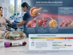
For metabolic evaluation, the clinician may combine established measures such as fasting glucose, A1C, lipids, liver enzymes, and sometimes a fasting insulin test. Each answers a different question; none can be replaced by an isolated GLP-1 result.
Interpreting Results and Next Steps
Interpretation should begin with the test design rather than the number. Before drawing conclusions, confirm the following:
- Was active, intact, or total GLP-1 measured?
- Was the person fasting, and for how long?
- Was the value collected before or after a glucose drink or mixed meal?
- What was the exact time point?
- Were DPP-4 and protease inhibitors required and used?
- Was the specimen chilled, separated, and frozen within the required interval?
- Which medications were taken in the preceding days?
- What were glucose, insulin, C-peptide, GIP, and glucagon doing at the same time?
- Does the laboratory provide a method-specific comparison interval or only research data?
- Do the findings match the person’s symptoms and reason for testing?
A result should be viewed as one part of a dynamic system. For example, a strong GLP-1 rise with excessive insulin, falling glucose, and post-meal neuroglycopenic symptoms after bariatric surgery has a different meaning from the same GLP-1 concentration in an asymptomatic research participant. A low active result from a mishandled sample has little clinical meaning even if it is below a study’s published average.
Follow-up depends on the original question. Possible next steps include repeating the test under a standardized protocol, reviewing the raw time points rather than only an AUC, checking for medication effects, assessing gastric emptying, or performing a supervised mixed-meal evaluation for suspected postprandial hypoglycemia. Routine diabetes concerns are better addressed with validated glucose-based tests. Suspected endogenous hyperinsulinemic hypoglycemia requires a properly timed hypoglycemia hormone panel during low plasma glucose rather than relying on GLP-1 alone.
Do not compare a personal result with a number from an online study unless the molecular form, assay, units, fasting state, meal, sample times, participant characteristics, and specimen processing all match. Even then, research distributions are not automatically clinical decision limits.
A well-designed GLP-1 test can illuminate incretin physiology, especially when paired with a standardized nutrient challenge and other metabolic markers. Its limitations are equally important: the hormone is unstable, assays differ, reference ranges are not harmonized, and most common clinical decisions do not require the measurement. Clear documentation of the protocol often adds more interpretive value than an extra decimal place in the result.
References
- The expanding incretin universe: from basic biology to clinical translation 2023 (Review)
- Glucagon-like peptide-1 secretion in people with versus without type 2 diabetes: a systematic review and meta-analysis of cross-sectional studies 2023 (Systematic Review)
- Methods paper: Performance characteristics of novel assays for circulating levels of proglucagon-derived peptides and validation in a placebo controlled cross-over randomized clinical trial 2022
- A UHPLC/MS/MS method for the analysis of active and inactive forms of GLP-1 and GIP incretins in human plasma 2022
- Altered GIP/GLP-1 Secretion Ratio is Associated With Impaired β Cell Function in Humans 2025
Disclaimer
This article provides general information about endogenous GLP-1 testing and does not diagnose diabetes, hypoglycemia, or another condition. GLP-1 results require method-specific interpretation by a qualified clinician, especially when testing follows bariatric surgery or is performed during symptomatic low glucose. Do not stop diabetes or weight-management medication for testing unless the prescribing clinician gives specific instructions.