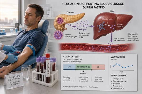
A glucagon blood test measures a pancreatic hormone that raises blood glucose when it begins to fall. Alpha cells release glucagon during fasting, exercise, and hypoglycemia. The hormone tells the liver to break down glycogen and make new glucose, helping protect the brain and other tissues between meals.
The test is specialized. It is most useful when doctors suspect a glucagon-secreting neuroendocrine tumor, called a glucagonoma, or when a specialist is studying unusual glucose regulation. A high value can also occur with diabetes, obesity, kidney or liver disease, stress, and assay cross-reactivity. A low value during hypoglycemia may indicate impaired counter-regulation, particularly in longstanding type 1 diabetes. Results depend heavily on fasting status, simultaneous glucose, specimen handling, and assay design. Glucagon resembles other peptides derived from proglucagon, and older or less specific immunoassays may detect related molecules. For that reason, serial measurements should use the same validated method.
- Glucagon raises blood glucose mainly by acting on the liver during fasting and hypoglycemia.
- One major laboratory uses a fasting adult reference value below 159 pg/mL, but ranges vary by assay.
- A high glucagon result alone does not diagnose glucagonoma; symptoms, glucose, nutritional findings, and imaging are required.
- Glucagon levels around 500–1,000 pg/mL with weight loss, diabetes, and necrolytic migratory erythema raise strong concern for glucagonoma.
- Low glucagon matters most when it fails to rise during hypoglycemia and contributes to recurrent severe low blood sugar.
- The specimen often requires an 8-hour fast, a prechilled EDTA tube, rapid cold processing, and immediate freezing.
Table of Contents
- What Glucagon Does
- When the Test Is Ordered
- Preparation and Sample Handling
- Normal Ranges and Assay Limits
- Causes of High Glucagon
- Low Glucagon and Hypoglycemia
- Glucagonoma Symptoms and Results
- Follow-Up and Glucose Management
What Glucagon Does
Glucagon is a 29-amino-acid peptide made mainly by alpha cells in the pancreatic islets. It comes from a larger precursor called proglucagon. Different tissues process proglucagon differently: pancreatic alpha cells produce glucagon, while intestinal cells produce related peptides such as GLP-1, GLP-2, oxyntomodulin, and glicentin.
Glucagon’s principal target is the liver. When blood glucose begins to fall, it binds to liver-cell receptors and increases cyclic AMP signaling. This leads to:
- Glycogenolysis: breakdown of stored liver glycogen into glucose
- Gluconeogenesis: production of glucose from amino acids, lactate, glycerol, and other substrates
- Ketogenesis during longer fasting: support for production of ketone bodies from fatty acids
- Amino-acid metabolism: coordination of the liver–alpha-cell axis that helps dispose of amino acids and regulate glucagon secretion
Glucagon does not directly release glucose from skeletal-muscle glycogen because muscle lacks the enzyme needed to export free glucose into the blood. Muscle glycogen supports local activity instead.
Insulin and glucagon work as a coordinated pair. After a carbohydrate-containing meal, insulin rises and glucagon is usually suppressed. During fasting or hypoglycemia, insulin falls and glucagon rises. The ratio between them helps determine whether the liver stores or releases fuel.
The relationship is more complex after a protein-rich meal. Amino acids can stimulate both insulin and glucagon. Insulin helps move amino acids into tissues, while glucagon prevents the associated insulin rise from causing hypoglycemia.
In diabetes, glucagon can become dysregulated. It may fail to suppress after meals, adding to high glucose, or fail to rise during hypoglycemia, increasing the danger of low glucose. A single fasting test captures only one moment and does not fully measure either dynamic response.
Injected glucagon is also used as a medicine for severe hypoglycemia and in some diagnostic procedures. A blood glucagon assay measures circulating hormone; it does not assess whether a rescue kit was administered correctly or whether liver glycogen stores are adequate.
When the Test Is Ordered
Glucagon is not part of routine diabetes screening. Fasting glucose, A1c, continuous glucose monitoring, insulin, and C-peptide are usually more useful for everyday care. The test is ordered for narrower questions.
Suspected glucagonoma
A glucagonoma is a rare pancreatic neuroendocrine tumor that secretes excessive glucagon. Testing is considered when a person has a compatible combination of:
- A characteristic recurring rash called necrolytic migratory erythema
- New or worsening diabetes or glucose intolerance
- Unexplained weight loss
- Anemia
- Inflamed mouth or tongue
- Diarrhea
- Blood clots
- A pancreatic mass or other neuroendocrine-tumor evidence
A high glucagon value supports the diagnosis but cannot confirm malignancy or locate the tumor.
Difficult hypoglycemia in diabetes
In longstanding type 1 diabetes, alpha cells may lose their normal response to falling glucose. A low or absent glucagon response can contribute to impaired awareness and severe episodes. Static fasting measurement has limited utility, but specialists may use dynamic protocols or research testing.
Research on diabetes and metabolism
Studies may measure fasting, post-meal, or amino-acid-stimulated glucagon to examine alpha-cell function, glucagon resistance, liver disease, obesity, or the effects of medicines. These results depend strongly on the protocol and are not interchangeable with a tumor evaluation.
Monitoring a known glucagon-secreting tumor
If glucagon was elevated before treatment, serial measurements may help follow response or recurrence along with symptoms and imaging. The same assay should be used whenever possible.
Glucagon is sometimes included in a pancreatic hormone test panel, but broad testing can produce incidental abnormalities. A focused order is preferable when the symptoms point toward one functional tumor.
Preparation and Sample Handling
Glucagon is vulnerable to preanalytical error. A current major-laboratory protocol requires an 8-hour fast, collection into a prechilled EDTA tube, cooling on wet ice, refrigerated centrifugation, immediate plasma separation, and freezing.
Follow the laboratory’s exact instructions. General preparation may include:
- Fast for the requested period, usually about 8 hours.
- Drink plain water unless told otherwise.
- Avoid food, caloric drinks, alcohol, gum, and candy during the fast.
- Avoid strenuous exercise shortly before collection unless the protocol is specifically studying exercise.
- Take or hold medicines only according to the clinician’s plan.
- Record recent hypoglycemia, glucose treatment, illness, and the time of the last meal.
Glucose should often be drawn at the same time. Without it, clinicians cannot judge whether glucagon is appropriately high, low, or suppressed.
Several factors can alter results:
- Insulin and other glucose-lowering medicines
- Recent glucagon rescue treatment
- Somatostatin analogs
- GLP-1-based medicines
- Acute stress, infection, surgery, or trauma
- Kidney or liver dysfunction
- Prolonged fasting or a recent high-protein meal
- Obesity, Cushing syndrome, or acromegaly
The sample type matters. Serum, room-temperature plasma, and properly chilled EDTA plasma may not yield equivalent results. Proteases can degrade glucagon, while related proglucagon fragments can interfere with nonspecific assays.
If testing is performed during hypoglycemia, safety takes priority. A critical sample should be collected promptly when feasible, but treatment should not be delayed in a person with confusion, seizure, loss of consciousness, or other severe symptoms.
The blood draw itself has the usual minor risks of soreness, bruising, and lightheadedness. The fasting requirement can create additional risk for someone using insulin or insulin-secretagogue medicine, so the plan should include glucose monitoring and instructions for treating low glucose.
Normal Ranges and Assay Limits
One major laboratory reports a fasting reference value below 159 pg/mL for people aged one year and older. Other laboratories use different upper limits, and older studies may report markedly different concentrations because of assay design.
A glucagon result is especially method-dependent. Proglucagon produces several peptides with overlapping sequences. A single-antibody assay may detect glucagon fragments, oxyntomodulin, glicentin, or other related molecules. Modern two-site assays use antibodies against both ends of intact glucagon and are generally more specific.
Important limitations include:
- Calibration differences: Two assays can give different numbers on the same sample.
- Cross-reactivity: Related gut peptides may falsely elevate a nonspecific result.
- Hook effect: Extremely high concentrations can rarely produce a falsely low immunoassay value.
- Heterophile antibodies: Patient antibodies can create false-high or false-low results.
- Sample degradation: Warm or delayed processing can lower the measured concentration.
- Biological timing: Fasting, meals, amino acids, and hypoglycemia alter secretion quickly.
The report’s reference interval should be used only for the same method and test conditions. Serial monitoring should remain with one laboratory whenever possible.
Interpretation also depends on glucose:
| Glucose state | Glucagon pattern | Possible meaning |
|---|---|---|
| Normal fasting glucose | Within range | Typical basal secretion |
| Hyperglycemia | Elevated or not suppressed | Diabetes-related alpha-cell dysregulation, stress, or another cause |
| Hypoglycemia | Appropriate rise | Preserved counter-regulation |
| Hypoglycemia | Low or absent rise | Impaired counter-regulation, especially in longstanding diabetes |
| No hypoglycemia | Markedly elevated | Consider glucagonoma, renal/hepatic disease, assay interference, or severe metabolic stress |
No universal “optimal” glucagon target exists for general metabolic health. Treatment goals focus on glucose patterns, nutrition, and the underlying disorder.
Causes of High Glucagon
High glucagon, or hyperglucagonemia, has several possible explanations. Glucagonoma is important but rare.
Diabetes and insulin deficiency
Insulin normally restrains alpha-cell secretion. In type 1 diabetes, insufficient insulin within the islet and systemic circulation can allow glucagon to rise, particularly during ketosis. In type 2 diabetes, glucagon may fail to suppress after meals and may be elevated fasting, contributing to excessive liver glucose production.
A high glucagon result does not diagnose diabetic ketoacidosis. DKA is diagnosed from glucose, ketones, bicarbonate, pH, anion gap, and clinical condition. Glucagon testing adds little to urgent management.
Obesity and metabolic liver disease
Some people with obesity, insulin resistance, or fatty liver have higher fasting glucagon. One proposed mechanism is glucagon resistance in the liver’s amino-acid pathways. If the liver responds poorly, amino acids rise and stimulate alpha cells to release more glucagon, creating a liver–alpha-cell feedback loop.
Kidney or liver dysfunction
The kidneys and liver contribute to peptide clearance. Reduced function can increase circulating glucagon or related immunoreactivity. Chronic liver disease also changes amino-acid metabolism and hormone response.
Stress and endocrine disorders
Acute illness, trauma, surgery, infection, and catecholamine release can raise glucagon. Cushing syndrome and acromegaly may also increase levels through insulin resistance and altered metabolic signaling.
Pancreatic and gastrointestinal conditions
Pancreatitis, pancreatic surgery, and altered gut anatomy can disrupt alpha-cell and proglucagon physiology. Certain neuroendocrine tumors outside the pancreas may produce glucagon-like immunoreactivity.
Assay interference
An unexpected moderate elevation without symptoms may reflect cross-reactivity with gut-derived peptides or heterophile antibodies. Confirmation with a specific two-site assay or mass-spectrometry method may be appropriate when the result would lead to imaging or invasive procedures.
High glucagon should be interpreted with glucose, amino-acid and nutritional status, liver and kidney function, symptoms, and the magnitude of elevation.
Low Glucagon and Hypoglycemia
Low glucagon is most important when blood glucose is falling. In a healthy counter-regulatory response, insulin secretion decreases first and glucagon rises quickly. Epinephrine, cortisol, and growth hormone provide additional support.
In type 1 diabetes, the glucagon response to hypoglycemia often becomes impaired within years of diagnosis. The alpha cells may still produce glucagon but fail to sense falling glucose correctly because local beta-cell insulin signaling is lost. Repeated hypoglycemia can further lower the glucose threshold at which counter-regulatory responses begin.
Consequences can include:
- More rapid or prolonged hypoglycemia
- Reduced warning symptoms
- Greater dependence on epinephrine responses
- Increased risk during sleep or exercise
- Severe events requiring help from another person
A low fasting glucagon during normal glucose does not establish this problem. Dynamic evaluation or review of continuous glucose data, symptom awareness, insulin dosing, and episode history is usually more useful.
Other causes of low or blunted glucagon may include extensive pancreatic damage, total pancreatectomy, chronic pancreatitis, autonomic dysfunction, and suppression by somatostatin analogs.
Management focuses on preventing hypoglycemia rather than raising a laboratory value. Strategies may include insulin-dose adjustment, continuous glucose monitoring, automated insulin delivery, higher temporary glucose targets, exercise planning, and ready access to rescue glucagon.
Modern rescue options include injectable kits, autoinjectors, prefilled syringes, and nasal glucagon. Family, coworkers, or caregivers should know where the product is and how to use it. After glucagon treatment, emergency assistance may still be needed, especially if the person does not respond promptly or has depleted liver glycogen from prolonged fasting, alcohol use, or severe illness.
A hypoglycemia hormone panel is aimed mainly at determining whether insulin is inappropriately present. Glucagon concentration usually plays a smaller role in that diagnostic workup.
Glucagonoma Symptoms and Results
Glucagonoma is a rare functional pancreatic neuroendocrine tumor. Most tumors arise in the pancreatic body or tail, and many are large or metastatic when recognized because early symptoms are nonspecific.
The classic syndrome includes:
Necrolytic migratory erythema
This distinctive rash often begins as red patches that blister, erode, crust, and heal with pigmentation. It commonly affects the groin, buttocks, lower abdomen, thighs, and areas around body openings. Lesions appear in waves and can be painful or itchy.
The rash is strongly associated with glucagonoma but can occur in other severe nutritional or liver conditions. Dermatology assessment and skin biopsy may support the diagnosis, although biopsy findings are not completely specific.
Metabolic and nutritional findings
Glucagon excess promotes liver glucose output and protein breakdown. Patients may develop:
- New diabetes or worsening glucose control
- Marked weight loss
- Low amino-acid levels
- Anemia
- Inflamed tongue, mouth sores, or cracked lips
- Diarrhea
- Muscle loss
Blood clots are an important complication. Depression and other neuropsychiatric symptoms may occur.
Interpreting the tumor marker
Current clinical guidance notes that a compatible syndrome plus plasma glucagon around 500–1,000 pg/mL is indicative of glucagonoma. Some tumors produce even higher levels. Lower elevations are nonspecific and occur in common metabolic conditions.
Biochemical evidence should be followed by tumor localization and staging with contrast-enhanced CT or MRI, endoscopic ultrasound in selected cases, and somatostatin-receptor imaging. A result does not show whether a tumor is malignant; spread to lymph nodes or liver determines staging.
Other laboratory tests may include glucose, A1c, complete blood count, amino acids, albumin, zinc, liver tests, and other neuroendocrine markers. Patients with pancreatic glucagonoma may also be assessed for MEN1 when history or tumor features suggest it.
Treatment can include surgery, somatostatin analogs, nutritional and amino-acid support, glucose treatment, targeted therapies, liver-directed treatment, and other oncology approaches. Falling glucagon may accompany response, but symptoms and imaging remain essential.
Follow-Up and Glucose Management
The next step after an abnormal result depends on its magnitude, glucose context, and symptoms.
For a mild or moderate elevation without classic symptoms, clinicians may:
- Confirm fasting status and proper cold processing.
- Repeat the test with the same or a more specific assay.
- Review glucose, A1c, kidney and liver function, body weight, and medicines.
- Assess for recent illness, stress, or high-protein intake.
- Avoid tumor imaging based solely on a borderline, unconfirmed result.
For a marked elevation with rash, weight loss, or diabetes, prompt referral to endocrinology, gastroenterology, dermatology, and a neuroendocrine-tumor team is appropriate. Biochemical confirmation and imaging should proceed together.
For low glucagon in a person with recurrent hypoglycemia, the practical response is a detailed safety review. Examine insulin timing, meal patterns, exercise, alcohol, kidney function, hypoglycemia awareness, and continuous glucose data. Repeated episodes should trigger less aggressive glucose targets until awareness and counter-regulation improve.
Do not use a glucagon blood test to decide whether a person needs a rescue prescription. Anyone at meaningful risk of severe hypoglycemia from insulin or certain insulin-releasing medicines should discuss rescue glucagon and training with a clinician.
Seek urgent care for severe hypoglycemia, seizure, unconsciousness, diabetic ketoacidosis symptoms, a rapidly spreading painful rash with systemic illness, signs of a blood clot, or severe dehydration. Those conditions require immediate treatment regardless of the laboratory result.
Glucagon testing is most useful at the extremes: a markedly elevated level in a person with a compatible tumor syndrome, or specialized assessment of impaired counter-regulation. For routine glucose management, established glucose measures provide more actionable information.
Interpreting Discordant Glucagon Results
A glucagon result deserves extra scrutiny when the concentration and clinical picture do not agree. Mild elevation in a person with diabetes, obesity, kidney impairment, liver disease, recent stress, or a protein-rich meal is far more common than glucagonoma. Confirm fasting conditions, review renal and hepatic function, and repeat the measurement with meticulous cold handling before escalating to tumor imaging.
Assay specificity is particularly important because circulating proglucagon-derived peptides can resemble one another. Some immunoassays may detect fragments or related molecules from the intestine, producing an apparent elevation that is not equivalent to pancreatic glucagon bioactivity. A specialist laboratory using a well-characterized method can help resolve a result that is unexpectedly high or inconsistent across platforms.
The glucose concentration at the same draw also matters. High glucagon during low glucose may be physiologically appropriate, while failure to rise can suggest impaired counter-regulation. High glucagon during hyperglycemia may reflect the dysregulated alpha-cell response seen in diabetes, but it does not by itself identify the cause of diabetes or determine treatment. Serial values are most useful when collection conditions and assay methods remain unchanged.
A marked, reproducible elevation becomes more concerning when it accompanies necrolytic migratory erythema, weight loss, anemia, low amino-acid levels, venous thrombosis, or newly worsening diabetes. Even then, imaging should follow biochemical and clinical confirmation. Conversely, a normal result obtained from a warm, delayed, or poorly preserved specimen cannot reliably exclude a secreting tumor.
For recurrent hypoglycemia, low measured glucagon should be interpreted alongside insulin, C-peptide, beta-hydroxybutyrate, cortisol, medication exposure, and the timing of symptoms. The clinically important issue is whether counter-regulation protects the person during falling glucose, not whether one fasting concentration sits below a printed interval.
References
- GLP – Overview: Glucagon, Plasma 2026 (Official Test Catalog)
- Neuroendocrine Tumors 2026 (Clinical Guidance)
- Advances in the clinical measurement of glucagon 2024 (Review)
- Glucagon: Physiological and Pharmacological Functions and Pathophysiological Significance in Type 2 Diabetes 2024 (Review)
- Advances in clinical research on glucagon 2024 (Review)
- Glucagon Resistance in Individuals With Obesity and Metabolic Dysfunction–Associated Steatotic Liver Disease 2024 (Research Study)
Disclaimer
A glucagon result cannot diagnose a glucagonoma, explain diabetes, or measure hypoglycemia risk by itself. A qualified healthcare professional should interpret it with simultaneous glucose, symptoms, fasting status, specimen handling, assay method, kidney and liver function, medicines, and follow-up imaging or dynamic testing when indicated.