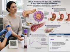
A pancreatic hormone test panel measures selected hormones made by islet cells, commonly insulin, C-peptide, proinsulin, and glucagon. These tests can help investigate documented hypoglycemia, assess endogenous insulin production, characterize unusual hyperglycemia, or evaluate symptoms suggesting a functional pancreatic neuroendocrine tumor. They do not form a universal screening panel for pancreatic cancer, pancreatitis, diabetes, or “pancreatic health.” Each marker has different physiology, specimen requirements, reference intervals, and diagnostic uses. Timing is especially important: insulin, C-peptide, and proinsulin are most informative for unexplained hypoglycemia when drawn together during low laboratory plasma glucose, while glucagon testing is usually reserved for a compatible syndrome such as unexplained weight loss, diabetes, and necrolytic migratory erythema. Kidney and liver function, food intake, medications, assay cross-reactivity, specimen handling, and acute illness can alter results. The panel should be selected and interpreted by a clinician who begins with the symptoms and uses the hormone pattern to answer a defined question rather than treating one high or low value as a diagnosis.
- Insulin, C-peptide, and proinsulin must be interpreted against the glucose concentration at the same blood draw.
- C-peptide and proinsulin reflect pancreatic beta-cell secretion; injected insulin contains neither marker.
- Glucagon comes mainly from alpha cells and is not routinely included in diabetes screening.
- A pancreatic hormone panel does not detect most nonfunctional pancreatic tumors and cannot replace imaging or tissue diagnosis when those are indicated.
- Results from different laboratories may not be comparable because immunoassays, units, and reference intervals vary.
Table of Contents
- What the Panel Measures
- When the Panel Is Ordered
- Preparation and Specimen Collection
- Insulin, C-Peptide, and Proinsulin Results
- Glucagon Results
- Common Result Patterns
- Limitations and Diagnostic Pitfalls
- Follow-Up Testing and Next Steps
What the Panel Measures
The endocrine pancreas contains clusters of cells called islets. Beta cells make insulin and its precursors, alpha cells make glucagon, delta cells make somatostatin, and pancreatic-polypeptide cells make pancreatic polypeptide. A four-analyte panel focuses on beta-cell secretion and alpha-cell activity.
Insulin
Insulin lowers blood glucose by promoting glucose uptake and storage, limiting liver glucose production, and suppressing fat breakdown and ketone formation. Its concentration changes rapidly after food and during hypoglycemia. The liver removes a large share of newly secreted insulin before it reaches peripheral blood, and circulating insulin has a short half-life.
An insulin result cannot be labeled high or low without the paired glucose. An insulin value that looks ordinary on a fasting reference interval may be abnormally high during severe hypoglycemia, because normal beta cells should nearly stop secretion. Conversely, a high post-meal insulin may be a normal response to rising glucose.
C-peptide
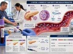
C-peptide is released when proinsulin is split into insulin and C-peptide inside beta-cell granules. The pancreas releases insulin and C-peptide in equal numbers of molecules, but C-peptide is cleared more slowly and mainly through the kidneys. It therefore gives a steadier indication of endogenous insulin production.
Injected insulin does not contain C-peptide. A high insulin concentration with low C-peptide during low glucose can support exogenous insulin exposure, provided the insulin assay detects the preparation. C-peptide is also used to estimate residual beta-cell function in diabetes, especially when a person is receiving insulin and direct insulin measurement would mix endogenous and injected hormone.
Proinsulin
Proinsulin is the precursor from which insulin and C-peptide are made. Healthy beta cells convert most proinsulin before secretion, so circulating concentrations are lower than insulin concentrations. Beta-cell stress, insulin resistance, and insulin-secreting tumors may increase absolute proinsulin or the proportion released incompletely processed.
During hypoglycemia, an inappropriately detectable or elevated proinsulin supports continued endogenous beta-cell secretion. A fasting proinsulin level obtained when glucose is normal has different meaning and cannot substitute for a critical sample.
Glucagon
Glucagon is an opposing hormone that helps maintain glucose between meals by stimulating liver glycogen breakdown and glucose production. It also promotes ketone formation during fasting. Glucagon normally rises when glucose is low and is suppressed after carbohydrate intake, although the response is complex in diabetes.
Glucagon testing is not usually needed for ordinary diabetes diagnosis. It is most useful when a clinician suspects a glucagon-secreting pancreatic neuroendocrine tumor or is conducting specialized research or dynamic testing. The assay is technically challenging because multiple glucagon-related peptides can interfere and the hormone is unstable unless the specimen is processed correctly.
Together, these measurements describe related but distinct pathways. They do not create a single validated score. The ordering clinician should specify whether the goal is hypoglycemia evaluation, diabetes classification, tumor investigation, or another focused purpose.
When the Panel Is Ordered
The most established use of insulin, C-peptide, and proinsulin together is the investigation of unexplained hypoglycemia. In adults without diabetes, clinicians generally look for Whipple’s triad: compatible symptoms, low plasma glucose measured by a reliable method, and improvement when glucose rises. Symptoms include sweating, tremor, hunger, palpitations, confusion, abnormal behavior, weakness, blurred vision, seizure, or loss of consciousness.
A critical sample is collected during the low glucose and commonly includes:
- Laboratory plasma glucose
- Insulin
- C-peptide
- Proinsulin
- Beta-hydroxybutyrate
- Sulfonylurea and meglitinide screen
Glucagon concentration is not always part of that sample, although intravenous glucagon may be given after collection to assess the glucose response. A full hypoglycemia hormone test panel is broader than the four-hormone panel in this article.
Clinicians may also order C-peptide, with or without insulin and glucose, to evaluate endogenous insulin production in diabetes. Questions include whether a person has severe insulin deficiency, whether a diabetes phenotype may be type 1 rather than type 2, whether pancreatic function remains after years of disease, or whether a person might safely use therapies that depend on residual beta-cell capacity. Stimulated C-peptide after a mixed meal or glucagon may be more informative than a fasting value in selected cases.
A pancreatic neuroendocrine tumor evaluation is symptom-directed. Functional tumors are classified by the hormone syndrome they produce:
- Insulinoma: recurrent fasting or exercise-related hypoglycemia
- Glucagonoma: diabetes or glucose intolerance, weight loss, characteristic rash, anemia, thrombosis, or mouth inflammation
- Gastrinoma: recurrent peptic ulcers, reflux, or secretory diarrhea
- VIPoma: profuse watery diarrhea, low potassium, and dehydration
- Somatostatinoma: diabetes, gallbladder disease, diarrhea or steatorrhea, and weight loss
Measuring insulin, C-peptide, proinsulin, and glucagon indiscriminately is less effective than choosing tests that match the syndrome. Most pancreatic neuroendocrine tumors are nonfunctional and may not produce diagnostic hormone elevations. Common pancreatic adenocarcinoma is not diagnosed with this panel.
The panel is not a screening test for pancreatitis. Pancreatitis is evaluated with symptoms, lipase, imaging, and clinical findings. It is also not a comprehensive assessment of exocrine pancreatic function, which involves digestive enzymes and may require fecal elastase, imaging, or direct function testing.
Preparation and Specimen Collection
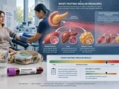
Preparation depends on the question. For a routine fasting metabolic assessment, the laboratory may request an overnight fast, commonly 8–12 hours, with water permitted. For hypoglycemia, the goal is not simply fasting; the goal is to collect the sample when glucose is actually low and before treatment changes the result.
A supervised fast may be used when spontaneous episodes cannot be captured. It can continue for up to 72 hours under a defined protocol, with progressively frequent glucose checks and immediate access to treatment. This is not safe as a home experiment. For symptoms after meals, a supervised mixed-meal test may reproduce the usual trigger. An oral glucose tolerance test is generally not preferred for ordinary postprandial hypoglycemia because a pure glucose load can create low readings that do not reflect real-life meals.
The patient should provide a complete medication and supplement list. Important agents include:
- Prescribed insulin and the exact analog
- Sulfonylureas and meglitinides
- GLP-1 receptor agonists and dual incretin medicines
- Corticosteroids
- Beta blockers
- Biotin in high-dose supplements
- Drugs associated with hypoglycemia or altered glucose metabolism
Medications should not be stopped unless the clinician directs it. In suspected gastrinoma or other separate syndromes, changing acid-suppressing therapy without supervision can be dangerous; that issue is not solved by this panel.
Specimen handling matters. Glucose falls in unseparated blood because cells continue consuming it. Insulin can be degraded by enzymes released during hemolysis. C-peptide may also be affected by hemolysis and is influenced by kidney clearance. Proinsulin and glucagon often require rapid separation, freezing, or special tubes. Glucagon is particularly vulnerable to degradation and immunoassay interference from related peptides.
For a critical sample, record the exact times of symptoms, bedside and laboratory glucose, blood collection, food, dextrose, glucagon, and medication. Insulin and C-peptide should come from the same blood draw if a molar ratio is calculated. Results collected after glucose treatment may no longer represent the cause of the event.
Fasting status, body mass index, kidney function, liver function, and recent illness should accompany interpretation. A single value outside the reference interval may be a physiological response to the collection conditions rather than disease.
Insulin, C-Peptide, and Proinsulin Results
The beta-cell markers should be interpreted as a coordinated pattern.
During hypoglycemia
When plasma glucose is sufficiently low, normal beta cells suppress insulin, C-peptide, and proinsulin. The important abnormality is failure to suppress, not necessarily a value above the ordinary fasting range.
Traditional adult criteria supporting endogenous hyperinsulinemia during a supervised fast include plasma glucose below about 55 mg/dL with insulin at least 3 µIU/mL, C-peptide at least 0.6 ng/mL, and proinsulin at least 5 pmol/L. These are not universal stand-alone cutoffs. Modern assays differ, kidney function changes C-peptide, and the full pattern—including ketones and drug screening—must be considered.
High or detectable insulin with high C-peptide and proinsulin suggests the pancreas is secreting insulin. Causes include insulinoma, sulfonylurea or meglitinide exposure, post-bariatric hypoglycemia, and rarer beta-cell disorders. A negative drug screen is required before attributing the pattern to a tumor.
High insulin with low C-peptide and low proinsulin suggests injected insulin. Interpretation can fail if the insulin immunoassay does not recognize the specific analog. An insulin/C-peptide ratio may provide supporting evidence after both concentrations are converted to molar units, but it is not definitive in the presence of insulin antibodies, kidney dysfunction, or assay mismatch.
Low insulin, C-peptide, and proinsulin with appropriately high beta-hydroxybutyrate suggests a non-insulin cause such as prolonged fasting, malnutrition, alcohol-related impaired glucose production, adrenal insufficiency, or severe illness. Low ketones despite suppressed insulin markers raises consideration of IGF-mediated tumor hypoglycemia or a fatty-acid oxidation disorder.
In diabetes assessment
C-peptide is usually more useful than insulin for estimating endogenous production in a person using insulin therapy. A low fasting value may reflect low glucose at the draw, so a paired glucose is necessary. Stimulated testing can show reserve that a fasting sample misses.
High insulin and C-peptide with normal or high glucose may occur with insulin resistance, obesity, early type 2 diabetes, corticosteroid effects, or other counter-regulatory hormone excess. These results do not diagnose insulin resistance by themselves. Standard glucose and A1C criteria diagnose diabetes, while clinical features and validated risk markers guide treatment.
Low C-peptide with hyperglycemia supports severe insulin deficiency, as can occur in type 1 diabetes, advanced type 2 diabetes, pancreatic destruction, or after pancreatectomy. Autoantibodies, clinical history, age, ketosis, and disease course help determine the cause.
Proinsulin may rise with beta-cell stress and has research value in predicting diabetes risk, but routine treatment targets are not established. A high fasting proinsulin is not equivalent to insulinoma unless it is inappropriately present during hypoglycemia.
Glucagon Results
Glucagon reference intervals are laboratory- and method-specific. Results may be reported in picograms per milliliter or picomoles per liter. The value should be compared only with the interval from the performing laboratory under the stated fasting conditions.
A markedly elevated glucagon concentration in a person with a compatible syndrome can support glucagonoma. Clinical features may include:
- Necrolytic migratory erythema, often involving groin, buttocks, lower abdomen, or perioral skin
- New or worsening diabetes or glucose intolerance
- Weight loss despite adequate intake
- Anemia
- Glossitis or stomatitis
- Diarrhea
- Venous thrombosis
- Depression or neuropsychiatric symptoms
Very high concentrations, historically in the range of 500–1,000 pg/mL or greater, are more concerning when the phenotype is present. A lower elevation is nonspecific and can occur with kidney failure, liver disease, pancreatitis, severe stress, prolonged fasting, uncontrolled diabetes, infection, or assay cross-reactivity. Some glucagonomas produce only modest elevations, so no single cutoff replaces clinical judgment.
Low glucagon is rarely a useful isolated diagnosis. People with long-standing type 1 diabetes may have an impaired glucagon response to hypoglycemia even if a fasting concentration is not clearly low. Measuring a baseline hormone does not fully assess dynamic counter-regulation.
Glucagon assays face important analytical problems. Proglucagon is processed into several related peptides in the pancreas and intestine. Some immunoassays cross-react with these fragments or detect different molecular forms. Collection in an appropriate tube, rapid chilling and separation, protease inhibition when specified, and frozen transport may be required.
A high glucagon result should be repeated or confirmed if it conflicts with the presentation. Imaging for a pancreatic tumor is more persuasive after biochemical and clinical confirmation. Cross-sectional imaging, somatostatin-receptor imaging, and endoscopic ultrasound may be used depending on the suspected tumor and local expertise.
Glucagon concentration should not be confused with the glucagon stimulation test. In that test, pharmaceutical glucagon is administered and glucose or C-peptide responses are measured. The purpose and interpretation are different from measuring fasting endogenous glucagon.
Common Result Patterns
| Clinical setting and pattern | Possible interpretation |
|---|---|
| Low glucose; insulin, C-peptide, and proinsulin not suppressed; ketones low; drug screen negative | Endogenous hyperinsulinism, including insulinoma |
| Low glucose; insulin high; C-peptide and proinsulin low; ketones low | Exogenous insulin, subject to analog assay limitations |
| Low glucose; insulin, C-peptide, and proinsulin not suppressed; drug screen positive | Sulfonylurea or meglitinide effect |
| High glucose; insulin and C-peptide high | Insulin resistance or compensatory hyperinsulinemia; interpret with metabolic context |
| High glucose; C-peptide very low | Severe endogenous insulin deficiency |
| Marked glucagon elevation plus characteristic rash, weight loss, and diabetes | Glucagonoma syndrome; confirm biochemistry and localize |
| Mild glucagon elevation without characteristic syndrome | Nonspecific; review fasting, kidney, liver, illness, and assay factors |
Patterns are more reliable than isolated flags. For example, an elevated insulin result during hyperglycemia can be an appropriate compensatory response, while the same number during hypoglycemia can be dangerous. A normal C-peptide in kidney failure may actually represent reduced secretion because impaired clearance should have raised it. A high proinsulin with normal glucose may reflect insulin resistance rather than a tumor.
The panel also does not distinguish every functional tumor. Gastrinoma, VIPoma, and somatostatinoma require their own hormone measurements. Pancreatic polypeptide and chromogranin A may be used in selected neuroendocrine tumor evaluations, but both have limited specificity. Proton pump inhibitors, kidney dysfunction, and assay variation can raise chromogranin A.
Multiple endocrine neoplasia type 1 should be considered in selected patients with pancreatic or duodenal neuroendocrine tumors, especially with young age, multiple tumors, family history, hyperparathyroidism, or pituitary disease. Other hereditary associations include von Hippel-Lindau disease, neurofibromatosis type 1, and tuberous sclerosis complex. Genetic counseling should be targeted rather than triggered by one mildly abnormal hormone.
Limitations and Diagnostic Pitfalls
Several common errors reduce the panel’s value.
Ordering without a clinical question: A broad hormone panel can generate incidental abnormalities that lead to unnecessary imaging. Symptoms should determine the tests.
Missing the critical sample: Insulin, C-peptide, and proinsulin collected after hypoglycemia resolves cannot determine what caused the event. A normal glucose at the draw removes the essential context.
Using home-device glucose as the only proof: Meters and continuous monitors are helpful but less accurate at low concentrations. Laboratory confirmation is important for rare spontaneous hypoglycemia disorders.
Ignoring assay detection of insulin analogs: Some insulin methods recognize human insulin but detect lispro, aspart, glargine, detemir, or degludec inconsistently. A low reported insulin does not always exclude injection.
Ignoring renal clearance: Kidney dysfunction raises C-peptide and can raise insulin and proinsulin. Glucagon may also increase. Reference cutoffs derived in normal renal function become less reliable.
Hemolysis or delayed processing: Hemolysis can degrade insulin and affect C-peptide. Delayed glucose separation can create falsely low glucose. Poor glucagon handling can produce unreliable values.
Biotin and antibody interference: High-dose biotin affects some immunoassays. Heterophile antibodies, insulin antibodies, and antibodies to assay components can cause unexpected results. The laboratory should be consulted when values conflict with physiology.
Equating a tumor marker with cancer screening: Functional hormone tests detect syndromes, not all tumors. Normal results do not rule out nonfunctional pancreatic neuroendocrine tumors or pancreatic adenocarcinoma.
Imaging before biochemical confirmation: Small pancreatic lesions are common enough to mislead, while insulinomas can be too small for initial imaging. Confirming the hormone syndrome first reduces unnecessary procedures.
Applying one laboratory’s range to another: Methods, calibrators, units, fasting requirements, and reference populations differ. Online “optimal ranges” should not replace the report.
Follow-Up Testing and Next Steps
Follow-up is driven by the pattern and the original question.
For unexplained hypoglycemia, an endocrinologist may review the original specimen, repeat testing during a spontaneous event, conduct a supervised fast, or perform a mixed-meal test. Beta-hydroxybutyrate, medication screening, insulin antibodies, cortisol, and organ-function tests help classify the mechanism. Once endogenous hyperinsulinism is established, pancreatic imaging and specialized localization procedures may follow.
For diabetes classification, follow-up may include fasting or stimulated C-peptide with paired glucose, pancreatic autoantibodies, A1C, ketones, and review of pancreatitis, surgery, cystic fibrosis, hemochromatosis, or other pancreatic disease. Treatment should respond to glucose control and risk of insulin deficiency, not to an isolated insulin value.
For suspected glucagonoma, clinicians evaluate the skin findings, diabetes, anemia, amino-acid or nutritional deficiency, liver metastases, and thrombosis risk. A repeat fasting glucagon with careful processing may be appropriate before imaging. Dermatologic biopsy can support necrolytic migratory erythema but is not specific by itself.
For a confirmed pancreatic neuroendocrine tumor, care may involve endocrinology, oncology, gastroenterology, radiology, nuclear medicine, genetics, surgery, nutrition, and dermatology. Monitoring uses the hormone that was clearly elevated at baseline, imaging, tumor grade, stage, and treatment response. An analyte that was normal before treatment is usually a poor surveillance marker.
Urgent care is required for severe hypoglycemia, persistent vomiting or diarrhea with dehydration, marked electrolyte abnormalities, or symptoms of thrombosis. Diagnostic testing should never delay emergency glucose, fluids, potassium replacement, or other stabilization.
The central rule is to interpret each hormone in its physiological context. Insulin, C-peptide, and proinsulin answer whether beta-cell secretion is present and appropriate for glucose. Glucagon answers a different question about alpha-cell secretion and a rare tumor syndrome. Used selectively, the panel can reveal important disorders; used as an untargeted wellness screen, it is more likely to confuse than clarify.
References
- Neuroendocrine Tumors | Choose the Right Test 2026 (Laboratory guidance)
- INS – Overview: Insulin, Serum 2026 (Laboratory test information)
- CPR – Overview: C-Peptide, Serum 2026 (Laboratory test information)
- Pancreatic Neuroendocrine Tumors—Diagnostic Pitfalls of the Most Common Functional Pancreatic Neuroendocrine Tumors 2025 (Review)
- Glucagonoma and Glucagonoma Syndrome: An Updated Review 2025 (Review)
Disclaimer
This article is general education and cannot diagnose hypoglycemia, diabetes type, or a pancreatic neuroendocrine tumor. Results require interpretation with the paired glucose, collection conditions, medications, kidney and liver function, assay method, and symptoms. Severe hypoglycemia, dehydration, seizure, unconsciousness, or suspected thrombosis requires urgent medical care.