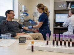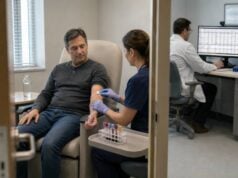
A growth hormone stimulation test evaluates whether the pituitary can release an adequate GH surge when challenged. It is used when a child has convincing growth failure or an adult has a strong risk of pituitary-related GH deficiency. Because GH is secreted in pulses, a random low level is expected and cannot diagnose deficiency. During stimulation testing, a medicine or controlled metabolic change triggers GH release, and blood is collected repeatedly to identify the peak. The result is not interpreted from one universal cutoff. The stimulating agent, age, puberty, body mass index, assay, sampling schedule, and pretest probability all matter. Insulin-induced hypoglycemia, glucagon, arginine, clonidine, and macimorelin have different protocols and risks. A low peak supports deficiency only when the clinical history, IGF-1, other pituitary hormones, and imaging findings are consistent and confounding conditions have been addressed.
- The test measures GH reserve, not the average daily GH concentration.
- Multiple blood samples are needed to capture the stimulated peak.
- Pediatric and adult cutoffs are different and assay-specific.
- Obesity can lower GH peaks without true pituitary deficiency.
- Some patients with strong structural evidence do not need provocative testing.
- Safety screening is essential, especially for insulin and glucagon protocols.
Table of Contents
- Why Stimulation Testing Is Used
- Who Should Have the Test
- Test Agents and Procedures
- Preparation, Safety, and Side Effects
- Peak Levels and Cutoffs
- Interpreting Low, Borderline, and Normal Results
- Next Steps and Questions
Why Stimulation Testing Is Used
Growth hormone is released in brief pulses, especially during deep sleep. Between pulses, the serum concentration can be near zero in a healthy person. A stimulation test creates a standardized challenge and asks whether the hypothalamic-pituitary system can generate a sufficient peak.
The test is most useful when the chance of GH deficiency is neither negligible nor already certain. In children, the decision begins with accurate growth measurements. Reduced height velocity, downward crossing of percentiles, delayed bone age, a low IGF-1 standard deviation score, or a pituitary risk factor may justify testing after common causes of poor growth are excluded.
In adults, nonspecific symptoms such as fatigue, increased abdominal fat, reduced exercise capacity, or low bone density are not enough. Most adult-onset cases occur after a pituitary or hypothalamic tumor, surgery, radiation, traumatic brain injury, infiltrative disease, or congenital disorder. Dynamic testing confirms the diagnosis when treatment is being considered.
The test measures reserve rather than spontaneous secretion. A normal response shows that the pituitary can release GH under that stimulus. It does not prove that every aspect of spontaneous secretion is normal. Conversely, a low response can reflect true deficiency or a false-positive result caused by obesity, delayed puberty, poor test conditions, or an inappropriate cutoff.
IGF-1 is measured before dynamic testing because it reflects integrated GH action. A low IGF-1 supports the diagnosis but is not specific; undernutrition, liver disease, hypothyroidism, inflammation, and poorly controlled diabetes can lower it. A normal IGF-1 makes severe deficiency less likely but does not exclude it in a high-risk adult or a child with organic pituitary disease.
Testing is not generally appropriate as an anti-aging assessment, fitness screen, or explanation for isolated tiredness. Normal age-related decline in GH secretion is not a disease requiring replacement.
Who Should Have the Test
Children are considered for stimulation testing when their growth pattern suggests impaired GH action and alternative causes have been evaluated. Concerning findings include height below about −2 standard deviations, growth velocity below the expected range, height far below the family target, or an inadequate pubertal growth spurt.
Strong pediatric risk factors include:
- congenital pituitary or midline abnormalities;
- neonatal hypoglycemia, prolonged jaundice, or micropenis;
- pituitary or brain tumor;
- cranial surgery or radiation;
- significant head injury;
- multiple pituitary hormone deficiencies;
- genetic variants affecting pituitary development or GH secretion.
Short stature alone is not enough. Familial short stature and constitutional delay are common. Celiac disease, hypothyroidism, kidney disease, inflammatory illness, poor nutrition, glucocorticoid exposure, skeletal dysplasia, and genetic growth-plate disorders must be considered.
Adults should usually have a clear hypothalamic-pituitary risk factor. Childhood-onset GH deficiency is retested after final height unless a permanent genetic, structural, or multiple-hormone cause makes persistence highly likely. Some individuals recover normal reserve during transition to adult care.
Dynamic testing may be unnecessary when the evidence is conclusive. Examples include a known genetic defect that causes permanent GH deficiency or an organic pituitary lesion accompanied by several additional pituitary hormone deficits and a low IGF-1. The exact exception depends on guideline and local policy.
Testing should be deferred when untreated hypothyroidism, severe systemic illness, significant undernutrition, or uncontrolled diabetes is likely to confound the result. In adults, sex-hormone status and oral estrogen use are considered because they affect IGF-1 and GH physiology.
A growth failure hormone test panel should precede pediatric stimulation, while adults need a complete pituitary review. Testing one axis in isolation can miss cortisol or thyroid deficiencies that require priority treatment.
Test Agents and Procedures
No single stimulus is ideal for every patient. The center selects an agent based on age, risk, availability, regulatory approval, and local validation.
Insulin Tolerance Test
The insulin tolerance test, or ITT, uses intravenous insulin to lower blood glucose, usually to below 40 mg/dL or to a level that produces clear neuroglycopenic symptoms. Hypoglycemia is a powerful stimulus for GH and cortisol release. Blood is collected before and at intervals after insulin, commonly over 90 to 120 minutes.
The ITT is a reference test in adults because it evaluates both GH and ACTH reserve. It requires experienced staff, continuous observation, and immediate access to glucose. It is avoided in seizure disorders, ischemic heart disease, some cerebrovascular conditions, and patients in whom severe hypoglycemia would be unsafe.
Glucagon Stimulation Test
Intramuscular glucagon stimulates GH through mechanisms involving glucose and neuroendocrine responses. Samples are collected for up to three or four hours because the GH peak is often delayed. Glucose and sometimes cortisol are measured simultaneously.
The glucagon test is widely used when ITT is contraindicated. Nausea and vomiting are common, and late hypoglycemia can occur. Older or frail patients and those with very low body weight need careful risk assessment. Cutoffs vary with body mass index and pretest probability.
Arginine Test
Intravenous arginine suppresses hypothalamic somatostatin and permits GH release. It is often combined with another stimulus in children or used where local protocols support it. Blood is sampled over approximately 90 to 120 minutes.
Arginine may cause nausea, flushing, headache, or local infusion discomfort. Severe reactions are uncommon. Arginine alone may be less powerful than combined tests, so the center must use a matching cutoff.
Clonidine Test
Oral clonidine stimulates GH through alpha-adrenergic pathways and is used mainly in children. Blood pressure and heart rate are monitored because drowsiness, dizziness, and hypotension can occur. Samples are commonly collected for two hours.
Macimorelin Test
Macimorelin is an oral ghrelin-receptor agonist used for adult GH deficiency in some countries. The patient drinks a weight-based solution after fasting, and GH is measured at defined times, commonly 30, 45, 60, and 90 minutes. The procedure is shorter and avoids deliberate hypoglycemia.
Drug interactions and QT prolongation risk must be reviewed. The validated cutoff is assay- and label-specific. Macimorelin is not a routine pediatric test.
Some centers perform two stimuli on the same day in children. Others require two separate failed tests for idiopathic isolated deficiency. A single test may be accepted when organic disease raises pretest probability. The report should identify every agent, dose, time point, and result.
Preparation, Safety, and Side Effects
Patients commonly fast overnight for 8 to 12 hours. Water instructions vary. The center may restrict vigorous exercise, alcohol, nicotine, and certain medicines beforehand. Children should receive age-appropriate preparation because repeated blood draws and fasting can be stressful.
An intravenous cannula is placed before most protocols so repeated samples do not require repeated needle sticks. Baseline glucose and GH are collected, the stimulus is administered, and samples are drawn at scheduled intervals. Vital signs and symptoms are recorded.
Medications and conditions to disclose include:
- insulin, glucose-lowering drugs, or a history of severe hypoglycemia;
- glucocorticoid replacement or suspected adrenal insufficiency;
- thyroid hormone and untreated hypothyroidism;
- estrogen, testosterone, puberty blockers, or fertility medicines;
- recombinant GH or long-acting GH;
- anticonvulsants and seizure history;
- heart disease, stroke, pregnancy, and severe liver or kidney disease;
- medicines that prolong the QT interval before macimorelin.
Hormone replacement should not be stopped without instructions. A person with known ACTH deficiency must take required glucocorticoid coverage. The ITT may be unsafe if cortisol reserve is inadequate or the patient cannot tolerate hypoglycemia.
Typical side effects are protocol-specific:
| Stimulus | Common effects | Major safety concern |
|---|---|---|
| Insulin | Sweating, tremor, hunger, confusion | Severe or prolonged hypoglycemia, seizure |
| Glucagon | Nausea, vomiting, abdominal discomfort | Delayed hypoglycemia, aspiration risk |
| Arginine | Nausea, flushing, headache | Infusion reaction, electrolyte concerns in selected patients |
| Clonidine | Sleepiness, dry mouth, dizziness | Hypotension or bradycardia |
| Macimorelin | Altered taste, dizziness, headache | QT prolongation or drug interaction |
After the test, food is given once it is safe, glucose is stable, and nausea has settled. Driving or returning immediately to school or work may be discouraged after sedating or hypoglycemic protocols.
A valid test requires an adequate stimulus. For ITT, glucose must fall enough. For glucagon, the full observation window is needed because peaks may occur late. Missing the expected sampling window can create a false low result.
Sex-steroid priming is considered in some older prepubertal children with delayed puberty. A short estrogen or testosterone course before testing may reduce false-positive low peaks. Practices differ because priming can also conceal a mild defect; the rationale should be documented.
Peak Levels and Cutoffs
The peak is the highest GH concentration measured during the series. GH is reported in ng/mL or µg/L, which are numerically equivalent. There is no single cutoff that applies to every agent and assay.
Pediatric Cutoffs
A peak below 10 ng/mL was historically used to diagnose childhood GH deficiency. Modern monoclonal assays often read lower, and many centers use thresholds of 7, 6, or 5 ng/mL. The correct cutoff is the value validated for the assay and stimulus.
A peak far below the cutoff in a child with severe growth failure, low IGF-1, and a structural pituitary abnormality is more convincing than a peak just below the line in a healthy prepubertal child with delayed bone age. The result exists on a continuum.
Adult ITT Cutoffs
A peak below approximately 3 ng/mL is commonly used for severe adult GH deficiency during an adequate ITT. Some guidance uses a higher cutoff in transition-age patients or selected high-probability groups. The achieved glucose nadir and symptoms must confirm adequate hypoglycemia.
Adult Glucagon Cutoffs
Glucagon interpretation may use 3 ng/mL in lean adults and those with high pretest probability, with a lower cutoff around 1 ng/mL in obesity or low-probability settings. Current recommendations differ, and recent studies continue to refine BMI- and age-specific performance.
Macimorelin Cutoffs
The original approved cutoff was a peak at or below 2.8 ng/mL with the validated assay. Some evidence supports a higher threshold around 5.1 ng/mL in patients with high pretest probability to improve sensitivity. The test must follow the labeled dose, timing, and assay.
| Setting | Illustrative low peak | Important qualifier |
|---|---|---|
| Child, older historical standard | Below 10 ng/mL | May overdiagnose with modern assays |
| Child, modern center | Below approximately 5–7 ng/mL | Agent- and assay-specific |
| Adult ITT | Below approximately 3 ng/mL | Requires adequate hypoglycemia |
| Adult glucagon | Below 1–3 ng/mL | BMI and pretest probability matter |
| Adult macimorelin | At or below 2.8 ng/mL | Assay and label-specific; higher cutoffs studied |
These values are educational examples. The laboratory’s validated protocol and endocrine center’s criteria control the diagnosis.
Body mass index is a major modifier. Obesity suppresses spontaneous and stimulated GH. Using a lean cutoff in a person with obesity can create a false diagnosis. Age, sex, oral estrogen, puberty, and recent nutritional status also affect peaks.
Assay evolution explains much disagreement. Older polyclonal assays measured multiple GH forms and often produced higher results. Modern assays may be calibrated to international standards and preferentially detect the 22-kDa form. A published cutoff must match the measurement method.
Interpreting Low, Borderline, and Normal Results
A low peak is interpreted as evidence, not a diagnosis by itself. The clinician checks whether the stimulus was adequate, every sample was collected, the correct cutoff was used, and confounders were addressed.
A convincing deficiency pattern includes:
- a high-risk pituitary history or clearly abnormal growth;
- low age-adjusted IGF-1 after excluding nutrition and systemic illness;
- a substantially low stimulated peak on a valid test;
- other pituitary hormone deficits;
- a compatible pituitary MRI or genetic finding.
A borderline result calls for caution. In children, constitutional delay, absent priming, obesity, or a single imperfect test may explain a peak just below threshold. Options include longitudinal growth follow-up, repeat testing with a different stimulus, review of IGF-1 SDS, and reassessment during puberty.
In adults, a borderline glucagon or macimorelin result is weighed against pretest probability. A person with multiple pituitary deficits and a structural lesion is different from someone with nonspecific fatigue and no pituitary disease. Testing low-probability populations produces more false positives.
A normal peak makes clinically significant GH deficiency less likely, but it does not explain the symptoms or poor growth. The evaluation then returns to nutrition, thyroid disease, celiac disease, chronic inflammation, skeletal and genetic disorders, sex-hormone status, sleep, medication, and psychosocial factors.
Discordant IGF-1 and stimulation results are common. Low IGF-1 with a normal peak may reflect undernutrition, liver disease, hypothyroidism, inflammation, or GH resistance. Normal IGF-1 with a low peak may reflect obesity, a false-positive test, or partial deficiency. Severe organic disease can still produce a normal-range IGF-1.
MRI findings increase or decrease confidence but do not replace physiology. Pituitary stalk interruption, ectopic posterior pituitary, pituitary hypoplasia, or a tumor supports organic disease. A mildly small pituitary can be incidental. Imaging should be reviewed by clinicians familiar with age-related pituitary anatomy.
For children treated after a borderline diagnosis, first-year growth response is informative but should not be used as the sole retrospective proof. A poor response prompts review of adherence, dose, thyroid status, nutrition, diagnosis, and other causes. Those with isolated idiopathic childhood deficiency are often retested at final height.
When a Test Should Be Repeated
Repeating a stimulation test is most useful when the original study had a correctable limitation. Examples include inadequate hypoglycemia during an ITT, vomiting before oral medication was absorbed, loss of intravenous access during the expected peak, premature termination of a glucagon test, or use of a cutoff that does not match the assay. The repeat should correct that problem or use a different validated stimulus.
A second test may also be required by pediatric diagnostic standards when isolated idiopathic deficiency is suspected. Agreement between two different stimuli increases confidence, but two tests can share the same confounders. Obesity, delayed puberty, untreated hypothyroidism, and poor nutrition should be addressed before simply repeating the procedure.
Repetition is less helpful when the pretest probability is extremely low. A person with nonspecific fatigue, normal IGF-1, no pituitary history, and one borderline result may be harmed by a cycle of additional testing and inappropriate treatment. Reassessing whether the test was indicated is part of good interpretation.
Conversely, a normal test may be reconsidered if pituitary disease is evolving. Cranial radiation can cause progressive GH loss years later, and a child’s growth velocity may deteriorate after an initially adequate result. New structural findings, additional pituitary deficits, or a clearly changed growth pattern justify a fresh evaluation rather than assuming that reserve remains permanently normal.
Next Steps and Questions
After confirmed deficiency, recombinant GH may be offered when clinically appropriate. Treatment is individualized and monitored with growth response in children, symptoms and body composition in adults, IGF-1, glucose, thyroid function, and adverse effects.
Before treatment, the team ensures that adrenal and thyroid replacement are adequate. Starting GH can increase cortisol metabolism and alter thyroid hormone conversion, revealing previously unrecognized deficiencies. Multiple pituitary axes must therefore be followed together.
Useful questions after a stimulation test include:
- Which stimulus and dose were used?
- Was the physiologic stimulus adequate, especially the glucose nadir during ITT?
- Were all planned samples collected through the expected peak window?
- Which GH assay and cutoff does this center use?
- Was the cutoff adjusted for age, puberty, body mass index, and pretest probability?
- What was the IGF-1 SDS, and were nutrition, thyroid, liver, and systemic disease considered?
- Does the MRI or another pituitary deficit support the result?
- Is a second test required before treatment?
- If the result is borderline, would observation or a different stimulus be safer?
- Will childhood-onset deficiency be retested at final height?
Seek urgent medical attention after testing for persistent confusion, seizure, fainting, chest pain, breathing difficulty, or symptoms of prolonged hypoglycemia. Contact the testing center for repeated vomiting, inability to eat, or severe dizziness after glucagon or clonidine.
During GH therapy, severe headache with vomiting or visual change, new hip or knee pain, marked swelling, or symptoms of high glucose require prompt review. These are not reasons to interpret the original peak differently, but they affect treatment safety.
A GH peak is only as reliable as the question, protocol, assay, and patient selection behind it. The best diagnosis emerges when dynamic testing confirms a coherent clinical and biochemical pattern rather than when one value barely crosses an arbitrary line.
When results are close to the cutoff, the report should identify the stimulating agent, assay, sampling schedule, peak time, and whether sex-steroid priming was used. Those details can change the conclusion.
References
- Diagnosis and testing for growth hormone deficiency across the ages: a global view of the accuracy, caveats, and cut-offs for diagnosis 2023 (Review)
- A 2024 Update on Growth Hormone Deficiency Syndrome in Adults: From Guidelines to Real Life 2024 (Review)
- Once upon a time: the glucagon stimulation test in diagnosing adult GH deficiency 2024 (Review)
- Growth Hormone Stimulation Tests in Assessing Adult Growth Hormone Deficiency 2023 (Clinical Review)
- Consensus and controversies about diagnosing GH deficiency: a Delphi survey by the GH Research Society 2025 (Consensus)
- Accuracy of Glucagon Testing Across Transition in Young Adults With Childhood-Onset Growth Hormone Deficiency 2025 (Research Article)
Disclaimer
This article provides general education and cannot diagnose growth hormone deficiency or select a stimulation protocol. Dynamic testing can cause hypoglycemia, hypotension, nausea, and other adverse effects and must be performed under medical supervision. Peak cutoffs are assay-, agent-, age-, and BMI-specific and require specialist interpretation.