A growth hormone suppression test evaluates whether oral glucose can lower GH appropriately. It is used mainly when acromegaly is suspected or when biochemical findings are uncertain after treatment. In people without autonomous GH excess, rising blood glucose suppresses pituitary GH secretion. In acromegaly, GH often fails to fall below the assay-specific nadir. The test is usually performed as a 75-gram oral glucose tolerance test with GH and glucose samples collected over about two hours. Interpretation begins with age-adjusted IGF-1, because IGF-1 is the primary screening marker and reflects integrated GH action more reliably than one random GH value. Modern assays can measure very low GH concentrations, so historical cutoffs of 1.0 ng/mL and stricter values near 0.4 ng/mL are not interchangeable. Body mass index, sex, estrogen, diabetes, nutrition, liver or kidney disease, assay calibration, and treatment status all affect results.
- The test asks whether glucose suppresses GH, not whether glucose tolerance is normal.
- IGF-1 is usually checked first and repeated when unexpectedly high.
- GH is measured before and after a standardized glucose drink.
- A failed suppression result must match the assay and protocol.
- Diabetes and incomplete glucose ingestion can complicate interpretation.
- Pituitary MRI follows biochemical confirmation, not an isolated random GH result.
Table of Contents
- Why Glucose Suppresses GH
- When the Test Is Ordered
- Preparation and Procedure
- GH Nadir Levels and Cutoffs
- Abnormal, Normal, and Discordant Results
- Use After Acromegaly Treatment
- Next Steps, Safety, and Questions
Why Glucose Suppresses GH
Growth hormone is released by anterior pituitary somatotroph cells in pulses. The hypothalamus regulates secretion through growth hormone-releasing hormone, somatostatin, ghrelin, sleep, nutrition, and metabolic signals. Because secretion is intermittent, a random GH level may be low in acromegaly or transiently high in a healthy person.
Oral glucose normally suppresses GH secretion. Rising glucose and insulin alter hypothalamic and pituitary signaling, causing GH to fall to a low nadir. Autonomous secretion from a GH-producing pituitary adenoma is less responsive, so the hormone remains above the expected threshold.
Acromegaly develops when excess GH raises IGF-1 after the growth plates have closed. Before closure, the same process can cause gigantism. Most cases arise from a pituitary adenoma. Rarely, ectopic growth hormone-releasing hormone stimulates pituitary somatotrophs.
IGF-1 is produced mainly in the liver and changes less rapidly than GH. An age-adjusted elevation is the key biochemical marker. Current consensus states that typical clinical features plus IGF-1 above approximately 1.3 times the upper limit of normal can confirm acromegaly in an appropriate patient. The suppression test is especially valuable when the phenotype or IGF-1 is equivocal, when mild disease is suspected, or when postoperative remission is being assessed.
The test is sometimes called an oral glucose GH suppression test, glucose tolerance test for acromegaly, or OGTT with GH. It differs from an ordinary diabetes OGTT because GH is the principal outcome, although glucose is measured to confirm the physiologic challenge and may reveal impaired glucose tolerance.
A general GH test must be interpreted according to context. A low value after glucose may be reassuring, while the same low value during a stimulation test could be abnormal.
When the Test Is Ordered
Testing begins with clinical suspicion and IGF-1. Acromegaly often develops gradually, so changes may be attributed to aging or weight gain for years. Comparing current and older photographs, ring size, shoe size, and dental changes can be informative.
Features that may prompt IGF-1 testing include:
- enlargement of hands or feet;
- coarser facial features or jaw growth;
- increased spacing between teeth;
- excessive sweating or oily skin;
- headaches or visual symptoms;
- snoring or obstructive sleep apnea;
- carpal tunnel syndrome;
- joint pain and reduced mobility;
- hypertension, diabetes, or cardiomyopathy;
- menstrual change, erectile dysfunction, or low libido;
- a pituitary mass found on imaging.
Not every patient needs a suppression test. When IGF-1 is clearly elevated on a reliable age-adjusted assay and the phenotype is typical, diagnosis may be sufficiently established for imaging and specialist management. When IGF-1 is only mildly elevated, repeating it under stable conditions is often the first step.
The test is useful when:
- IGF-1 remains elevated but the clinical picture is subtle;
- random GH and IGF-1 are discordant;
- laboratory or physiologic causes of an elevated IGF-1 have been reviewed;
- postoperative biochemical status is uncertain;
- a specialist wants prognostic information from the GH nadir;
- mild or recurrent acromegaly is being considered.
Conditions that can complicate IGF-1 include adolescence, pregnancy, liver or kidney disease, malnutrition, poorly controlled diabetes, oral estrogen, hypothyroidism, and assay interference. These do not all raise IGF-1 in the same direction, but they can make the result unreliable or discordant.
A suppression test is not used to investigate GH deficiency. Deficiency requires a GH stimulation test, which asks the opposite physiologic question.
Routine screening of healthy people is not recommended. The low prevalence of acromegaly means indiscriminate testing creates false-positive results, anxiety, and unnecessary imaging.
Preparation and Procedure
The test is usually performed in the morning after an overnight fast of 8 to 12 hours. The center provides instructions about water, medications, smoking, exercise, and diabetes treatment. The patient should not change insulin or other prescribed medicines without a specific plan.
Factors to report beforehand include:
- known diabetes or episodes of hypoglycemia;
- pregnancy;
- severe nausea, vomiting, or gastrointestinal surgery;
- current somatostatin analogue, pegvisomant, dopamine agonist, or paltusotine therapy;
- oral or transdermal estrogen;
- recent pituitary surgery or radiation;
- liver, kidney, or acute systemic illness;
- high-dose biotin or supplements that may interfere with immunoassays.
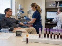
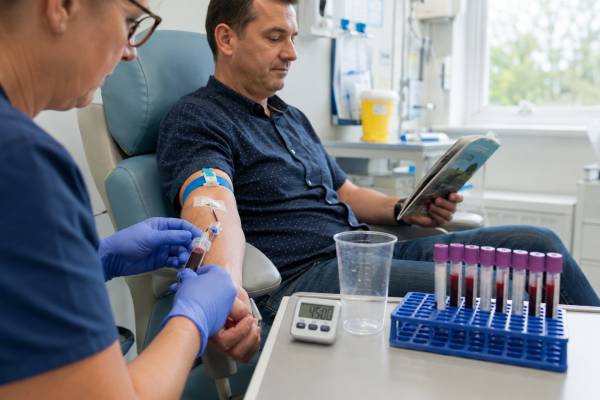
At baseline, blood is collected for GH and glucose. IGF-1 may be measured separately or at the same visit. The patient then drinks 75 grams of glucose dissolved in water, usually within five minutes. Pediatric protocols may use a weight-based dose up to the adult maximum.
Blood is collected at defined intervals. A common schedule is 0, 30, 60, 90, and 120 minutes, although some centers use fewer or different time points. The lowest GH value is the nadir. Glucose results show whether the drink was absorbed and whether an adequate metabolic challenge occurred.
The test takes approximately two to three hours including preparation and recovery. The patient remains seated or resting. Eating, smoking, and exercise are avoided because they can change glucose and GH.
Common symptoms include sweetness-related nausea, bloating, headache, thirst, lightheadedness, sweating, or fatigue. Reactive hypoglycemia can occur later, especially in people with strong insulin responses. Staff should be told immediately about severe dizziness, confusion, vomiting, or faintness.
Vomiting before the drink is absorbed can invalidate the study. Delayed gastric emptying, prior bariatric surgery, or malabsorption may alter glucose delivery and make the standard protocol less reliable. The team may choose an alternative diagnostic approach.
Diabetes does not automatically make testing impossible, but poor glycemic control can impair GH suppression and complicate interpretation. Some specialists rely more heavily on repeated IGF-1 and other evidence when hyperglycemia is severe.
After the final sample, the patient is usually given food and can leave once clinically well. Someone prone to hypoglycemia may need additional observation or instructions for the trip home.
GH Nadir Levels and Cutoffs
The GH nadir is the lowest value after glucose. Results are reported in ng/mL or µg/L, which are numerically equivalent. The cutoff must match the assay and clinical purpose.
Historically, suppression below 1.0 ng/mL was considered normal. With modern ultrasensitive assays, many healthy people suppress below 0.4 ng/mL, and this stricter threshold is often used. Some recent protocols apply BMI- or sex-specific values because healthy suppression differs among populations.
| Post-glucose GH nadir | General interpretation |
|---|---|
| Below 1.0 ng/mL | Traditional evidence of suppression |
| Below about 0.4 ng/mL | Stricter suppression with sensitive assays |
| Above the validated threshold | Supports autonomous GH secretion when IGF-1 and clinical findings agree |
| Near the threshold | Requires assay, BMI, glucose response, and IGF-1 review |
These values are examples, not universal rules. A laboratory that cannot measure accurately at very low concentrations should not use an ultrasensitive cutoff.
Several variables affect the nadir:
- Assay: antibodies, calibration, and detected GH isoforms differ.
- Body mass index: obesity often lowers spontaneous and post-glucose GH.
- Sex and estrogen: women, especially with oral estrogen exposure, may have different GH patterns.
- Age: normal secretion declines over adulthood.
- Glucose response: inadequate glucose rise or severe uncontrolled diabetes changes the challenge.
- Nutrition and liver function: alter the GH–IGF-1 relationship.
The 2023 diagnostic consensus places greater emphasis on IGF-1 than on a mandatory OGTT in every patient. This reflects improved IGF-1 assays and recognition that rigid GH cutoffs can misclassify mild or treated disease.
A high baseline GH that suppresses normally does not establish acromegaly. Stress, fasting, and a physiologic pulse can elevate baseline GH. Conversely, a low baseline value does not exclude disease because secretion is pulsatile.
The glucose curve is not merely incidental. If glucose barely rises, the challenge may have been inadequate. If fasting and post-load glucose meet diabetes criteria, that finding needs separate clinical follow-up, but it does not automatically explain an elevated IGF-1.
Nadir timing varies. Some patients reach the lowest value at 30 or 60 minutes, while others suppress later. Stopping the test too early may miss the true nadir. The full protocol should be completed unless safety requires otherwise.
Abnormal, Normal, and Discordant Results
A failed suppression result supports acromegaly when IGF-1 is persistently elevated and clinical findings fit. The next step is pituitary MRI, not repeated random GH sampling. Other pituitary hormones and visual fields are assessed according to tumor size and symptoms.
A normal suppression result with normal IGF-1 makes acromegaly very unlikely. If symptoms remain unexplained, clinicians reconsider sleep apnea, thyroid disease, medication effects, osteoarthritis, metabolic disease, and other causes rather than continuing GH testing indefinitely.
Discordant patterns require more care.
High IGF-1 With Normal GH Suppression
Possible explanations include assay or reference-interval error, physiologic elevation, recovery from malnutrition, pregnancy, adolescence, or a very mild early disease state. The IGF-1 result should be repeated using a high-quality age-adjusted method after reversible factors are addressed. Many patients with repeatedly elevated IGF-1 but convincing GH suppression do not progress to acromegaly, but follow-up is individualized.
Normal IGF-1 With Incomplete Suppression
This pattern may reflect physiologic variability, assay sensitivity, low BMI, estrogen effects, stress, or diabetes. Active acromegaly is unlikely when IGF-1 is repeatedly normal and there is no strong clinical progression, but treated patients and rare circumstances require specialist judgment.
Mildly High IGF-1 and Borderline Nadir
Repeat testing is often appropriate. The same laboratory should be used when possible, and the exact upper limit, IGF-1 ratio, BMI, glucose response, and GH assay should be documented. Decisions should not turn on a hundredth of a nanogram per milliliter near an uncertain cutoff.
High GH With Low IGF-1
This does not fit acromegaly. Undernutrition, uncontrolled type 1 diabetes, liver disease, critical illness, and GH resistance can produce high GH with low IGF-1. The systemic condition should be addressed.
Biotin interference and heterophile antibodies can distort immunoassays. When the biochemical pattern is physiologically impossible, the laboratory can repeat testing on another platform or perform interference studies.
A pituitary incidentaloma does not prove that a borderline result represents acromegaly. Small nonfunctioning adenomas are common. Biochemical diagnosis should precede attributing symptoms to the lesion.
Laboratory Quality and Repeat Strategy
When a result is surprising, the first repeat should improve the evidence rather than simply reproduce the same uncertainty. IGF-1 is repeated after confirming the patient’s age, the laboratory’s upper limit, and whether the assay changed. A report should provide the absolute value and the ratio to the upper limit of normal; an IGF-1 only a few percent above the limit is different from one severalfold elevated.
The GH suppression report should list every sample time, GH value, glucose value, and the assay’s lower quantification limit. A nadir reported as “less than” a detection threshold may be adequate for one cutoff but not another. If a sample was hemolyzed, delayed, mislabeled, or omitted at the likely nadir time, the test may need repetition.
Biotin can produce falsely high or low results depending on assay design. Patients should tell the laboratory about high-dose hair, skin, or nail supplements and follow the laboratory’s recommended washout. Heterophile antibodies and unusual GH-binding effects are less common but should be considered when results remain incompatible with the clinical picture.
When two laboratories disagree, serial monitoring should generally continue with one well-validated method. Converting values mathematically between assays is unreliable. The endocrine team may ask the laboratory to compare platforms or send a sample to a reference center.
Special Populations
During adolescence, IGF-1 is physiologically high and must be interpreted with exact age and pubertal stage. Gigantism is suspected from excessive growth velocity and height, not from applying an adult IGF-1 range to a teenager. Pediatric glucose dosing and supervision follow local protocols.
Pregnancy changes the GH–IGF-1 axis because placental GH becomes prominent and routine assays vary in cross-reactivity. Standard diagnostic suppression testing is generally deferred unless a specialist identifies an exceptional reason. Tumor symptoms such as visual change are evaluated clinically and with pregnancy-appropriate imaging.
In older adults, age-adjusted IGF-1 ranges are lower and acromegaly can be subtle. In people with obesity, normal post-glucose GH may suppress to very low concentrations, so BMI-specific research cutoffs can be stricter. However, obesity does not explain a clearly high age-adjusted IGF-1 with progressive acromegaly features.
Borderline suppression should be treated as a question to resolve, not as a diagnosis by itself. The clinician may repeat IGF-1 after correcting reversible factors, confirm that the glucose rise was adequate, review the GH assay and cutoff, and repeat dynamic testing if the result would change treatment. A nadir just above a laboratory threshold carries different weight from persistently high GH throughout the test. Discordance is also more common after surgery or during medical therapy, when GH secretion patterns and IGF-1 normalization may recover at different speeds.
Comparing serial tests requires the same glucose load, sample schedule, assay platform, and clinical state whenever possible. Puberty, pregnancy, uncontrolled diabetes, liver or kidney disease, malnutrition, estrogen use, and severe illness can alter GH or IGF-1. These factors should be recorded rather than explained away after the result returns.
Use After Acromegaly Treatment
After transsphenoidal surgery, GH can fall quickly, while IGF-1 may take weeks to months to stabilize. Early random GH provides prognostic information, but formal remission is usually assessed after an appropriate interval using IGF-1 and, when needed, an OGTT.
Suppressed GH after glucose is associated with a favorable long-term outcome. Current therapeutic-outcome consensus recognizes the prognostic value of a nadir below 1.0 ng/mL and, with sensitive assays, lower thresholds. Exact timing and targets depend on the center and treatment type.
Postoperative discordance is common. IGF-1 may remain mildly elevated while GH suppresses, or IGF-1 may normalize while the nadir remains above a strict cutoff. Possible reasons include delayed IGF-1 normalization, assay differences, age, estrogen, obesity, or residual low-level disease. Trends and clinical course are more informative than one early pair.
The test is not equally useful during every medical therapy. Somatostatin receptor ligands suppress GH secretion and lower IGF-1. Dopamine agonists can also affect both. Paltusotine and other oral somatostatin receptor agonists alter the same pathway.
Pegvisomant blocks the GH receptor, causing IGF-1 to fall while serum GH may rise. GH measurements, including suppression testing, are not used to monitor biochemical control on pegvisomant. IGF-1 is the appropriate marker.
After radiation, hormone levels can decline gradually over years. Monitoring includes IGF-1 and other pituitary axes because radiation can produce hypopituitarism. An OGTT is used selectively rather than automatically at every visit.
Recurrence can occur after apparent remission. Rising IGF-1, renewed symptoms, or tumor change prompts reassessment. Comparing values requires awareness of assay changes over time.
Next Steps, Safety, and Questions
If the test supports acromegaly, pituitary MRI defines tumor size and relationship to the optic pathways and cavernous sinus. Visual-field testing is performed when the lesion approaches the optic chiasm. Prolactin, cortisol, thyroid, and gonadal axes are reviewed.
Treatment may include pituitary surgery, medical therapy, and radiation. Choice depends on tumor anatomy, biochemical severity, symptoms, comorbidities, patient preference, and local expertise. Biochemical control aims to normalize IGF-1 and reduce GH-related risk, not merely improve appearance.
Associated conditions require active assessment. These may include sleep apnea, hypertension, diabetes, cardiomyopathy, valve disease, joint disease, carpal tunnel syndrome, colon polyps, thyroid nodules, and vertebral fractures. The testing plan is individualized.
Questions to ask about the result include:
- Was IGF-1 elevated on more than one age-adjusted measurement?
- Which GH assay and post-glucose cutoff were used?
- What was the lowest GH value, and at what time did it occur?
- Did glucose rise enough to confirm an adequate challenge?
- Could diabetes, BMI, estrogen, pregnancy, nutrition, liver disease, kidney disease, or medication affect the findings?
- Are the IGF-1 and GH results concordant?
- Should either test be repeated before MRI or treatment?
- Is the test being used for diagnosis, postoperative remission, or medical-therapy monitoring?
- Does my current treatment make GH measurement unreliable?
Seek urgent care for sudden severe headache, visual loss, double vision, vomiting, confusion, fainting, or weakness because pituitary apoplexy can present this way. During the test, severe dizziness, confusion, or loss of consciousness requires immediate staff attention.
The test is generally safe, but it can uncover diabetes or provoke symptomatic glucose swings. The glucose result deserves appropriate follow-up independent of the GH interpretation.
The most defensible conclusion uses persistent age-adjusted IGF-1 elevation, a properly performed suppression test when indicated, a matching clinical phenotype, and pituitary imaging. One borderline nadir without that framework should not carry the weight of an acromegaly diagnosis.
A repeat test is most useful when it is likely to resolve a specific discordance and when the same validated protocol can be reproduced.
References
- Consensus on criteria for acromegaly diagnosis and remission 2024 (Consensus)
- Consensus on acromegaly therapeutic outcomes: an update 2025 (Consensus)
- Acromegaly: Diagnostic Challenges and Individualized Treatment 2025 (Review)
- The Biochemical Diagnosis of Acromegaly 2021 (Review)
- Interpreting growth hormone and IGF-I results using modern assays and reference ranges for the monitoring of treatment effectiveness in acromegaly 2023 (Review)
- Insulin-Like Growth Factor 1 as a Pillar in Acromegaly: From Diagnosis to Long-Term Management 2024 (Review)
Disclaimer
This article provides general education and cannot diagnose acromegaly or define remission. GH suppression cutoffs are assay- and protocol-specific and must be interpreted with age-adjusted IGF-1, glucose response, treatment status, and clinical findings. Do not change pituitary medication or diabetes treatment for testing without direct instructions.