
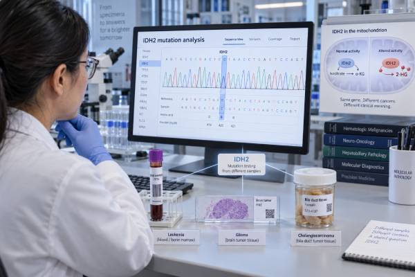
An IDH2 mutation test looks for specific changes in the IDH2 gene that can help classify a tumor, explain part of its biology, and sometimes guide treatment. IDH2 mutations are most clinically important in acute myeloid leukemia (AML), adult diffuse gliomas, and intrahepatic cholangiocarcinoma, but the meaning is not the same in each disease. In AML, an IDH2 mutation can identify a target for an IDH2 inhibitor in certain treatment settings. In glioma, an IDH2 mutation supports the broader category of an IDH-mutant diffuse glioma and must be interpreted with markers such as 1p/19q codeletion and ATRX. In cholangiocarcinoma, IDH2 can help define the molecular profile, although currently approved IDH-directed treatment is centered on IDH1 rather than IDH2. The most useful interpretation therefore combines the exact variant, tumor type, specimen quality, other molecular findings, and the patient’s clinical situation.
- A positive IDH2 result means a pathogenic or likely pathogenic IDH2 variant was detected; it does not mean the same thing in every cancer.
- Common IDH2 hotspot mutations occur at codons R140 and R172 and create an abnormal enzyme that produces the oncometabolite 2-hydroxyglutarate.
- In AML, an IDH2 mutation may have direct treatment relevance, including eligibility for an IDH2-targeted inhibitor in appropriate settings.
- In adult diffuse glioma, IDH2 positivity supports an IDH-mutant diagnosis, but 1p/19q, ATRX, TP53, and other findings are needed for complete classification.
- A negative result does not always exclude an IDH-driven tumor if the assay is limited, tumor content is low, or the alteration lies outside the regions tested.
Table of Contents
- What the IDH2 Mutation Test Measures
- How IDH2 Testing Is Done
- Positive, Negative, and Uncertain Results
- Meaning in Acute Myeloid Leukemia
- Meaning in Glioma and Cholangiocarcinoma
- Treatment, Prognosis, and Monitoring
- Limitations and Next Steps
What the IDH2 Mutation Test Measures
The IDH2 mutation test detects DNA changes in the isocitrate dehydrogenase 2 gene. IDH2 encodes a mitochondrial enzyme that normally helps convert isocitrate to alpha-ketoglutarate. Certain cancer-associated mutations change the enzyme’s activity. Instead of performing its normal reaction, mutant IDH2 produces large amounts of D-2-hydroxyglutarate, often shortened to 2-HG.
2-HG is called an oncometabolite because it can interfere with enzymes that regulate DNA and histone methylation. The result is widespread epigenetic change that can block normal cell differentiation and contribute to cancer development. This altered biology is why an IDH2 mutation can function as a disease-defining or treatment-relevant biomarker rather than simply being one more incidental genetic change.
The most common pathogenic IDH2 changes affect arginine 140 (R140) or arginine 172 (R172). In AML, R140 variants are especially common, while R172 also occurs. In adult diffuse gliomas, IDH2 variants are much less common than the classic IDH1 R132H mutation, but they are still important because a glioma can be IDH-mutant even when the common IDH1 immunostain is negative. In intrahepatic cholangiocarcinoma, IDH1 mutations are more common than IDH2 mutations, but both belong to the same metabolic pathway and can help define a molecular subgroup.
A laboratory report may list the result at several levels of detail. For example, it may say “IDH2 mutation detected,” give a protein notation such as p.R140Q, and provide a DNA-level notation and variant allele frequency (VAF). VAF is the fraction of sequencing reads that contain the variant. It is affected by tumor purity, copy number, clonality, and specimen type, so it should not be interpreted as a simple percentage of cancer cells.
IDH2 testing is usually somatic tumor testing, meaning the laboratory is looking for a mutation acquired by the cancer. This is different from hereditary cancer testing. An IDH2 mutation found in leukemia cells or a solid tumor generally does not imply that the patient inherited that mutation or that relatives need testing for the same change.
How IDH2 Testing Is Done
IDH2 testing can be performed with several molecular methods. The best method depends on the disease, the amount of available material, and whether the clinician needs one gene or a broader molecular profile.
For AML and other myeloid neoplasms, laboratories often use a next-generation sequencing (NGS) myeloid panel that evaluates IDH2 together with genes such as NPM1, FLT3, DNMT3A, RUNX1, TP53, and others. A rapid targeted PCR or other focused assay may be used when a faster answer is needed for a known hotspot. Bone marrow aspirate is commonly tested, although peripheral blood can be suitable when circulating blasts or leukemic cells are present in adequate numbers.
For brain tumors and cholangiocarcinoma, testing is usually performed on formalin-fixed, paraffin-embedded tumor tissue from a biopsy or resection. NGS is useful because these tumors require interpretation of several markers at once. In adult diffuse glioma, an integrated workup may include IDH1/IDH2 sequencing, 1p/19q status, ATRX, TP53, CDKN2A/B, and other molecular features. In cholangiocarcinoma, broader profiling can also detect FGFR2 rearrangements, BRAF variants, ERBB2 changes, KRAS alterations, and other potentially relevant findings.
No special fasting or medication preparation is needed for the molecular test itself. Preparation depends on how the sample is collected. Bone marrow biopsy and tumor biopsy have their own procedural instructions, bleeding precautions, and sedation considerations.
Tumor content and assay sensitivity matter
A negative test can occur because there is no IDH2 mutation, but it can also occur when the specimen contains too few tumor cells or the assay does not cover the relevant region. Pathologists often estimate the percentage of tumor in the tested material and may select or enrich a tumor-rich area before DNA extraction.
NGS assays commonly detect variants present at low single-digit allele fractions, but the exact lower limit varies by laboratory. Some assays are designed for broad profiling and others for highly sensitive monitoring. The report’s limit of detection is therefore important when comparing a diagnostic result with a later follow-up result.
Liquid biopsy using circulating tumor DNA can sometimes supplement tissue testing in solid tumors, especially when tissue is unavailable, but a negative blood result cannot reliably exclude a mutation in every patient. Tumor DNA shedding varies by cancer type, disease burden, and treatment status.
Positive, Negative, and Uncertain Results
A useful interpretation begins with the exact result category rather than with the gene name alone.
Positive for a pathogenic or likely pathogenic IDH2 mutation
A positive result means the laboratory detected an IDH2 variant with evidence that it alters protein function and is relevant to cancer. In the right tumor type, this can support diagnosis, classification, or treatment selection. The report should be read for the specific codon, amino-acid change, VAF, specimen, and assay method.
A positive result does not automatically tell you the cancer stage, whether it has spread, or whether treatment will work. It also does not by itself establish a diagnosis of AML, glioma, or cholangiocarcinoma. Diagnosis still requires the appropriate clinical, morphologic, imaging, and laboratory context.
Negative or “not detected”
A negative result means the assay did not identify a reportable IDH2 mutation in the tested material. This may support an IDH2-wild-type interpretation, but it does not necessarily mean the entire IDH pathway is normal. A tumor can have an IDH1 mutation instead, and some assays test only common hotspots rather than the full coding region.
In glioma, a negative IDH1 R132H immunostain is a common reason to perform sequencing for noncanonical IDH1 and IDH2 variants, especially in younger adults or when the tumor’s morphology and other markers raise suspicion for an IDH-mutant diffuse glioma.
Variant of uncertain significance
A variant of uncertain significance (VUS) is a change for which available evidence is insufficient to classify it as cancer-driving or benign. A VUS should not be treated as equivalent to a known IDH2 hotspot mutation. It generally should not be used alone to select targeted therapy or make a major diagnostic classification unless additional evidence establishes its significance.
If the report uses terms such as “low-level,” “subclonal,” or “below validated reporting threshold,” the laboratory may need to clarify whether the finding is analytically reliable and clinically reportable. In leukemia, very low VAF results can also raise questions about residual disease, clonal hematopoiesis, specimen contamination, or an emerging subclone, depending on timing and context.
Meaning in Acute Myeloid Leukemia
In AML, an IDH2 mutation is a recognized recurrent molecular abnormality with both biologic and therapeutic importance. IDH2 mutations occur in a meaningful minority of AML cases, most often at R140 and less often at R172. They promote accumulation of 2-HG and interfere with normal myeloid differentiation.
The result is interpreted as part of a larger AML profile. Modern AML diagnosis and risk assessment integrate cytogenetics and multiple genes rather than assigning prognosis from IDH2 alone. Co-mutations can strongly influence the disease. NPM1, DNMT3A, SRSF2, RUNX1, FLT3, and other abnormalities may coexist, and the overall pattern helps define disease biology and treatment planning.
Treatment relevance in AML
The clearest practical consequence of an IDH2-positive AML result is that the tumor may be susceptible to IDH2-directed therapy. Enasidenib is a selective mutant-IDH2 inhibitor used in appropriate patients with IDH2-mutated AML. Rather than acting like conventional chemotherapy, it can promote differentiation of leukemic cells.
That mechanism creates a treatment-specific complication called differentiation syndrome. Symptoms can include fever, breathing difficulty, rapid weight gain, swelling, low blood pressure, kidney problems, and lung infiltrates. It can become serious and requires prompt recognition and treatment. The presence of an IDH2 mutation identifies the molecular target, but the decision to use an inhibitor depends on treatment history, disease status, age, fitness, other mutations, available regimens, and current regulatory guidance.
An IDH2 result can also matter when AML returns. Retesting may be useful because the clonal composition of leukemia can change over time. A mutation present at diagnosis may persist, shrink, disappear, or coexist with new resistance-associated clones.
Prognosis is not determined by IDH2 alone
It is a mistake to label every IDH2-mutated AML as “good risk” or “bad risk.” Prognostic effects vary across studies and depend heavily on co-mutations and treatment context. Current AML risk frameworks use a panel of genetic and cytogenetic findings. The treating hematologist therefore interprets IDH2 together with the complete profile, response to induction or lower-intensity therapy, measurable residual disease results, and transplant considerations.
Meaning in Glioma and Cholangiocarcinoma
The same IDH2 mutation has a different role in solid tumors.
Adult diffuse glioma
In adult diffuse gliomas, the crucial question is usually whether the tumor is IDH-mutant or IDH-wild-type. IDH1 mutations are far more common, but an IDH2 mutation can establish IDH-mutant status when it is a recognized pathogenic hotspot.
That classification has major diagnostic importance. Adult diffuse gliomas are divided into molecularly defined entities that include astrocytoma, IDH-mutant and oligodendroglioma, IDH-mutant and 1p/19q-codeleted. Therefore, finding IDH2 is only one step. If an IDH mutation is present, 1p/19q testing helps determine whether the tumor meets criteria for oligodendroglioma. ATRX and TP53 findings can support an astrocytic lineage, while additional molecular features contribute to grading and prognostic assessment.
IDH2 mutations are particularly associated with a subset of oligodendrogliomas. Because the common IDH1 R132H antibody does not detect IDH2 mutations, sequencing is important when an IDH-mutant tumor remains clinically or morphologically plausible despite negative immunohistochemistry.
In general, IDH-mutant adult diffuse gliomas have a different biology and often a more favorable natural history than IDH-wild-type glioblastoma, but grade, age, extent of resection, 1p/19q status, CDKN2A/B status, treatment, and other factors still matter. IDH2 positivity should therefore be viewed as a classification anchor, not a complete prognosis.
Intrahepatic cholangiocarcinoma
IDH1 and IDH2 mutations occur predominantly in intrahepatic rather than extrahepatic cholangiocarcinoma and are especially associated with the small-duct molecular phenotype. Their presence supports molecular characterization but is not, by itself, diagnostic of cholangiocarcinoma.
This distinction matters for treatment. IDH1-mutated cholangiocarcinoma has an established targeted therapy pathway with an IDH1 inhibitor in appropriate advanced disease. An IDH2 mutation is not interchangeable with an IDH1 mutation, and an IDH1-specific drug should not be assumed to work against IDH2 simply because the genes are related. For IDH2-positive cholangiocarcinoma, clinicians generally rely on the overall disease setting, other actionable alterations, standard systemic therapy, and clinical-trial options.
The prognostic effect of IDH1/2 mutations in cholangiocarcinoma remains less certain than their biologic significance. Some studies suggest distinct outcomes, while others do not show an independent survival advantage after accounting for stage and other variables.
Treatment, Prognosis, and Monitoring
IDH2 testing can influence care in three different ways: it can identify a target, define a tumor class, or contribute to a broader molecular profile.
In AML, the mutation can directly affect treatment options. In glioma, its greatest value is usually integrated diagnosis and classification. In cholangiocarcinoma, it contributes to molecular profiling but currently does not create the same approved targeted-treatment pathway as an IDH1 mutation.
Monitoring also differs by disease. In AML, a known mutation may be followed with sensitive molecular methods in selected settings, but IDH2 is not universally used as a stand-alone measurable residual disease marker. Persistent mutations after treatment can be difficult to interpret because some myeloid mutations may remain in preleukemic or clonal hematopoietic cells. The significance depends on the gene, VAF trend, co-mutations, blood counts, marrow findings, and the assay used.
In glioma and cholangiocarcinoma, routine follow-up usually relies more on imaging, clinical status, and standard disease-specific markers than on repeated tissue IDH2 testing. Repeat molecular profiling may become useful at progression when resistance mechanisms or new therapeutic opportunities are being considered.
A practical way to read an IDH2 report is to ask four questions:
- What disease and specimen were tested? Bone marrow AML, a brain tumor, and a liver biopsy require different interpretation.
- What exact IDH2 variant was found? A known hotspot such as R140 or R172 carries stronger evidence than an unusual VUS.
- What other molecular findings are present? Co-mutations can change diagnosis, prognosis, and treatment options.
- Is the result being used for diagnosis, therapy selection, or follow-up? The same positive result can serve different purposes at different points in care.
Limitations and Next Steps
IDH2 testing is highly useful, but it is not self-interpreting. The most common mistakes come from treating a molecular result as though it were independent of specimen quality, assay design, and tumor type.
A false-negative result is more likely when tumor cellularity is low, DNA is degraded, the sample is small, or the assay covers only selected hotspots. Decalcified specimens can be especially challenging because some processing methods damage nucleic acids. A negative plasma liquid-biopsy result can also miss a mutation when a solid tumor sheds little DNA into the bloodstream.
A positive result can also be misunderstood. VAF is not the same as tumor percentage, and the presence of IDH2 does not prove that every tumor cell carries the alteration. In AML, clonal evolution can create subclones. In solid tumors, spatial heterogeneity and treatment pressure can alter the molecular landscape over time.
When an IDH2 result is unexpected, the best next step is usually to review the full pathology report and ask whether the finding fits the diagnosis. If a brain tumor is suspected to be IDH-mutant but initial IDH1 immunohistochemistry is negative, sequencing for noncanonical IDH1 and IDH2 variants may be appropriate. If AML is IDH2-positive, the clinician should review the full myeloid mutation panel, cytogenetics, treatment history, and eligibility for targeted therapy. If cholangiocarcinoma is being profiled, the report should be checked for other actionable alterations rather than assuming IDH2 has the same implications as IDH1.
Patients should also know whether the test was performed on tumor tissue or as a germline hereditary-cancer test. Most IDH2 findings in the cancers discussed here are somatic. If a report raises concern for a possible inherited condition, that question should be addressed with a genetics professional and a properly validated germline specimen rather than inferred from tumor sequencing alone.
References
- Diagnosis and management of AML in adults: 2022 recommendations from an international expert panel on behalf of the ELN 2022 (Guideline)
- IDH2 mutations in acute myeloid leukemia 2023 (Review)
- IDH-mutant diffuse gliomas: tips and tricks in the era of genomic tumor classification 2023 (Review)
- Molecular profiling in cholangiocarcinoma: A practical guide to next-generation sequencing 2023 (Review)
- Biology of IDH mutant cholangiocarcinoma 2022 (Review)
- Practical considerations for pathological diagnosis and molecular profiling of cholangiocarcinoma: an expert review for best practices 2024 (Review)
Disclaimer
IDH2 results must be interpreted with the tumor type, pathology findings, other molecular results, and treatment history. This article is educational and does not replace diagnosis or treatment advice from a hematologist, oncologist, neurologist, pathologist, or genetics professional who has reviewed the complete medical record.





