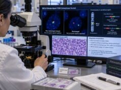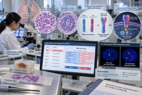
An ALK fusion test looks for a cancer-driving rearrangement involving the ALK gene, most often in non-small cell lung cancer (NSCLC). The classic example is an EML4-ALK fusion, in which part of ALK becomes joined to another gene and produces an abnormal kinase that sends continuous growth signals. Finding an ALK fusion matters because it can define a distinct molecular subtype of lung cancer and can make a patient eligible for highly effective ALK-targeted therapy. Testing may use next-generation sequencing, fluorescence in situ hybridization (FISH), or immunohistochemistry (IHC), and the best method depends on the specimen and laboratory workflow. A positive result does not describe cancer stage or tumor size, but it can strongly affect treatment choices. A negative result is meaningful only in the context of the method used, sample quality, tumor content, and whether the assay was designed to detect uncommon fusion partners.
- A positive ALK fusion result means a rearrangement capable of activating ALK was detected or strongly supported by a validated companion test.
- ALK fusions occur in roughly 3%–7% of advanced NSCLC, especially lung adenocarcinoma, and EML4 is the most common fusion partner.
- ALK-positive NSCLC is treated differently from tumors driven by EGFR, ROS1, BRAF, KRAS, RET, MET, or NTRK alterations.
- RNA-based NGS can identify the exact fusion transcript and uncommon partners, while FISH detects rearrangement and IHC detects ALK protein expression.
- No fasting is needed for molecular testing; the main practical issue is having enough good-quality tumor tissue or, when appropriate, circulating tumor DNA.
Table of Contents
- What an ALK Fusion Is
- When ALK Fusion Testing Is Ordered
- How ALK Fusions Are Detected
- What Positive, Negative, and Discordant Results Mean
- ALK-Positive Lung Cancer and Treatment
- ALK Fusions Outside Lung Cancer
- Questions to Ask After an ALK Test
What an ALK Fusion Is
An ALK fusion is a structural DNA change that joins part of the ALK gene to another gene, creating an abnormal growth-driving signal. ALK stands for anaplastic lymphoma kinase. In normal adult tissues, ALK activity is limited. In certain cancers, a rearrangement places the kinase portion of ALK under the control of a partner gene, causing the resulting fusion protein to remain active.
In lung adenocarcinoma, EML4-ALK is by far the best-known fusion. EML4 can join ALK at different breakpoints, creating several EML4-ALK variants. Other ALK fusion partners also exist. The exact partner and breakpoint can be reported by sequencing, although in routine care the key first question is usually whether a functional ALK fusion is present.
This type of alteration is different from a small point mutation. A point mutation changes one or a few DNA letters. A fusion is a larger structural rearrangement involving pieces of two genes. That difference matters because some test methods are better at finding fusions than others.
ALK fusions activate downstream signaling pathways that promote cell growth and survival, including RAS-MAPK, PI3K-AKT, and JAK-STAT signaling. Because the tumor may depend heavily on this abnormal kinase, ALK becomes a therapeutic target.
ALK is one of several major driver alterations assessed in modern lung-cancer testing. A broad lung cancer biomarker panel can evaluate ALK together with EGFR, ROS1, BRAF, MET, RET, NTRK, KRAS, HER2, and other markers. These drivers are important because they can define different treatment pathways even when the tumors look similar under the microscope.
When ALK Fusion Testing Is Ordered
ALK testing is a standard part of biomarker evaluation for many patients with non-squamous NSCLC, especially advanced lung adenocarcinoma. Current molecular practice favors broad profiling because several targetable drivers may need to be assessed from a limited biopsy specimen.
ALK fusions are found in a minority of NSCLC cases—commonly around 3% to 7% in advanced disease—but identifying that minority has major treatment consequences. ALK-positive tumors are often adenocarcinomas and are more common in people who are younger and have never smoked or smoked lightly. Those clinical patterns can raise suspicion, but they should not be used to decide that testing is unnecessary in someone who does not fit the typical profile.
Testing is commonly performed:
- At diagnosis of advanced or metastatic non-squamous NSCLC.
- In selected squamous tumors when clinical or pathologic features suggest a driver alteration may be present.
- After complete resection of eligible early-stage NSCLC when adjuvant targeted therapy could be considered.
- When a previous limited panel did not include ALK or used a method with uncertain sensitivity.
- At progression when resistance testing or confirmation of the original driver could affect the next treatment decision.
An ALK IHC test may be used as a rapid and tissue-sparing approach in some laboratories. Other centers use RNA-based NGS up front so that ALK and many other fusions can be tested together.
ALK testing is not a screening test for people without cancer. It does not estimate the chance of developing lung cancer, and a tumor ALK fusion is generally somatic, meaning it arose in the cancer rather than being inherited from a parent.
How ALK Fusions Are Detected
ALK status can be established by several validated methods, but each method measures a different feature of the alteration. Understanding those differences helps explain why reports may use terms such as “fusion detected,” “rearranged,” or “ALK positive.”
| Method | What it detects | Main strength | Important limitation |
|---|---|---|---|
| RNA-based NGS | Expressed ALK fusion transcripts | Can identify exact partner and breakpoint; strong for diverse fusions | RNA quality can be poor in old or small formalin-fixed samples |
| DNA-based NGS | Genomic rearrangements involving ALK | Profiles many mutation types in one assay | Large introns and complex breakpoints can make some fusions harder to detect |
| FISH | Separation of ALK break-apart probe signals | Established rearrangement assay; does not require knowing the partner | Does not directly identify the fusion transcript or partner and can produce borderline patterns |
| IHC | Abnormal ALK protein expression | Fast, tissue-efficient, and clinically validated in NSCLC | Protein expression is an indirect marker of the underlying fusion |
A lung cancer NGS panel is increasingly useful because it can examine multiple driver genes at once. RNA sequencing is especially valuable for fusions because it demonstrates that a rearranged transcript is actually expressed. Some comprehensive assays combine DNA and RNA components to improve coverage.
FISH uses fluorescent probes that bind on opposite sides of the ALK locus. In an intact gene, the signals remain close together. In a rearranged gene, the signals separate or show another qualifying abnormal pattern. The laboratory applies a validated scoring system and minimum number of tumor cells.
IHC uses an antibody, commonly the D5F3 clone in a validated companion-diagnostic format, to detect ALK protein. Strong characteristic staining can establish ALK-positive status in the appropriate assay. IHC is not the same as sequencing: it tells the pathologist that ALK protein is abnormally expressed, not which partner gene created the fusion.
No special preparation is needed for the molecular test itself. Tissue may come from a core biopsy, fine-needle specimen, surgical resection, or cytology cell block if adequately validated. Liquid biopsy can sometimes detect ALK fusions in circulating tumor DNA, but fusion detection in plasma is technically challenging and depends on tumor shedding. A negative plasma result may need tissue testing if clinically feasible.
Small specimens require careful tissue management because the same sample may also be needed for histology, PD-L1 testing, and other molecular markers. Reflex comprehensive testing can reduce repeated sectioning and preserve material compared with ordering many single-gene assays one after another. Laboratories also report quality-control failures when DNA or RNA quantity is too low; a failed test is not a negative result.
What Positive, Negative, and Discordant Results Mean
A confirmed positive ALK result in NSCLC identifies a targetable oncogenic driver; a negative result means no ALK alteration was found by the assay used. The wording of the report matters because different methods support the conclusion in different ways.
A sequencing report may say:
- EML4-ALK fusion detected, sometimes with the exact exons joined.
- ALK fusion detected, with another partner named.
- ALK rearrangement detected, when the genomic structure is clear but the functional transcript may not be fully characterized.
A FISH report usually states ALK rearrangement positive or negative. An IHC report may state ALK positive or negative based on protein staining.
A positive result generally means the tumor belongs to the ALK-driven molecular subtype and should be evaluated for ALK-targeted therapy. In advanced NSCLC, this finding usually takes priority over immune-checkpoint therapy as the initial systemic strategy because targeted ALK inhibitors can produce deep and durable responses, including activity in the brain.
A negative result does not have an absolute meaning unless you know the test’s coverage. For example, a negative DNA-only panel may be less reassuring if the assay has limited intronic coverage for ALK. A negative plasma assay may reflect low circulating tumor DNA. When the clinical picture strongly suggests a driver and the original assay was limited, reflex RNA testing or another method can be reasonable.
What if ALK tests disagree?
Discordance can occur—for example, FISH positive but IHC and RNA NGS negative, or IHC positive with an equivocal molecular result. Causes include unusual rearrangement patterns, technical artifacts, low tumor content, sample heterogeneity, RNA degradation, or a genomic rearrangement that does not create an expressed oncogenic fusion.
When results conflict, the most useful next step is often orthogonal confirmation with a method that measures a different feature. A questionable FISH result may be clarified by RNA NGS; an unexpected focal IHC result may be assessed with sequencing. The pathology and molecular teams should integrate all available evidence rather than treating every isolated abnormal signal as equally predictive.
ALK-Positive Lung Cancer and Treatment
ALK positivity can directly determine treatment in both advanced and selected resected NSCLC. This is the central clinical reason ALK testing is performed.
For advanced ALK-positive NSCLC, several generations of ALK tyrosine kinase inhibitors have been developed. These drugs bind the ALK kinase and block the abnormal growth signal. Later-generation agents were designed to improve potency, activity against resistance mutations, and penetration into the central nervous system.
Modern first-line options can achieve long disease control. In the phase 3 CROWN study, long-term follow-up showed markedly prolonged progression-free survival with lorlatinib compared with crizotinib, with strong protection against intracranial progression. Alectinib and brigatinib are also established ALK-directed therapies in advanced disease. The best initial drug depends on the clinical setting, regulatory approvals, toxicity profiles, brain metastases, comorbidities, and local guidelines.
ALK status now also matters after surgery. In 2024, the U.S. FDA approved adjuvant alectinib after complete tumor resection for patients with ALK-positive NSCLC meeting the approved stage criteria. The ALINA trial showed a substantial disease-free survival benefit compared with platinum-based chemotherapy in the studied population.
Because treatment depends on the driver, ALK results need to be interpreted alongside other molecular findings. An EGFR mutation test, for example, identifies a different targetable subtype. Likewise, a ROS1 fusion test identifies another rearranged kinase with its own drug options. A tumor should not be assumed to follow the ALK pathway simply because the patient’s age, smoking history, or pathology resembles a typical ALK-positive case.
Resistance after ALK-targeted therapy
ALK-positive cancer can develop resistance over time. Some resistant tumors acquire secondary mutations in the ALK kinase domain; others activate bypass pathways or undergo broader cellular changes. The specific resistance mechanism can influence subsequent treatment, although treatment choice also depends on which ALK inhibitor was used first and where the cancer is progressing.
A repeat tissue biopsy or plasma NGS may sometimes be used at progression to look for resistance alterations. However, a negative resistance test does not mean resistance is absent; not every mechanism is detectable in circulating DNA, and some resistance is not caused by a new ALK mutation.
ALK Fusions Outside Lung Cancer
ALK rearrangements are not unique to lung cancer, but their meaning changes with the tumor type. The gene was originally recognized through an ALK fusion in anaplastic large cell lymphoma, and ALK alterations also occur in inflammatory myofibroblastic tumors and several rarer cancers.
In anaplastic large cell lymphoma, ALK is commonly fused with NPM1 or another partner, and ALK status contributes to disease classification and prognosis. An ALK test for anaplastic large cell lymphoma therefore answers a different clinical question from ALK testing in lung adenocarcinoma.
In inflammatory myofibroblastic tumor, ALK rearrangements can also act as oncogenic drivers and may have treatment implications. Other rare ALK fusions have been described across solid tumors, but evidence and approved therapies vary by histology.
This is why the phrase “ALK positive” is not enough by itself. The report must be interpreted with the diagnosis. An ALK fusion in lung adenocarcinoma, an ALK rearrangement in lymphoma, and ALK expression in another tumor do not automatically carry the same prognosis or treatment recommendation.
The exact fusion partner can sometimes matter biologically. In lung cancer, different EML4-ALK variants have been studied for possible differences in resistance patterns and outcomes, but routine treatment decisions are still driven mainly by the presence of a clinically validated ALK fusion, disease setting, and prior therapy rather than by fusion variant alone.
Questions to Ask After an ALK Test
The next step after testing is to confirm what was measured and how the result changes treatment. These questions can make a molecular report easier to use:
- Was the result based on RNA NGS, DNA NGS, FISH, IHC, or more than one method?
- If sequencing was positive, what is the fusion partner and breakpoint?
- Is the finding considered a functional, targetable ALK fusion?
- Was the sample tumor tissue or plasma, and was the specimen adequate?
- If the result is negative, did the assay have strong fusion coverage, or would RNA testing add value?
- Were all other guideline-relevant lung cancer biomarkers tested at the same time?
- If the tests disagree, should the sample be reviewed or confirmed with another method?
- Does the ALK result make me eligible for an approved ALK inhibitor in my stage and treatment setting?
- If the cancer has progressed on an ALK inhibitor, would repeat molecular testing help choose the next therapy?
A positive ALK test can be one of the most treatment-changing findings in lung cancer, but the value comes from accurate detection and correct clinical context. Broad molecular profiling, careful pathology review, and confirmation of unusual or discordant findings help prevent both missed ALK-positive cancers and false-positive interpretations.
A practical point is to keep the original molecular report. ALK testing can use IHC, FISH, DNA NGS, or RNA NGS, and later treatment decisions may depend on knowing exactly which method was positive. If resistance develops after an ALK inhibitor, repeat tissue or plasma profiling can identify secondary ALK mutations or bypass mechanisms that may influence the next therapy. A new negative liquid-biopsy result does not erase a previously proven ALK fusion, especially when circulating tumor DNA is low.
References
- Identification and therapeutic evaluation of ALK rearrangements in non-small-cell lung cancer 2022
- New update to the guidelines on testing predictive biomarkers in non-small-cell lung cancer: a National Consensus of the Spanish Society of Pathology and the Spanish Society of Medical Oncology 2023 (Guideline)
- Advancements of ALK inhibition of non-small cell lung cancer: a literature review 2023 (Review)
- Alectinib in Resected ALK-Positive Non-Small-Cell Lung Cancer 2024 (RCT)
- FDA approves alectinib as adjuvant treatment for ALK-positive non-small cell lung cancer 2024
- Lorlatinib Versus Crizotinib in Patients With Advanced ALK-Positive Non-Small Cell Lung Cancer: 5-Year Outcomes From the Phase III CROWN Study 2024 (RCT)
Disclaimer
This article is for general education and does not replace diagnosis or treatment advice from a lung-cancer specialist, pathologist, or molecular laboratory. ALK results must be interpreted with the tumor type, stage, testing method, specimen quality, other biomarkers, and current treatment approvals. Do not change cancer treatment based on an ALK report without discussing it with your oncology team.