
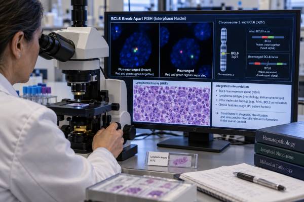
A BCL6 rearrangement test looks for a structural chromosome change involving the BCL6 gene, usually with fluorescence in situ hybridization (FISH). BCL6 is a transcriptional regulator that normally helps germinal-center B cells mature. In lymphoma, a rearrangement can place BCL6 under the control of another gene’s regulatory elements and disturb normal B-cell development. BCL6 rearrangements are found in a subset of diffuse large B-cell lymphomas (DLBCL), high-grade B-cell lymphomas, follicular lymphomas, and other mature B-cell neoplasms. The result is most useful when interpreted with the biopsy appearance, immunohistochemistry, and FISH for MYC and BCL2. A positive BCL6 rearrangement is not the same as positive BCL6 protein staining, and it does not by itself establish a diagnosis, stage, or treatment plan. Its prognostic meaning also depends on the disease context, especially whether MYC or BCL2 is rearranged and how the lymphoma is classified under current WHO and International Consensus Classification systems.
- A positive BCL6 FISH result means a qualifying structural rearrangement involving BCL6 was detected in the tested tumor cells.
- BCL6 rearrangement is different from BCL6 protein expression on immunohistochemistry; the two tests measure different biological features.
- In aggressive B-cell lymphoma, BCL6 is often tested together with MYC and BCL2 because the combination of abnormalities affects classification.
- MYC/BCL6-rearranged lymphoma is not equivalent to the current WHO entity defined by MYC and BCL2 rearrangements.
- The result must be interpreted with morphology, immunophenotype, clinical stage, and other molecular findings rather than used as a stand-alone prognosis marker.
Table of Contents
- What BCL6 Rearrangement Means
- When BCL6 Rearrangement Testing Is Used
- How BCL6 FISH Testing Works
- How to Interpret a BCL6 FISH Result
- BCL6 in DLBCL and High-Grade Lymphoma
- MYC, BCL2, BCL6, and Double-Hit Classification
- What Happens After BCL6 Testing
What BCL6 Rearrangement Means
A BCL6 rearrangement is a chromosome-level change that moves or breaks the BCL6 locus at 3q27 and places it next to a new regulatory partner. BCL6 normally encodes a transcriptional repressor that is highly active in germinal-center B cells. It helps these cells tolerate the temporary DNA damage that occurs while antibodies are being refined, then must be switched off as B cells complete differentiation.
In lymphoma, a rearrangement can uncouple BCL6 from its normal controls. Unlike some fusions that create a single characteristic fusion protein, BCL6 can rearrange with many partners. Immunoglobulin genes are common partners, but numerous non-immunoglobulin partners also occur. This partner diversity is one reason a break-apart FISH probe is widely used.
BCL6 rearrangement is not specific to one lymphoma. It occurs in a meaningful subset of DLBCL and can be seen in high-grade B-cell lymphoma, follicular lymphoma, marginal zone lymphoma, and other mature B-cell tumors. The same FISH abnormality can therefore have different meaning in different biopsies.
A BCL6 IHC test measures BCL6 protein expression, not gene structure. Many germinal-center-derived lymphomas express BCL6 protein even when the gene is not rearranged. Conversely, a gene rearrangement may occur with variable protein staining. A pathology report should specify whether “BCL6 positive” refers to IHC or FISH because the terms are not interchangeable.
BCL6 rearrangements usually arise as acquired changes in the lymphoma clone. They are not ordinarily interpreted as inherited cancer-predisposition variants, and they do not imply that relatives should undergo germline testing.
When BCL6 Rearrangement Testing Is Used
BCL6 FISH is most often ordered during classification of an aggressive mature B-cell lymphoma, particularly when MYC and BCL2 rearrangements are also clinically relevant. The decision is usually made after a hematopathologist reviews the biopsy morphology and immunophenotype.
Common reasons for testing include:
- Newly diagnosed DLBCL or high-grade B-cell lymphoma in a laboratory or center that uses a broad rearrangement-testing strategy.
- A tumor with high-grade morphology, very high proliferation, germinal-center markers, or other features that raise concern for rearrangement-defined disease.
- A lymphoma in which MYC rearrangement has already been found and the pathologist needs BCL2 and BCL6 results to complete classification.
- A follicular or other indolent B-cell lymphoma with unusual morphology, transformation, or molecular features that need clarification.
- A prior equivocal, failed, or incomplete cytogenetic study when classification still depends on the result.
A double-hit lymphoma FISH panel commonly evaluates MYC, BCL2, and BCL6 in parallel. The exact testing algorithm varies by laboratory. Some institutions screen aggressive B-cell lymphomas broadly, while others use morphology and immunophenotype to decide which cases receive FISH. Current classification increasingly emphasizes complete genetic assessment when the result can move a case into a different diagnostic group.
No special fasting, medication change, or preparation is required for the FISH study itself. Testing is usually performed on tissue already obtained by lymph node biopsy, core biopsy, bone marrow biopsy, or another procedure. The quality and tumor content of that specimen matter more than patient preparation.
How BCL6 FISH Testing Works
BCL6 break-apart FISH uses fluorescent DNA probes on opposite sides of the BCL6 locus to show whether the gene has been structurally separated. In a normal nucleus, the probe signals remain together or close enough to appear fused. A rearrangement separates the probes beyond the laboratory’s validated distance threshold.
Because BCL6 has many possible partner genes, break-apart FISH is efficient: it answers “Is BCL6 rearranged?” without requiring the partner to be known in advance. However, a positive break-apart study usually does not identify which partner gene is involved or the exact DNA breakpoint.
The laboratory examines a defined number of nuclei and compares the percentage with a validated positivity cutoff. A report may state, for example, that a certain percentage of scored nuclei show a split-signal pattern. This percentage is not a direct measure of tumor burden, stage, or treatment response. It is a laboratory observation used to call the rearrangement positive or negative.
Several technical issues can affect interpretation. Formalin fixation can damage DNA. Tissue sections can truncate nuclei, creating signal loss. Overlapping nuclei can mimic extra signals. Strong-acid decalcification of bone marrow may reduce assay quality. A small tumor focus can also be diluted by benign cells. For these reasons, equivocal or technically failed results deserve pathologist review rather than being treated as simple negatives.
FISH can also reveal unusual signal patterns, such as extra BCL6 copies in addition to a rearrangement. Copy-number gain is not the same biological event as a rearrangement. The report should distinguish them whenever possible.
Next-generation sequencing may detect BCL6 rearrangements on appropriately designed DNA- or RNA-based panels, but sensitivity depends on assay design and breakpoint coverage. Targeted FISH remains common because it is rapid, works on routine paraffin tissue, and has well-established interpretation in lymphoma classification.
If the broader workup includes a MYC rearrangement test and a BCL2 rearrangement test, the three results should be read together rather than as unrelated laboratory findings.
How to Interpret a BCL6 FISH Result
A BCL6 FISH report is usually interpreted as positive, negative, equivocal, or technically unsuccessful, but the clinical meaning depends on the lymphoma diagnosis.
| Result | What it supports | What it does not prove |
|---|---|---|
| BCL6 rearrangement positive | A structural BCL6 rearrangement is present in a qualifying proportion of nuclei | It does not diagnose a specific lymphoma by itself |
| BCL6 rearrangement negative | No rearrangement was detected by that assay | It does not exclude DLBCL, HGBL, or BCL6 protein expression |
| Equivocal or borderline | The signal pattern is near the cutoff or difficult to classify | It should not be overcalled without review or repeat testing |
| Technically unsuccessful | The specimen did not produce an interpretable assay | It is not a true negative result |
A positive result should prompt the question: What else is present? If BCL6 is rearranged but MYC is not, the finding may contribute to the molecular description of DLBCL without defining a special “double-hit” category. If MYC is also rearranged, classification depends on morphology and on whether BCL2 is rearranged.
A negative result also requires context. The tumor can still strongly express BCL6 protein and still be a germinal-center B-cell lymphoma. A negative BCL6 break-apart assay only means that the specific assay did not detect a qualifying BCL6 rearrangement.
The word “positive” should not be translated into a numerical prognosis. The fraction of abnormal nuclei does not map directly to survival, and BCL6 rearrangement is not generally used as a serial molecular marker. Once the diagnosis is established, routine response assessment relies on clinical evaluation, imaging, blood tests, marrow studies when appropriate, and disease-specific criteria rather than repeated BCL6 FISH.
BCL6 in DLBCL and High-Grade Lymphoma
In DLBCL, BCL6 rearrangement is a recurrent molecular event, but its prognostic effect is not sufficiently consistent to be interpreted in isolation. DLBCL is not one disease at the molecular level. Cell-of-origin, MYC/BCL2 status, TP53 alterations, structural variants, clinical stage, International Prognostic Index, age, performance status, and treatment all shape outcome.
Historically, some studies associated BCL6 rearrangement with adverse outcomes, while others found little independent effect after accounting for other variables. This inconsistency is clinically important. A report that simply says “BCL6 rearrangement detected” should not be converted into a fixed risk percentage or a predetermined treatment choice.
BCL6 can be rearranged in both germinal-center B-cell-like and activated B-cell-like molecular backgrounds, depending on the partner and broader genomic profile. This adds to the heterogeneity. In practical terms, the rearrangement is most informative as part of a complete diagnostic framework rather than as a single prognostic biomarker.
The same is true in high-grade B-cell lymphoma. Morphology still matters. A pathologist must distinguish DLBCL, high-grade B-cell lymphoma not otherwise specified, Burkitt lymphoma, lymphoblastic lymphoma, blastoid mantle cell lymphoma, and other mimics before the FISH result is assigned its final significance.
BCL6 protein staining is also used as part of germinal-center immunophenotyping, often alongside CD10 and MUM1/IRF4. That classification is related to, but not identical with, the genomic event detected by FISH. A tumor can be BCL6-IHC positive and BCL6-FISH negative, which is common and not contradictory.
The rearrangement partner can add biological information even though routine break-apart FISH does not identify it. BCL6 can partner with immunoglobulin loci or with many non-immunoglobulin genes, and these events can occur at different stages of B-cell evolution. Specialized sequencing or partner-specific FISH may be considered in unusual cases, but most routine classification decisions can be made from a validated break-apart result combined with the rest of the pathology workup.
Another useful distinction is rearrangement versus copy-number gain. Extra BCL6 signals can reflect chromosome gain or amplification without a true break-apart event. These findings may be biologically relevant, but they do not satisfy a rearrangement-based diagnostic definition. The same principle applies to MYC and BCL2: extra copies, protein overexpression, and translocations are separate test categories.
MYC, BCL2, BCL6, and Double-Hit Classification
The most clinically important interpretation of BCL6 rearrangement occurs when MYC and BCL2 results are known, because modern lymphoma classifications no longer treat every MYC/BCL6 combination as the same entity as MYC/BCL2 disease.
Older terminology often grouped lymphomas with MYC plus BCL2 and/or BCL6 rearrangements under one “double-hit” or “triple-hit” umbrella. The 2022 fifth edition of the World Health Organization classification changed that approach. Its distinct rearrangement-defined large B-cell/high-grade B-cell lymphoma entity is centered on MYC and BCL2 rearrangements, with or without an additional BCL6 rearrangement.
By contrast, a lymphoma with MYC and BCL6 rearrangements but no BCL2 rearrangement is classified according to its morphology, commonly within DLBCL, not otherwise specified, or high-grade B-cell lymphoma, not otherwise specified. The International Consensus Classification differs somewhat by recognizing MYC/BCL6-rearranged high-grade B-cell lymphoma as a provisional category. This difference is why the exact classification system named in the pathology report matters.
Recent clinicopathologic studies reinforce that MYC/BCL6-rearranged tumors are heterogeneous. Some have aggressive features, and a subset with particular genomic architecture may behave poorly, but they do not form the same uniformly defined biological group as MYC/BCL2-rearranged lymphoma. A 2025 series of 60 MYC/BCL6-rearranged cases, for example, found substantial heterogeneity and suggested that germinal-center phenotype may identify a more aggressive subset. That evidence supports careful stratification rather than a one-size-fits-all label.
“Double hit” must also be separated from double expression. Double-expressor lymphoma refers to increased MYC and BCL2 protein by IHC. It does not require a rearrangement of either gene and cannot be diagnosed from BCL6 FISH. Protein expression and gene rearrangement answer different questions.
Treatment is therefore based on the final integrated diagnosis, disease extent, patient fitness, and current lymphoma guidelines. A BCL6 rearrangement can influence classification, but there is no universal BCL6-targeted standard therapy selected solely because this FISH test is positive.
What Happens After BCL6 Testing
After BCL6 testing, the pathologist and oncology team combine the result with MYC/BCL2 studies, immunophenotyping, morphology, and staging to establish the final lymphoma category and treatment plan. A single FISH line should never be read apart from the integrated pathology diagnosis.
For a newly diagnosed aggressive B-cell lymphoma, common next steps include completing MYC and BCL2 testing if not already done, reviewing BCL6 and other IHC markers, and staging with imaging and laboratory studies. Bone marrow evaluation, cerebrospinal-fluid assessment, or additional molecular testing may be used in selected patients based on the final subtype and clinical risk.
If the BCL6 result is equivocal or technically inadequate, the pathologist may repeat FISH on another block, test a different specimen, or use a complementary molecular method. Repeat testing is most useful when the result could change the formal diagnosis or management.
In follicular or marginal zone lymphoma, a BCL6 rearrangement may be one piece of evidence among many. The pathologist will weigh architecture, grade, CD10/BCL6 expression, BCL2 status, and other molecular changes. In transformed disease, the aggressive biopsy is generally the most informative material for current classification.
Patients reading a report can ask three practical questions: Was BCL6 rearranged? Were MYC and BCL2 also tested? What final lymphoma diagnosis did the pathologist assign after combining all results? Those answers are more clinically useful than the rearrangement percentage alone.
BCL6 FISH is fundamentally a classification tool. Its greatest value comes from showing how the BCL6 locus fits into the tumor’s broader genetics—not from acting as a stand-alone prediction of survival. When the pathology report integrates the gene result with the rest of the biopsy, it can clarify a complicated lymphoma diagnosis and help the oncology team choose the most appropriate evidence-based management pathway.
For patients, the most important line on the report is usually the integrated diagnosis rather than the raw FISH percentage. If wording from an older biopsy uses a previous “double-hit” category, contemporary pathology review may relabel the tumor under newer WHO or ICC criteria without implying that the biology itself changed. The terminology changed because evidence improved; treatment decisions should use the current classification together with the patient’s actual disease course.
References
- The 5th edition of the World Health Organization Classification of Haematolymphoid Tumours: Lymphoid Neoplasms 2022 (Review)
- Large B-Cell Lymphomas in the 5th Edition of the WHO-Classification of Haematolymphoid Neoplasms-Updated Classification and New Concepts 2023 (Review)
- The high-grade B-cell lymphomas: double hit and more 2024 (Review)
- How I diagnose high-grade B-cell lymphoma 2025 (Review)
- Diffuse Large B-Cell Lymphoma/High-Grade B-Cell Lymphoma With MYC and BCL6 Rearrangements: A Study of 60 Cases 2025 (Study)
- The predictive value of immunohistochemistry for MYC, BCL2, and BCL6 in fluorescence in situ hybridization in aggressive mature B-cell lymphomas and re-evaluation according to the 5 th edition of the World Health Organization classification: A single-center 9-year experience 2026 (Study)
Disclaimer
This article is for general educational information and does not replace individualized medical advice, pathology interpretation, or treatment planning. Lymphoma classification and the meaning of a BCL6 rearrangement can change as diagnostic standards evolve, so results should be reviewed with a hematopathologist or oncology team familiar with the full biopsy and clinical history.





