BCL6 immunohistochemistry (IHC) is a tissue stain used mainly to identify germinal-center differentiation in lymphoid cells. The BCL6 protein is a nuclear transcription factor that is normally expressed in germinal-center B cells and in a subset of specialized T cells. In lymphoma diagnosis, BCL6 helps classify follicular lymphoma, diffuse large B-cell lymphoma (DLBCL), and other germinal-center–derived neoplasms. A positive result is useful, but it is not specific for one lymphoma and does not by itself prove malignancy.
Pathologists interpret BCL6 according to the cells that stain, the tissue architecture, and companion markers such as CD10, BCL2, MUM1/IRF4, CD20, MYC, and Ki-67. BCL6 protein expression is also different from a BCL6 gene rearrangement. IHC shows protein in the nucleus; FISH or another genetic method is required to identify a rearrangement. That distinction matters especially in aggressive B-cell lymphomas, where modern classifications use genetics as well as morphology and immunophenotype.
- BCL6 IHC detects nuclear BCL6 protein and is commonly used as a germinal-center marker in lymphoma pathology.
- Normal reactive germinal centers are BCL6 positive, so positivity alone does not mean lymphoma.
- Follicular lymphoma and many DLBCLs can express BCL6, usually together with other B-cell or germinal-center markers.
- BCL6 protein positivity is not the same as a BCL6 gene rearrangement; genetic testing is needed to detect rearrangements.
- There is no blood reference range or universal positive percentage for BCL6 IHC; interpretation depends on tumor type and the diagnostic panel.
Table of Contents
- What BCL6 IHC Detects
- Normal BCL6 Staining Pattern
- BCL6 in Follicular Lymphoma
- BCL6 in Diffuse Large B-Cell Lymphoma
- BCL6 Protein Versus Gene Rearrangement
- How to Interpret BCL6 Results
- What Happens After BCL6 Testing
What BCL6 IHC Detects
BCL6 is a transcriptional repressor that helps germinal-center B cells undergo the rapid proliferation and DNA remodeling needed to create high-affinity antibodies. Because BCL6 is tightly linked to germinal-center biology, its expression is a useful clue to the developmental stage of a lymphoma cell.
The test is performed on tissue rather than blood. A laboratory applies an antibody to a section of formalin-fixed, paraffin-embedded biopsy tissue. Positive cells show nuclear staining because BCL6 functions in the nucleus. Cytoplasmic color without convincing nuclear labeling is not the expected diagnostic pattern.
Pathologists use BCL6 to answer questions such as:
- Does a follicular lymphoid proliferation show germinal-center differentiation?
- Does a DLBCL have a germinal-center–type immunophenotype?
- Does the immunophenotype fit Burkitt lymphoma or another germinal-center neoplasm?
- Are the BCL6-positive cells part of the tumor or only residual reactive follicles?
- Should genetic testing for BCL6, MYC, or BCL2 rearrangements be considered?
BCL6 is usually part of a larger tumor IHC panel. The pathologist combines it with morphology, B-cell lineage markers, other germinal-center markers, and disease-specific genetic studies. A single BCL6-positive nucleus is not diagnostically meaningful without knowing which cell population it belongs to.
Normal BCL6 Staining Pattern
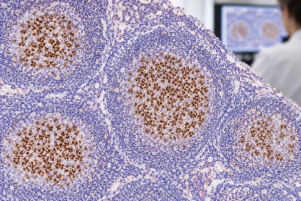
In a reactive lymph node, BCL6 strongly labels germinal-center B cells. This normal expression is expected and provides a built-in map of follicular architecture. The surrounding mantle-zone B cells are generally negative, helping the germinal center stand out.
BCL6 can also label T-follicular helper cells, a specialized T-cell population within germinal centers. Those cells are far fewer than the B cells and are interpreted with other T-follicular helper markers when a T-cell lymphoma is suspected.
The fact that normal germinal centers are BCL6 positive creates both value and a potential pitfall. In a lymphoma that partially replaces a node, residual benign follicles may stain brightly even if the tumor itself is BCL6 negative. The pathologist therefore compares the BCL6 slide with the hematoxylin-and-eosin section and other IHC stains to ensure the correct cells are being scored.
BCL6 contrasts with BCL2 in reactive follicles. Normal germinal-center B cells usually express BCL6 but suppress BCL2. This opposite pattern is useful when distinguishing reactive follicular hyperplasia from conventional follicular lymphoma, in which neoplastic follicles often co-express BCL6 and abnormal BCL2.
Because BCL6 is a normal germinal center protein, it is especially useful for mapping follicular architecture. In a reactive node, BCL6 highlights germinal center B cells but not the mantle zones. That pattern helps the pathologist see whether follicles are appropriately shaped and distributed. In follicular lymphoma, BCL6-positive neoplastic cells often fill crowded follicles and may extend beyond them when the tumor has a diffuse component.
BCL6 should not be confused with BCL2 simply because both names begin with “BCL.” BCL6 is a transcriptional repressor associated with germinal center differentiation, whereas BCL2 is an anti-apoptotic protein normally suppressed in reactive germinal centers. Their contrasting normal patterns are one reason the combination is diagnostically useful.
BCL6 in Follicular Lymphoma
Follicular lymphoma is a mature B-cell neoplasm derived from germinal-center cells, so BCL6 is commonly positive in the neoplastic follicles. The staining is nuclear and often involves many of the centrocytes and centroblasts that make up the tumor.
A CD10 IHC test is often paired with BCL6. Both support germinal-center differentiation, but they are not identical. Some follicular lymphomas, particularly higher-grade, unusual, or extranodal examples, may lose CD10 while retaining BCL6. That can make BCL6 especially helpful when the phenotype is incomplete.
BCL2 provides another complementary pattern. In reactive follicles, germinal centers are usually BCL2 negative; in conventional follicular lymphoma they are often abnormally BCL2 positive. A BCL2 IHC result therefore helps distinguish abnormal follicular survival signaling from the normal germinal-center pattern.
The typical follicular lymphoma panel may include CD20, CD10, BCL6, BCL2, Ki-67, and additional markers depending on the case. FISH for BCL2 or BCL6 rearrangements can be useful when morphology and IHC are unusual. Modern classifications recognize that follicular lymphomas are genetically diverse; not every case carries the classic IGH::BCL2 rearrangement.
BCL6 positivity does not grade follicular lymphoma. Grading and classification depend on morphology and the specific disease entity. Nor does strong BCL6 staining by itself predict prognosis. Its primary value in this setting is demonstrating germinal-center phenotype.
Some follicular-pattern lymphomas are BCL6 positive but CD10 weak or negative, and some have unusual BCL2 findings. In those cases, the pathologist may add other germinal center markers, examine the follicular dendritic cell meshwork, and use FISH when a BCL2 or BCL6 rearrangement would clarify classification. Site and age matter as well because pediatric-type follicular lymphoma, primary cutaneous follicle center lymphoma, and conventional nodal follicular lymphoma have different expected profiles.
BCL6 positivity therefore supports a germinal center program but does not by itself prove conventional follicular lymphoma. A reactive follicle is also BCL6 positive, and several aggressive lymphomas retain BCL6. Architecture, cytology, B-cell clonality, and companion markers determine whether the positive cells are normal, neoplastic, or part of another germinal center-derived lymphoma.
BCL6 in Diffuse Large B-Cell Lymphoma
DLBCL is biologically heterogeneous. Some tumors resemble germinal-center B cells, while others resemble later activated B cells. BCL6 is one of the IHC markers used to approximate this cell-of-origin biology in routine pathology.
A common algorithm uses CD10, BCL6, and MUM1/IRF4 to classify DLBCL as germinal-center B-cell–like or non-germinal-center by IHC. In simplified terms, CD10 positivity supports a germinal-center phenotype; in CD10-negative cases, BCL6 and MUM1 help refine the assignment. These algorithms are practical but are only surrogates for gene-expression profiling.
The pathologist usually estimates the percentage of large tumor cells with BCL6 nuclear staining. Published algorithms often use thresholds such as 30%, but laboratories should follow validated methods and the interpretive system specified in the report. A percentage used for one algorithm should not be treated as a universal biologic definition.
A MUM1 IHC test is especially useful because MUM1 marks later B-cell or plasma-cell differentiation and can overlap with BCL6 in some DLBCLs. Co-expression reflects the complexity of lymphoma biology rather than an impossible result.
BCL6 also enters the genetic workup of aggressive B-cell lymphoma. Rearrangements of BCL6 can occur in DLBCL, either alone or with MYC or other alterations. Current WHO and International Consensus Classification schemes treat MYC/BCL6-rearranged tumors differently from MYC/BCL2-rearranged tumors, which is why FISH results must be read using current classification criteria rather than older “double-hit” shorthand alone.
In DLBCL, BCL6 is commonly interpreted with CD10 and MUM1/IRF4 in IHC cell-of-origin algorithms. A frequently used approach applies a 30% cutoff to these markers, but the algorithm is only a surrogate for gene-expression profiling. A BCL6-positive result can contribute to either the germinal-center or non-germinal-center branch depending on the CD10 and MUM1 results, so the marker cannot be read in isolation.
This is also why a pathology report may contain two different kinds of “BCL6” information: an IHC percentage and a FISH result. They answer different questions. The IHC result describes protein expression and differentiation; the FISH result looks for a structural gene rearrangement. Neither result automatically predicts the other.
BCL6 Protein Versus Gene Rearrangement
BCL6 IHC detects protein expression. BCL6 FISH detects a structural rearrangement of the gene. These findings overlap only partially.
A lymphoma can express BCL6 protein because it retains a germinal-center differentiation program without having a BCL6 rearrangement. Conversely, a rearranged tumor may show variable protein expression. The IHC result therefore cannot be used as a substitute for genetic testing when the genetic alteration itself affects classification.
| Test | What it measures | Main use |
|---|---|---|
| BCL6 IHC | Nuclear BCL6 protein | Germinal-center phenotype and DLBCL IHC algorithms |
| BCL6 break-apart FISH | BCL6 gene rearrangement | Genetic classification of selected aggressive B-cell lymphomas |
| Broad molecular profiling | Multiple mutations, rearrangements, or expression features | Complex or research-level molecular subclassification |
This distinction is especially important when a report contains both “BCL6 positive” and “BCL6 rearrangement not detected.” Those statements are not contradictory. One describes protein; the other describes chromosome structure.
The same principle applies to MYC and BCL2. Protein co-expression by IHC and gene rearrangements by FISH define different phenomena. Current aggressive B-cell lymphoma classification increasingly depends on integrating both.
BCL6 rearrangements are not confined to a single lymphoma diagnosis. They occur in subsets of DLBCL and can be present in other B-cell neoplasms. Their meaning depends on the partner alteration, morphology, and the current classification scheme. This is especially important when MYC and BCL2 rearrangements are also being evaluated, because modern categories define some high-grade B-cell lymphomas by specific genetic combinations rather than by BCL6 protein staining.
For the reader, the practical rule is simple: BCL6 IHC positivity is not the same as a positive BCL6 FISH test. If the report lists both, each should be read on its own terms before the final integrated diagnosis is considered.
BCL6 can also be expressed in lymphomas of T-follicular helper origin because normal T-follicular helper cells participate in germinal center reactions. In that setting, a BCL6-positive cell is not automatically a B cell. CD3, CD4, PD-1, ICOS, CXCL13, and other markers are used to establish the lineage and specialized phenotype of the tumor. This is a useful reminder that even a classic “germinal center marker” must be assigned to the correct cell population.
In small core biopsies, architecture may be difficult to assess and BCL6-positive reactive germinal centers may coexist with tumor. Serial sections and comparison with CD20, CD3, CD10, and follicular dendritic cell markers help determine whether the BCL6-positive cells belong to the neoplasm or to residual normal tissue.
How to Interpret BCL6 Results
A useful interpretation begins with the final diagnosis and the identity of the stained cells. BCL6 positivity in a normal germinal center is expected. BCL6 positivity in a neoplastic follicle supports germinal-center lymphoma. BCL6 positivity in a diffuse large-cell tumor may contribute to a cell-of-origin algorithm.
Common result patterns include:
- BCL6 positive in well-defined reactive germinal centers: normal pattern.
- BCL6 positive in abnormal crowded follicles: supports germinal-center differentiation and may fit follicular lymphoma.
- BCL6 positive in many large diffuse B cells: common in a subset of DLBCL and relevant to IHC classification algorithms.
- BCL6 negative in a suspected follicular lymphoma: does not automatically exclude the diagnosis; phenotype, grade, site, and technical factors matter.
- BCL6 positive by IHC but FISH negative: possible because protein expression does not require gene rearrangement.
A CD20 IHC test often confirms that the BCL6-positive tumor belongs to the B-cell lineage. Ki-67 IHC can then show proliferative activity, but a high Ki-67 score and BCL6 positivity answer different biologic questions.
Technical quality matters as well. Poor fixation, decalcification, crush artifact, or scant viable tumor may weaken nuclear staining. If the stain is unexpectedly negative, the pathologist checks internal controls and may repeat the assay on another tissue block.
A negative BCL6 stain requires the same discipline as a positive one. It may be genuinely absent in a lymphoma that has moved away from a germinal center phenotype, but poor fixation, decalcification, scant viable tumor, or difficult nuclear staining can also reduce the signal. Internal reactive germinal centers, when present, are useful controls because they should show clear nuclear positivity.
The clinical meaning of BCL6 is also disease-specific. In follicular lymphoma it primarily supports lineage and architecture; in DLBCL it contributes to phenotypic classification; in high-grade B-cell lymphoma it may prompt genetic testing. There is no universal statement that “BCL6 positive is good” or “BCL6 negative is bad.” Prognosis is determined by the final lymphoma entity, molecular findings, clinical risk factors, stage, and response to therapy.
For serial or relapse biopsies, BCL6 expression may also change as a lymphoma evolves. A transformed component does not always reproduce the exact phenotype of an earlier low-grade lesion. Comparing current and prior tissue helps the pathologist decide whether a different staining pattern reflects progression, sampling, therapy-related selection, or a separate lymphoma.
What Happens After BCL6 Testing
After BCL6 IHC, further testing depends on the suspected lymphoma. A follicular lymphoma workup may add BCL2 FISH or other studies if the morphology or phenotype is atypical. A DLBCL workup may include MYC and BCL2 IHC, EBV studies, and FISH for MYC, BCL2, and BCL6 rearrangements when indicated.
The final report integrates morphology, immunophenotype, genetics, and clinical information. BCL6 helps define where the tumor sits along the B-cell differentiation pathway, but it rarely provides a complete diagnosis by itself.
For patients, useful questions include: Were the tumor cells or only normal follicles BCL6 positive? Was the stain used to support follicular lymphoma or to classify DLBCL? Was BCL6 genetic testing performed separately? Does the final diagnosis depend on a rearrangement, an IHC phenotype, or both?
Those questions prevent the most common misunderstanding: treating “BCL6 positive” as if it were a specific lymphoma name or a gene-mutation result. In pathology, BCL6 is most powerful when its nuclear staining pattern is interpreted alongside the architecture and the rest of the panel.
For patients, the key is to read BCL6 as one part of the final diagnosis. Ask whether the positive cells are the lymphoma cells, whether CD10 and MUM1 support the same phenotype, and whether FISH was needed. Those details explain what BCL6 actually contributed without overinterpreting a single stain.
References
- The International Consensus Classification of Mature Lymphoid Neoplasms: a report from the Clinical Advisory Committee 2022 (Consensus Classification)
- The fifth edition of the WHO classification of mature B-cell neoplasms: open questions for research 2024 (Review)
- A Diagnostic Approach in Large B-Cell Lymphomas According to the Fifth World Health Organization and International Consensus Classifications and a Practical Algorithm in Routine Practice 2024 (Review)
- Expression and Clinical Significance of Ki-67, CD10, BCL6, MUM1, c-MYC, and EBV in Diffuse Large B Cell Lymphoma Patients 2024
- High-grade B-cell lymphomas: Double hit and non-double hit. 2024 (Review)
Disclaimer
BCL6 IHC is a tissue marker used as part of lymphoma classification and cannot diagnose a lymphoma by itself. Results must be interpreted with morphology, other immunostains, genetic testing when indicated, and clinical findings. Discuss the complete pathology report with the treating hematologist, oncologist, or pathologist.