
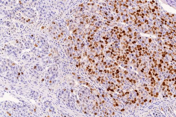
Ki-67 immunohistochemistry (IHC) estimates how actively tumor cells are dividing. A pathologist stains a tissue sample for Ki-67, a protein found in proliferating cells, then reports the percentage of tumor-cell nuclei that stain. That percentage is often called the Ki-67 index, Ki-67 labeling index, or proliferation index. In many cancers, a higher index is associated with faster growth or more aggressive behavior, but there is no single cutoff that makes a Ki-67 result “high” for every tumor. Interpretation depends on the cancer type, specimen, scoring method, and the clinical question. Ki-67 is especially important in grading many neuroendocrine tumors and is also used in breast cancer, lymphomas, brain tumors, and other neoplasms. It works best as one piece of a pathology diagnosis rather than as a stand-alone cancer test. The pathology report should be read together with tumor type, grade, stage, morphology, and other biomarkers.
- Ki-67 measures tumor-cell proliferation: the result is usually the percentage of tumor nuclei that show positive staining.
- A higher Ki-67 usually means faster cell turnover, but “high” is tumor-specific: a percentage cannot be interpreted reliably without knowing the cancer type and scoring context.
- In selected early ER-positive/HER2-negative breast cancers, values of 5% or less and 30% or more are the most reproducible clinical ranges; intermediate values are less definitive.
- For many gastroenteropancreatic neuroendocrine tumors, Ki-67 helps assign grade, commonly using under 3%, 3%–20%, and over 20% ranges together with morphology and mitotic activity.
- No fasting or medication change is usually needed for Ki-67 itself: it is performed on biopsy or surgical tissue already collected for pathology.
Table of Contents
- What the Ki-67 IHC Test Measures
- How Ki-67 Is Tested and Scored
- What a High or Low Ki-67 Means
- How Ki-67 Is Used by Tumor Type
- Limitations and Sources of Variation
- How to Read a Ki-67 Pathology Report
- Questions to Ask After a Ki-67 Result
What the Ki-67 IHC Test Measures
Ki-67 is a tissue marker of cell proliferation, not a blood tumor marker and not a direct measurement of how fast a tumor will grow in the body. The Ki-67 protein is present during active phases of the cell cycle and is largely absent from resting cells. Immunohistochemistry uses an antibody, commonly one directed against the Ki-67 antigen, to make positive nuclei visible under the microscope.
The pathologist usually reports the result as a percentage. A Ki-67 index of 20%, for example, means that about 20 of every 100 tumor cells counted showed nuclear staining by the method used. It does not mean that the tumor is growing 20% per day, that 20% of the tumor is cancer, or that there is a 20% chance of recurrence.
Ki-67 can answer several different pathology questions:
- How proliferative is this tumor compared with other tumors of the same type?
- Does the proliferation level support a particular tumor grade?
- Is the result consistent with the appearance of the tumor under the microscope?
- Does the index add prognostic information in a setting where Ki-67 has been validated?
- In selected cancers, can it contribute to a treatment decision or help assess response after therapy?
The result matters most when the tumor classification specifically incorporates proliferation. In many neuroendocrine neoplasms, for example, the Ki-67 index is part of formal grading. In other tumors, Ki-67 may be informative but not required for diagnosis or treatment selection.
Ki-67 should also be distinguished from mitotic count. A mitotic count records cells visibly undergoing mitosis in a defined tissue area, while Ki-67 identifies cells across several active phases of the cell cycle. The two measures often correlate, but they are not interchangeable. A pathologist may use both because discordance can provide useful diagnostic information.
How Ki-67 Is Tested and Scored
Ki-67 testing starts with a tissue specimen, usually a core biopsy, excision, resection, or cell block, and ends with a percentage based on nuclear staining in tumor cells. Patients normally do not need to fast, stop medications, or prepare specifically for the stain. The important preparation steps occur in the pathology laboratory.
After tissue is removed, it is fixed, processed, embedded in paraffin, cut into thin sections, and placed on slides. The laboratory applies an antibody that binds Ki-67 and uses a detection system that produces visible nuclear staining. The pathologist then identifies the relevant tumor and evaluates the fraction of positive nuclei.
Counting methods can differ
A Ki-67 score may be obtained by visual estimation, manual counting, image analysis, or a validated digital method. Depending on the tumor type and guideline, the pathologist may count a representative overall area, focus on a hot spot with the greatest labeling, or use a weighted approach that accounts for areas with different staining levels.
That distinction matters. Suppose one tumor has large areas around 10% but a small focus around 35%. A global average and a hot-spot method will produce different numbers even though both observers are looking at the same slide. The correct approach depends on the disease-specific scoring standard.
For a reliable count, the denominator should consist of tumor cells, not lymphocytes, stromal cells, endothelial cells, or other non-neoplastic cells that may also stain. Necrotic tissue and crushed or poorly preserved areas are generally avoided. Laboratories also use internal and external controls to confirm that the stain performed as expected.
Why the exact percentage is not perfectly precise
Ki-67 looks numerical, but the result has measurement uncertainty. Sampling, fixation, antibody performance, the selected microscopic field, and observer judgment can all affect the percentage. A reported value of 19% should therefore not automatically be treated as biologically different from 21% when a cutoff is 20%, especially if the specimen is small or the tumor is heterogeneous.
When a result lies near a clinically important boundary, the pathologist may count more cells, review another block, use a resection specimen when available, or interpret the value alongside mitoses and morphology. The goal is not to chase an artificial level of decimal-point precision; it is to place the tumor in the most defensible diagnostic category.
What a High or Low Ki-67 Means
A high Ki-67 index generally indicates that a larger fraction of tumor cells is actively cycling, while a low index indicates lower proliferative activity. That broad principle is useful, but a number becomes clinically meaningful only after it is matched to the tumor type.
There is no universal normal range. Healthy tissues have very different baseline proliferative rates, and tumors arise from many different cell types. A Ki-67 value that is high for one neoplasm may fall in an expected range for another. This is why internet charts that label, for example, “under 10% low, 10%–20% intermediate, over 20% high” across all cancers are misleading.
In a validated disease setting, a higher Ki-67 can be associated with one or more of the following:
- a higher histologic or biologic grade;
- a greater likelihood of rapid progression;
- a less favorable prognosis;
- a different treatment strategy;
- greater sensitivity to some therapies that target rapidly dividing cells, although this relationship is not universal.
A low Ki-67 can support a lower-grade or more slowly proliferative tumor, but it does not prove that the tumor is harmless. Some cancers grow slowly yet can recur or metastasize. Likewise, a high Ki-67 does not by itself predict an individual person’s survival or prove that a specific drug will work.
The most important question is therefore not “Is my Ki-67 high?” but “What does this Ki-67 mean for this exact tumor under the grading or treatment system being used?”
How Ki-67 Is Used by Tumor Type
The clinical weight of Ki-67 varies greatly by cancer type. In some tumors it is part of formal grading; in others it is supportive, prognostic, or mainly used in research. The same percentage can therefore have very different consequences.
| Tumor setting | Typical role of Ki-67 | Key interpretation point |
|---|---|---|
| Gastroenteropancreatic neuroendocrine tumors | Formal grading with proliferation measures | Common ranges are <3%, 3%–20%, and >20%, interpreted with morphology and mitotic count |
| Early ER-positive/HER2-negative breast cancer | Prognostic information in selected patients | Very low (≤5%) and high (≥30%) values are more reproducible than the intermediate zone |
| Lymphoma | Supports assessment of proliferative activity | Meaning depends strongly on lymphoma subtype and morphology |
| Brain and other solid tumors | Variable diagnostic or prognostic support | Disease-specific criteria matter; Ki-67 is not universally part of formal grading |
Neuroendocrine tumors
Ki-67 has one of its clearest roles in well-differentiated neuroendocrine tumors. For many gastrointestinal and pancreatic neuroendocrine tumors, the World Health Organization grading framework separates tumors into G1, G2, and G3 categories using proliferation. A commonly used Ki-67 framework is less than 3% for G1, 3%–20% for G2, and more than 20% for G3, although site-specific classification details must be followed.
A high Ki-67 does not automatically mean “neuroendocrine carcinoma.” Well-differentiated neuroendocrine tumor G3 and poorly differentiated neuroendocrine carcinoma are biologically distinct diagnoses. Morphology, mitotic activity, necrosis, and sometimes additional IHC or molecular findings help separate them. This distinction can materially affect treatment.
Sampling also matters. Metastatic sites can show a different proliferation rate from the primary tumor, and a small biopsy can miss the most proliferative area. If the clinical behavior seems much more aggressive than the reported grade, review or repeat sampling may be appropriate.
Breast cancer
Ki-67 is widely reported in breast pathology, but its use requires restraint because laboratories and scoring methods have historically shown substantial variability. The International Ki67 in Breast Cancer Working Group concluded that standardized assessment can be reproducible and that Ki-67 has clinical validity for prognosis in selected early-stage, estrogen receptor-positive, HER2-negative breast cancers.
In that specific setting, 5% or less and 30% or more are the ranges with the strongest reproducibility for identifying very low and clearly high proliferation. Values between those ranges should not be forced into a simple yes-or-no treatment rule. Other features, including tumor size, lymph-node status, grade, hormone receptor expression, HER2 status, genomic assays when indicated, age, and overall health, may be more important to a chemotherapy decision.
Lymphomas and other tumors
In lymphoid malignancies, Ki-67 can help characterize how proliferative a population is. Some aggressive lymphomas often show very high labeling, while indolent lymphomas typically show lower proliferation. However, overlap exists, and the value must be interpreted with the lymphoma subtype, architecture, cytology, immunophenotype, and genetic findings. A high index alone cannot diagnose a specific lymphoma.
Similar caution applies to sarcomas, melanocytic tumors, meningiomas, prostate tumors, and many other neoplasms. Ki-67 may add prognostic or grading information in selected contexts, but the validated cutoff and clinical purpose are disease-specific.
Limitations and Sources of Variation
Ki-67 is useful because it converts a biologic feature into a visible tissue measurement, but the measurement is sensitive to both biology and technique. Understanding the main sources of variation prevents overinterpretation of small differences.
First, tumors are heterogeneous. One region may have few proliferating cells while another contains a hot spot. A needle biopsy samples only part of the lesion, so its Ki-67 may not perfectly match a later resection. This is not necessarily a laboratory error; it can reflect genuine intratumoral variation.
Second, preanalytic handling can alter staining. Delayed fixation, underfixation, overfixation, tissue damage, and some decalcification methods may affect antigen preservation. Bone metastases that require strong acid decalcification can be particularly challenging for many IHC markers.
Third, laboratories may use different antibody clones, staining platforms, detection systems, and scoring procedures. Standardization and quality-assurance programs reduce these differences, but they do not eliminate them completely.
Fourth, pathologists must decide which cells count as tumor. Ki-67 can stain normal proliferating cells, especially lymphocytes. In a tumor with heavy inflammation, including these cells would falsely raise the index. Proper cell identification is therefore as important as counting.
Finally, a cutoff creates an artificial boundary in a continuous biological process. Two tumors with Ki-67 values of 19% and 21% may be more similar than the labels on either side of a 20% threshold suggest. Near a cutoff, clinicians should look for concordance with morphology, mitotic count, specimen quality, and the rules of the relevant classification system.
How to Read a Ki-67 Pathology Report
Read the Ki-67 number in the same paragraph as the diagnosis, tumor grade, and method whenever possible. A report that says “Ki-67 proliferation index approximately 40%” is incomplete without knowing what type of tumor was tested and how that percentage affects its classification.
A useful review follows a simple sequence:
- Confirm the exact diagnosis. Ki-67 should support an established or suspected tumor type, not replace it.
- Find the reported percentage and wording. The report may say “labeling index,” “proliferation index,” “hot-spot index,” or provide a range.
- Check whether the score changes the grade. This is especially important in neuroendocrine tumors.
- Look for the specimen type. A core biopsy, resection, lymph-node metastasis, or bone biopsy may each sample the disease differently.
- Compare Ki-67 with mitoses and morphology. Concordant findings strengthen the interpretation; major discordance may require review.
- Ask whether the result changes management. Some Ki-67 values are clinically actionable; others are descriptive or supportive.
A range can be more informative than a single precise number when staining is heterogeneous. Wording such as “10% overall, with focal hot spots up to 30%” alerts the care team that the tumor does not have uniform proliferation. In some diseases, the hot spot controls grading; in others, a global method is preferred.
If a report from another institution produces a very different Ki-67 score, compare the specimen, block, staining method, and scoring approach before assuming the tumor has biologically changed. Repeat testing may be reasonable when the difference would alter grade or treatment and there is enough appropriate tissue.
Questions to Ask After a Ki-67 Result
The most useful follow-up is to ask how the Ki-67 result affects the diagnosis or treatment of the specific tumor, not whether the percentage is “good” or “bad” in isolation. The pathology number should lead to a clinical interpretation, not anxiety over a cutoff taken out of context.
Consider asking the pathologist or treating clinician:
- What exact tumor type was stained, and is Ki-67 formally part of its grading system?
- Was the reported value based on a hot spot, an average, manual counting, or digital image analysis?
- Is the result close to a cutoff that could change the grade or management?
- Does the Ki-67 agree with the mitotic count and the way the tumor looks under the microscope?
- Could the biopsy have under-sampled a more proliferative part of the tumor?
- Would another specimen or pathology review change a treatment decision?
- Is this Ki-67 result prognostic, predictive of treatment benefit, or simply supportive of the diagnosis?
A Ki-67 result usually does not require urgent medical action by itself. Urgency comes from the overall diagnosis, symptoms, stage, and clinical condition. If the pathology report shows a high-grade or aggressive malignancy, the appropriate next step is timely discussion with the treating oncology team so that staging and treatment planning can proceed.
The central point is simple: Ki-67 is a context-dependent proliferation marker. It is most powerful when the laboratory uses a standardized method and the care team interprets the result under tumor-specific criteria. A high score can signal faster proliferation, but the percentage should never be translated into prognosis or treatment without the rest of the pathology and clinical picture.
References
- Assessment of Ki67 in Breast Cancer: Updated Recommendations From the International Ki67 in Breast Cancer Working Group 2021 (Guideline)
- Expression, assessment and significance of Ki67 expression in breast cancer: an update 2023 (Review)
- Diagnostic, Prognostic, and Predictive Role of Ki67 Proliferative Index in Neuroendocrine and Endocrine Neoplasms: Past, Present, and Future 2023 (Review)
- Overview of the 2022 WHO Classification of Neuroendocrine Neoplasms 2022 (Review)
- Peer-to-peer validation of Ki-67 scoring in a pathology quality circle as a tool to assess interobserver variability: are we better than we thought? 2024
Disclaimer
Ki-67 results must be interpreted by a qualified pathologist and treating clinician in the context of the exact tumor type, specimen, morphology, grade, and other biomarkers. A percentage or cutoff alone cannot diagnose cancer, predict an individual outcome, or determine treatment. This article is for general education and is not a substitute for medical advice or review of your pathology report.





