Tissue Tumor Markers and IHC
Home Tissue Tumor Markers and IHC
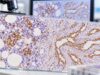
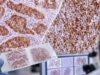
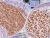
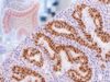
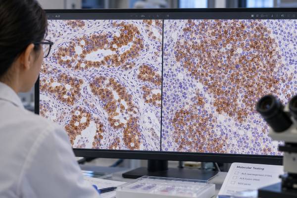
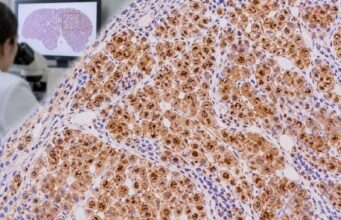
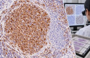
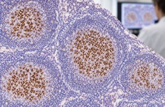
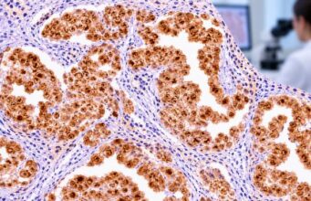
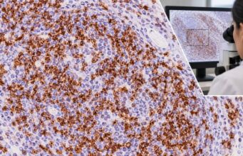
Tissue tumor markers and immunohistochemistry (IHC) help pathologists identify where a tumor originated, determine its cell lineage and differentiation, classify cancer subtypes, and evaluate markers that may influence prognosis or treatment. This category provides clear, practical guides to commonly used IHC tests and explains what positive, negative, abnormal, or characteristic staining patterns can mean in the context of a tissue biopsy.
Explore markers used across many cancer types, including ER, PR, HER2, Ki-67, and p53; lung markers such as TTF-1, Napsin A, and p40; lymphoma markers including CD3, CD10, CD15, CD20, CD30, BCL2, BCL6, and PAX5; and melanoma markers such as S100, SOX10, HMB-45, and Melan-A. Coverage also includes markers for gastrointestinal, liver, pancreatic, ovarian, neuroendocrine, mesothelial, germ cell, and soft-tissue tumors.
Each guide explains what the marker detects, typical staining patterns, diagnostic uses, limitations, and how results fit into broader IHC panels. The goal is to make complex pathology terminology and tumor-marker interpretation easier to understand without treating a single stain as a diagnosis on its own.