
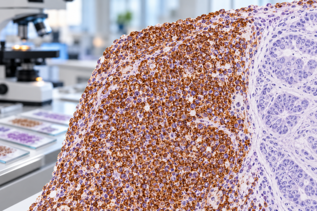
CDX2 immunohistochemistry (IHC) detects a nuclear transcription factor involved in intestinal epithelial differentiation. Pathologists use it most often to support gastrointestinal—especially colorectal—origin in an adenocarcinoma, including metastatic tumors found in the liver, lung, ovary, lymph nodes, or other sites. Most conventional colorectal adenocarcinomas are strongly and diffusely CDX2 positive, but CDX2 is not perfectly specific for colon and is not positive in every colorectal cancer. Gastric, small-intestinal, pancreaticobiliary, ovarian mucinous, and other tumors can express it, while poorly differentiated, medullary, MSI-high, or BRAF-mutated colorectal cancers may show reduced or absent staining. The strongest interpretation comes from a panel. CDX2 combined with SATB2, CK20, and CK7 can substantially refine the probability of lower-GI origin, while morphology and clinical imaging remain essential. A positive CDX2 result therefore supports intestinal differentiation; it does not by itself prove where a cancer began.
- Diffuse nuclear CDX2 positivity strongly supports intestinal differentiation and is common in colorectal adenocarcinoma.
- CDX2 is not colon-specific: upper-GI, pancreaticobiliary, and some ovarian or other tumors can also be positive.
- A colorectal cancer can be CDX2 negative, particularly in some poorly differentiated or molecularly distinct tumors.
- CDX2 is usually interpreted with SATB2, CK20, and CK7 when the goal is to identify the origin of a metastatic adenocarcinoma.
- CDX2 staining intensity is not a stand-alone treatment or staging test; molecular biomarkers and stage guide therapy.
Table of Contents
- What CDX2 IHC detects
- Normal expression and staining pattern
- CDX2 in colorectal cancer
- CDX2 in other tumors
- Using CDX2 to find tumor origin
- Negative results and diagnostic pitfalls
- How to read the report and next steps
- Practical CDX2 scenarios and diagnostic decisions
What CDX2 IHC detects
CDX2 is a homeobox transcription factor that regulates intestinal development and mature intestinal epithelial identity. Because it functions in the nucleus, true IHC positivity appears as nuclear staining. That clean localization makes CDX2 relatively straightforward to score compared with markers that have more variable cytoplasmic patterns.
The test is performed on a tissue section from a biopsy or surgical specimen. The pathologist evaluates whether the malignant cells show nuclear staining and whether it is diffuse or focal. Strong, diffuse labeling in most tumor cells is more characteristic of conventional colorectal adenocarcinoma than rare scattered positive nuclei.
CDX2 is a differentiation marker, not a mutation test. It does not measure a blood concentration and has no “normal range.” It also does not directly test for Lynch syndrome, microsatellite instability, RAS mutations, BRAF mutations, or another actionable molecular alteration. Those require separate assays.
Its main value is diagnostic pattern recognition. If a metastatic adenocarcinoma is found in the liver, a CDX2-positive result shifts the differential toward an intestinal primary. The pathologist then integrates CK7, CK20, SATB2, morphology, imaging, and clinical history before assigning origin.
Normal expression and staining pattern
Normal intestinal epithelium shows nuclear CDX2 expression, particularly in small bowel and colorectal mucosa. This provides a useful internal control when normal mucosa is present in the same specimen. Gastric epithelium can show CDX2 in areas of intestinal metaplasia, reflecting a change toward an intestinal phenotype.
In colorectal adenocarcinoma, the classic pattern is strong diffuse nuclear staining. The stain should line up with malignant gland nuclei on the corresponding H&E section. Cytoplasmic staining alone is generally not the expected specific pattern.
Extent is important. A tumor in which nearly all nuclei stain strongly provides more convincing intestinal evidence than one with only occasional weak positive cells. Many laboratories do not use a formal percentage cutoff because the diagnostic meaning depends on the differential diagnosis.
Technical variables can alter expression. Poor fixation, necrosis, decalcification, and scant viable tumor may reduce staining. If normal intestinal epithelium is present and positive, it supports adequate technical performance. When controls fail, an apparently negative tumor result requires caution.
CDX2 in colorectal cancer
Most conventional colorectal adenocarcinomas retain CDX2 expression, including many metastatic tumors. A recent meta-analysis estimated expression in a high proportion of both primary and metastatic colorectal cancers, supporting CDX2 as a sensitive marker in this setting.
However, colorectal cancer is biologically heterogeneous. Reduced or lost CDX2 expression is more frequent in poorly differentiated tumors, medullary-type carcinomas, and subsets associated with mismatch-repair deficiency, microsatellite instability, or BRAF V600E. That means CDX2 negativity should not automatically exclude colorectal origin when the clinical and morphologic evidence points strongly to the colon.
CDX2 has also been studied as a prognostic marker. Some studies associate loss of CDX2 with adverse clinicopathologic features or poorer outcomes, but the marker is not used as a universal stand-alone prognostic score in routine care. Stage, histologic features, mismatch-repair status, RAS/BRAF and other molecular findings, and the patient’s overall clinical picture remain more important.
For diagnosis, CDX2 becomes even more useful when paired with SATB2 IHC. SATB2 is relatively specific for lower-GI differentiation, so dual positivity can strongly support colorectal or appendiceal origin. CK20 adds another compatible epithelial pattern, while CK7 is often negative in conventional colorectal cancer.
CDX2 in other tumors
CDX2 is not restricted to colorectal cancer. Small-intestinal adenocarcinomas commonly express it. Gastric adenocarcinomas, especially those with intestinal differentiation, may also be positive. Pancreatic and biliary adenocarcinomas can show focal or variable expression, and some ovarian mucinous tumors have an intestinal-type phenotype with CDX2 positivity.
This overlap is why CDX2 should be described as an intestinal differentiation marker rather than a colon-specific marker. The anatomic site, staining extent, and other IHC results determine how much weight to give it.
In ovarian mucinous tumor differentials, for example, metastatic colorectal or appendiceal adenocarcinoma can mimic a primary ovarian mucinous neoplasm. Strong diffuse CDX2 and SATB2, especially with CK7 negativity, favor lower-GI origin, while diffuse CK7 and PAX8 support primary Müllerian differentiation in the appropriate setting. Tumor size, laterality, surface involvement, and growth pattern also matter.
Some non-GI carcinomas can show aberrant focal CDX2 expression. Rare lung adenocarcinomas and other tumors may express intestinal markers, particularly when they have enteric morphology. In such cases, lung markers such as TTF-1 and Napsin A, imaging, and clinical context prevent overcalling a GI primary.
Neuroendocrine tumors can also use CDX2 as a site-of-origin marker. In a metastatic well-differentiated neuroendocrine tumor, CDX2 positivity can support midgut or intestinal origin, but the interpretation differs from adenocarcinoma and uses a separate neuroendocrine panel.
Using CDX2 to find tumor origin
When a metastatic adenocarcinoma is discovered before the primary cancer is known, pathologists usually use a staged IHC approach. First, morphology and broad lineage markers establish that the tumor is a carcinoma. CK7 and CK20 may provide an initial pattern. Site-specific markers then refine the possibilities.
A classic lower-GI profile is CK7 negative, CK20 positive, CDX2 positive, and SATB2 positive. The CK20 IHC test is useful in this combination because colorectal cancers frequently express CK20, while SATB2 adds specificity. A tumor with CK7 positivity, only focal CDX2, and negative SATB2 may instead prompt consideration of upper-GI, pancreaticobiliary, or ovarian mucinous origin.
| Pattern | Common interpretation | Important caveat |
|---|---|---|
| CDX2 diffuse+, SATB2+, CK20+, CK7− | Strongly favors colorectal/appendiceal origin | Correlate with morphology and imaging |
| CDX2+, CK7+, SATB2− | Could fit upper-GI, pancreaticobiliary, or mucinous tumor | Not site-specific |
| CDX2− in a poorly differentiated carcinoma | Does not exclude colorectal origin | Use SATB2, molecular context, prior pathology |
| CDX2+ neuroendocrine tumor | Can support intestinal neuroendocrine origin | Interpret within a neuroendocrine panel |
No IHC panel is perfect. The final wording may be “consistent with colorectal origin” or “favored lower-GI primary,” especially when the tumor is poorly differentiated or markers conflict. That calibrated language reflects the probabilistic nature of tumor-origin assignment.
Negative results and diagnostic pitfalls
The most important pitfall is assuming CDX2 negativity rules out colorectal cancer. Loss of intestinal differentiation can accompany high-grade morphology and particular molecular pathways. If a patient has a known colorectal primary, a metastatic lesion with reduced CDX2 may still be compatible after direct comparison with the original tumor.
The reverse pitfall is treating any positivity as proof of colon. Upper-GI and other tumors can express CDX2, especially focally. The distribution and strength matter, and SATB2 can provide additional discriminatory value.
Another issue is tumor heterogeneity. A biopsy may sample only a poorly differentiated area with reduced expression. A resection or another block may show stronger staining. Pathologists generally avoid repeating stains without a reason, but discordance between morphology, clinical findings, and IHC may justify additional sampling.
Technical issues include poor fixation, extensive necrosis, and tissue processing. Normal positive controls are essential. The stain also cannot determine whether a colorectal tumor is microsatellite stable, mismatch-repair deficient, RAS mutated, or otherwise molecularly actionable.
Finally, a CDX2-positive metastasis does not tell whether the primary is in colon versus appendix versus small bowel with certainty. Clinical imaging and endoscopic evaluation remain important, especially when the exact primary site affects surgery or systemic treatment.
How to read the report and next steps
Read CDX2 in the context of the entire immunophenotype. “Strong diffuse nuclear CDX2” carries more diagnostic weight than “rare weak nuclear staining.” If SATB2 and CK20 are also positive and CK7 is negative, the report may confidently favor colorectal origin. If the markers are mixed, the wording may be more cautious.
For a confirmed colorectal primary, CDX2 is usually not the key test that guides therapy. The clinically important next biomarkers may include mismatch-repair proteins or MSI testing, KRAS/NRAS, BRAF, HER2 in selected metastatic cases, and other molecular targets depending on current treatment guidelines. Colorectal cancer biomarker testing addresses different questions from CDX2 IHC.
For a cancer of unknown primary, the next steps may include additional IHC, imaging, colonoscopy or upper endoscopy, comparison with prior specimens, and occasionally molecular tumor profiling. The pathologist usually selects these tests based on the most plausible sites rather than ordering every available stain.
Patients can ask whether CDX2 was diffuse or focal, whether SATB2 was performed, how CK7 and CK20 fit the pattern, and how confident the pathologist is about the proposed primary site. If the tumor is CDX2 negative but colorectal origin is still favored, ask what other evidence supports that conclusion.
The best use of CDX2 is as part of a coherent diagnostic story. Strong nuclear expression can be highly informative, but no single tissue marker should override contradictory morphology, clinical findings, or more specific evidence.
Practical CDX2 scenarios and diagnostic decisions
CDX2 becomes most useful when it answers a focused question rather than when it is treated as a universal “colon stain.” The same positive result can carry very different weight in a liver metastasis, an ovarian mucinous tumor, a poorly differentiated colon cancer, or a neuroendocrine neoplasm. Working through common scenarios shows how pathologists combine CDX2 with morphology and other markers.
Liver metastasis with gland formation. A metastatic adenocarcinoma in the liver often raises colorectal, pancreaticobiliary, upper-GI, lung, and other possibilities. Strong diffuse nuclear CDX2 together with SATB2 and CK20, especially in a CK7-negative tumor with dirty necrosis or other colorectal-type morphology, strongly favors lower-GI origin. The conclusion becomes less specific when CDX2 is focal, SATB2 is negative, and CK7 is diffuse. In that setting, upper-GI or pancreaticobiliary primaries may remain plausible, and imaging or endoscopy may be more decisive than another broad round of stains.
Mucinous tumor involving the ovary. Primary ovarian mucinous tumors and metastases from colorectal or appendiceal primaries can overlap substantially. Diffuse CDX2 is not enough by itself to call a metastasis because some primary ovarian mucinous neoplasms show intestinal differentiation. SATB2, CK7, PAX8, the pattern and extent of staining, tumor laterality, ovarian surface involvement, and growth pattern all contribute. A lower-GI pattern is most persuasive when multiple independent features point in the same direction.
Poorly differentiated colorectal cancer with weak or absent CDX2. Loss of CDX2 is a recognized diagnostic trap. High-grade tumors can lose the mature intestinal phenotype seen in better-differentiated colorectal adenocarcinoma. If the tumor arose in the colon or a prior colorectal primary is documented, negative CDX2 should trigger correlation rather than automatic rejection of colorectal origin. SATB2 may remain informative, but it too can be reduced in poorly differentiated disease. Comparing the metastasis directly with the original resection can solve discrepancies that no single stain can resolve.
A lung mass with enteric morphology. Some pulmonary adenocarcinomas can express CDX2, and tumors with enteric differentiation may resemble metastatic colorectal carcinoma. Here the diagnostic question is not whether CDX2 is “truly” positive but which lineage pattern is more coherent. TTF-1 and Napsin A support lung origin when present, while SATB2 and a classic lower-GI CK7/CK20 profile support colorectal origin. The distribution of disease on imaging and the presence or absence of a known colorectal tumor are essential parts of the final interpretation.
Neuroendocrine tumor in the liver. CDX2 can help localize a well-differentiated neuroendocrine tumor, particularly when an intestinal or midgut primary is suspected. That use is different from diagnosing adenocarcinoma. The tumor first needs convincing neuroendocrine differentiation with morphology and markers such as synaptophysin, chromogranin, or INSM1. Site-directed markers can then be layered onto that diagnosis. A CDX2-positive neuroendocrine tumor should not be described as colorectal adenocarcinoma simply because the same transcription factor is positive.
Mismatch-repair-deficient or molecularly unusual colorectal tumors. In established colorectal cancer, CDX2 does not replace molecular testing. A tumor may show reduced intestinal-marker expression yet still require mismatch-repair or MSI assessment, RAS and BRAF testing, and other treatment-directed biomarkers according to stage and clinical setting. Diagnostic IHC and predictive biomarkers answer different questions. Keeping those purposes separate prevents a useful lineage stain from being given treatment implications it does not have.
Tissue stewardship matters in each scenario. Small biopsies contain a finite amount of viable tumor, and broad “shotgun” panels can consume material needed for predictive testing. An efficient approach starts with morphology and clinical context, uses a small set of discriminating stains, and expands only when the first results leave a meaningful uncertainty. CDX2 is especially effective when paired with a marker that adds specificity, such as SATB2 for lower-GI origin, rather than repeated with multiple overlapping intestinal markers.
When a report uses cautious terms such as “favored,” “consistent with,” or “compatible with,” that wording often reflects real overlap between immunophenotypes rather than indecision. The most useful follow-up question is what evidence supports the proposed origin and what alternative remains plausible. That answer may include morphology, multiple stains, prior specimens, imaging, endoscopy, and molecular findings. CDX2 contributes strongly to that combined assessment, but its greatest value comes from being interpreted as one piece of a pattern.
A further clue is consistency across separate tumor samples. If a primary colon tumor and a later metastasis share the same unusual morphology and a similar CDX2/SATB2/CK7/CK20 profile, that concordance can be more persuasive than whether every marker follows the textbook pattern. Conversely, a new lesion with a sharply different phenotype may justify reconsidering a second primary cancer. Pathologists often resolve difficult origin questions by comparing slides side by side rather than interpreting each specimen in isolation.
CDX2 is also best interpreted with attention to extent and intensity, not merely a positive/negative checkbox. Diffuse strong nuclear staining across most viable tumor cells is more characteristic of a mature intestinal phenotype than rare weak nuclei at the edge of a specimen. Focal staining can still be real, but it carries less site-specific weight because several non-colorectal tumors can acquire partial intestinal differentiation. The report may therefore describe staining as diffuse, patchy, or focal. That wording is clinically useful: it tells the treating team how much confidence the marker should add to the proposed origin and whether the rest of the panel needs to do more of the diagnostic work.
This makes descriptive staining detail clinically meaningful rather than decorative.
References
- Diagnostic and Prognostic Roles of CDX2 Immunohistochemical Expression in Colorectal Cancers 2022 (Systematic Review)
- Use of SATB2 and CDX2 Immunohistochemistry to Characterize and Diagnose Colorectal Cancer 2024
- Cytokeratin 7 and cytokeratin 20 expression in cancer: A tissue microarray study on 15,424 cancers 2022
- Carcinoma of Unknown Primary Origin: Application of Immunohistochemistry With Emphasis to Different Cytokeratin 7 and 20 Staining Patterns. 2022
- Real-world histopathological approach to malignancy of undefined primary origin (MUO) to diagnose cancers of unknown primary (CUPs) 2023
- Diagnostic and Clinical Significance of the Special AT-Rich Sequence-Binding Protein 2 Deregulation in Gastrointestinal Malignancies. 2026 (Systematic Review)
Disclaimer
CDX2 IHC is a diagnostic marker of intestinal differentiation and cannot prove a primary tumor site by itself. Results should be interpreted with morphology, SATB2/CK7/CK20 and other stains, molecular findings, imaging, and the patient’s clinical history.





