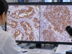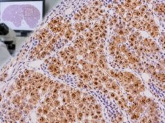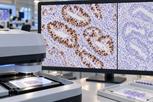
SATB2 immunohistochemistry (IHC) is a tissue stain that helps pathologists recognize colorectal and other lower gastrointestinal differentiation in a tumor. SATB2 is a nuclear transcription factor normally associated with tissues including the lower intestinal tract and bone. In diagnostic pathology, strong nuclear staining in an adenocarcinoma can support a colorectal or appendiceal origin, especially when SATB2 agrees with CDX2 and cytokeratin patterns. The test is particularly useful when cancer is found at a metastatic site, such as the liver, lung, ovary, peritoneum, or lymph node, and the primary site is uncertain. SATB2 is not perfectly sensitive or specific. Some colorectal cancers lose expression, and selected non-colorectal tumors—including tumors with osteoblastic differentiation—can be positive. The stain is therefore interpreted as part of a panel, with the tumor’s microscopic appearance, clinical history, imaging, and other IHC results guiding the final diagnosis.
- SATB2 positivity is usually nuclear and supports lower gastrointestinal differentiation in the right morphologic setting.
- Colorectal and appendiceal tumors are common SATB2-positive cancers, but not every colorectal cancer retains the marker.
- SATB2 is most useful in a panel with markers such as CDX2, CK20, and CK7 rather than as a stand-alone test.
- There is no blood reference range for SATB2 IHC. The report describes distribution and intensity of tumor-cell staining.
- A negative SATB2 result does not rule out colorectal origin, particularly in poorly differentiated or molecularly unusual tumors.
Table of Contents
- What the SATB2 IHC test detects
- What positive SATB2 staining means
- How SATB2 supports colorectal and lower-GI tumor origin
- Why a colorectal cancer can be SATB2 negative
- Important SATB2 pitfalls and non-colorectal positivity
- How SATB2 fits into an IHC panel
- How to use a SATB2 result in the pathology report
What the SATB2 IHC test detects
SATB2 IHC detects SATB2 protein in tissue sections. In a diagnostic tumor sample, pathologists assess nuclear staining in the neoplastic cells. The test is performed on fixed tissue from a biopsy or resection and does not produce a serum concentration or universal numeric “normal range.”
SATB2 is useful because many colorectal cancers preserve a lower-GI differentiation program. When a metastatic adenocarcinoma is SATB2 positive, the finding can shift the differential toward colorectal or appendiceal origin. An accompanying CDX2 IHC test often provides complementary evidence because CDX2 is highly sensitive for intestinal differentiation but can also be expressed in upper-GI and other tumors.
The stain is most informative after the pathologist has already evaluated the tumor’s architecture, mucin production, grade, and clinical distribution. A result that conflicts with morphology or other markers is a reason to broaden the workup, not to force the tumor into a colorectal category.
What positive SATB2 staining means
A positive SATB2 stain usually means that tumor-cell nuclei contain detectable SATB2 protein. Diffuse, strong nuclear labeling is more persuasive for lower-GI differentiation than weak or very focal staining. Pathologists may report a percentage or qualitative extent, but laboratories do not use one universal cutoff for every tumor type and diagnostic question.
In metastatic adenocarcinoma, concordant SATB2 and CDX2 positivity, particularly with CK20 expression and little or no CK7, is a classic pattern supporting colorectal origin. A CK20 IHC stain can strengthen that pattern, while CK7 may help identify alternative primaries.
The result must still fit the case. Appendiceal neoplasms also frequently express SATB2, and some small intestinal or other gastrointestinal tumors can be positive. SATB2 can also label osteoblastic tumors, so a SATB2-positive bone lesion should not automatically be called metastatic colorectal carcinoma without morphology and epithelial markers supporting that conclusion.
How SATB2 supports colorectal and lower-GI tumor origin
SATB2 is valuable in several recurring diagnostic problems. A liver metastasis with gland formation may be tested for SATB2 and CDX2 when colorectal cancer is in the differential. A mucinous ovarian tumor may require distinction between a primary ovarian neoplasm and metastasis from the colon or appendix. Peritoneal mucinous disease can raise a similar question.
For ovarian mucinous tumors, SATB2 can be especially useful when combined with CK7 and PAX8. Lower-GI metastases tend to favor SATB2 and CDX2, while primary ovarian mucinous tumors are more often CK7 positive and usually lack strong diffuse SATB2. No single combination is absolute, so laterality, tumor size, surface involvement, clinical imaging, and gross findings remain important.
The lower-GI association also means SATB2 can help when the primary lesion is tiny, previously removed, or clinically occult. In those cases, IHC suggests a likely lineage that can direct colonoscopy, imaging, or review of prior pathology.
Why a colorectal cancer can be SATB2 negative
Not all colorectal cancers retain SATB2. Loss or reduction of expression can occur in poorly differentiated tumors and in subsets with particular molecular or morphologic features. Tissue quality also matters: necrosis, decalcification, prolonged fixation, or scant viable tumor may reduce staining.
A negative result therefore lowers support for colorectal origin but does not exclude it. Pathologists look for other intestinal markers and for the broader immunophenotype. CDX2 may remain positive when SATB2 is reduced, and CK20 can be informative. Conversely, some colorectal carcinomas can show an atypical CK7 staining pattern, so even a familiar panel has exceptions.
When the clinical suspicion for colorectal origin is high, the pathology team may repeat SATB2 on a different block, add molecular testing, or correlate with endoscopy and radiology. The strongest diagnosis comes from converging evidence rather than a single negative stain.
Important SATB2 pitfalls and non-colorectal positivity
SATB2 is often described as a colorectal marker, but the label can be misleading if it is taken to mean “colorectal-specific.” Osteoblasts normally express SATB2, and osteosarcoma commonly shows nuclear staining. Some other tumors can show focal or variable positivity, and appendiceal tumors are expected to overlap with colorectal tumors because they share lower-GI differentiation.
Another pitfall is overcalling minimal staining. A few weak nuclei in a large tumor should not carry the same weight as a diffuse, intense pattern. The pathologist also needs to distinguish tumor-cell staining from staining in entrapped non-neoplastic cells.
The clinical site changes the meaning of the test. In a metastatic gland-forming carcinoma, SATB2 may be a tissue-of-origin clue. In a primary bone-forming tumor, the same nuclear marker supports osteoblastic differentiation. This is why the diagnosis and the full panel matter more than the isolated word “positive.”
How SATB2 fits into an IHC panel
A practical lower-GI panel often includes SATB2, CDX2, CK20, and CK7, with additional markers chosen for the competing diagnosis. PAX8 may be added when a Müllerian or renal primary is considered; TTF-1 and Napsin A when lung origin is a concern; GATA3 for breast or urothelial possibilities; and organ-specific markers for other sites.
Panels work because each stain answers a different question. SATB2 favors lower-GI differentiation, CDX2 indicates intestinal differentiation, and CK7/CK20 provide a broad epithelial pattern. When these results align with morphology and clinical findings, confidence rises substantially. When they conflict, the conflict itself is useful and may reveal an unusual phenotype.
The approach is similar to a formal tumor-origin IHC panel, in which stains are selected stepwise to preserve tissue and avoid misleading “shotgun” testing.
How to use a SATB2 result in the pathology report
For patients, the most important information is the final integrated diagnosis rather than the SATB2 result alone. If the report says “SATB2 positive,” look for the pathology comment explaining whether the overall profile supports colorectal, appendiceal, or another origin.
Useful questions include whether staining was diffuse or focal, whether CDX2 and CK20 were also positive, whether CK7 or PAX8 changed the interpretation, and whether the tumor’s location and imaging agree with the IHC profile. In an unknown-primary setting, the result may guide colonoscopy or targeted imaging. In a known colorectal cancer, it may help confirm that a new lesion is metastatic disease.
SATB2 IHC generally does not determine a specific systemic therapy by itself. Treatment decisions depend on the confirmed cancer type, stage, resectability, molecular biomarkers, and the patient’s overall clinical situation.
References
- Use of SATB2 and CDX2 Immunohistochemistry to Characterize and Diagnose Colorectal Cancer 2024 (Study)
- Diagnostic accuracy of SATB2 in identifying primary and metastatic colorectal carcinoma: a comparative immunohistochemical study 2022 (Study)
- Immunohistochemical expression and significance of SATB2 protein in colorectal cancer 2021 (Study)
- The utility of PAX8 and SATB2 immunohistochemical stains in distinguishing ovarian mucinous neoplasms from colonic and appendiceal mucinous neoplasm. 2020 (Study)
- SATB2 as an Immunohistochemical Marker for Colorectal Adenocarcinoma: A Concise Review of Benefits and Pitfalls 2017 (Review)
Disclaimer
SATB2 IHC is an ancillary tissue test and cannot establish a primary tumor site by itself. Results must be interpreted by a pathologist with morphology, the rest of the IHC panel, clinical history, and imaging. Individual tumors may show atypical staining patterns.