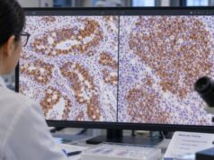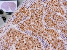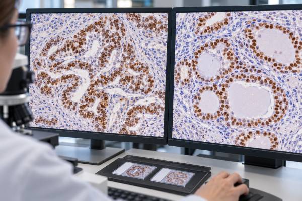
TTF-1 immunohistochemistry (IHC) is a nuclear tissue stain used mainly to support pulmonary or thyroid differentiation in a tumor. TTF-1, also called NKX2-1, is a transcription factor normally expressed in lung epithelial cells and thyroid follicular cells. In cancer diagnosis, nuclear TTF-1 staining is common in lung adenocarcinoma, many small-cell lung carcinomas and other pulmonary neuroendocrine tumors, and most differentiated thyroid carcinomas. The marker is especially useful when a metastatic tumor must be distinguished from a primary lung cancer or when a poorly differentiated tumor may be thyroid in origin. TTF-1 is highly useful but not perfectly specific: rare tumors outside the lung and thyroid can be positive, and some genuine lung adenocarcinomas are negative. Antibody clone and tumor subtype can also affect sensitivity. Pathologists therefore interpret TTF-1 with morphology and companion markers such as Napsin A, p40, PAX8, thyroglobulin, and organ-directed IHC panels.
- TTF-1 positivity is defined by nuclear staining in the tumor cells.
- Lung adenocarcinoma and thyroid follicular-derived carcinomas are common TTF-1-positive tumors.
- Many pulmonary small-cell and other neuroendocrine tumors can also express TTF-1.
- TTF-1 is not perfectly specific or sensitive, so a positive or negative result cannot establish tumor origin alone.
- Napsin A improves specificity for lung adenocarcinoma when both markers are positive, while thyroglobulin and PAX8 help assess thyroid origin.
Table of Contents
- What the TTF-1 IHC test detects
- TTF-1 in lung adenocarcinoma
- TTF-1 in small-cell and other neuroendocrine lung tumors
- TTF-1 in thyroid tumors
- False-positive and false-negative TTF-1 results
- Using TTF-1 in an IHC panel
- What the TTF-1 result means for next steps
What the TTF-1 IHC test detects
TTF-1 IHC detects the NKX2-1 transcription factor in fixed tissue. A diagnostically meaningful result is nuclear staining. Cytoplasmic staining does not carry the same established interpretation and should not be treated as equivalent to true nuclear positivity.
The stain is often used when a carcinoma is found in the lung, brain, bone, liver, pleura, or another metastatic site and the primary origin is uncertain. In a lung mass, TTF-1 helps distinguish pulmonary adenocarcinoma from squamous carcinoma and from many metastases. In a neck or metastatic lesion, it can raise the possibility of thyroid origin.
TTF-1 IHC is a protein-expression test, not a blood test or a test for a specific NKX2-1 mutation. There is no serum reference range and no patient preparation beyond obtaining the tissue sample used for routine pathology.
TTF-1 in lung adenocarcinoma
Most conventional lung adenocarcinomas express TTF-1, making it one of the most useful lineage markers in thoracic pathology. Recent large tissue studies confirm high sensitivity, but they also show that specificity is improved when TTF-1 is combined with Napsin A.
A Napsin A IHC stain is particularly useful because coexpression of TTF-1 and Napsin A strongly favors pulmonary adenocarcinoma in the appropriate setting. Neither marker is absolute, and mucinous lung adenocarcinomas are a well-known group that may show reduced or absent TTF-1.
For poorly differentiated non-small-cell carcinoma, TTF-1 is often paired with p40. TTF-1 positivity supports adenocarcinoma lineage, while strong p40 supports squamous differentiation. This limited panel helps preserve tissue for molecular testing.
TTF-1 in small-cell and other neuroendocrine lung tumors
TTF-1 is not limited to lung adenocarcinoma. A substantial proportion of pulmonary small-cell carcinomas and large-cell neuroendocrine carcinomas express the marker, and some carcinoid tumors can be positive as well.
This creates a useful but imperfect tissue-of-origin clue in metastatic neuroendocrine carcinoma. A TTF-1-positive small-cell carcinoma is compatible with pulmonary origin, but extrapulmonary small-cell carcinomas can occasionally express TTF-1. Synaptophysin, chromogranin, INSM1, morphology, imaging, and the patient’s clinical presentation remain central.
When a neuroendocrine tumor is found outside the lung, TTF-1 is therefore best interpreted as one probabilistic marker rather than proof of a lung primary.
TTF-1 in thyroid tumors
Normal thyroid follicular cells express TTF-1, and many follicular-derived thyroid cancers—including papillary and follicular thyroid carcinomas—retain nuclear expression. Medullary thyroid carcinoma may also express TTF-1, although its lineage is different and additional markers are required.
TTF-1 alone cannot distinguish lung from thyroid origin because both can be strongly positive. PAX8 and thyroglobulin help resolve that problem. A PAX8 IHC stain supports thyroid, renal, or Müllerian lineage depending on context, while thyroglobulin strongly supports follicular-cell thyroid differentiation.
For a TTF-1-positive metastasis, the practical question is therefore which companion markers and clinical findings fit best: Napsin A with a lung mass, or PAX8/thyroglobulin with a thyroid lesion.
False-positive and false-negative TTF-1 results
TTF-1 is highly useful but not absolutely specific. Large tumor surveys have documented positivity in a small fraction of gastrointestinal and other carcinomas, as well as certain non-epithelial tumors. This matters when a metastatic cancer has an unusual morphology or an enteric immunophenotype.
False-negative interpretation is also possible. A true lung adenocarcinoma may lack TTF-1, particularly in mucinous or poorly differentiated subsets. Technical factors, small samples, treatment effect, and antibody clone can change sensitivity. Some antibody clones are more sensitive but may stain a broader range of nonpulmonary tumors.
Pathologists therefore avoid the shortcut “TTF-1 positive equals lung” or “TTF-1 negative excludes lung.” The result shifts probability and must agree with the rest of the case.
Using TTF-1 in an IHC panel
For suspected lung adenocarcinoma, TTF-1 and Napsin A are common core markers. For squamous differentiation, p40 and CK5/6 may be used. For a metastatic lesion, the panel expands according to competing sites: PAX8 for thyroid/renal/Müllerian tumors, GATA3 for breast or urothelial carcinoma, CDX2 and SATB2 for intestinal origin, and other site-specific markers as needed.
When thyroid origin is considered, thyroglobulin is especially informative. When neuroendocrine differentiation is present, synaptophysin, chromogranin, and INSM1 are assessed independently of TTF-1.
This targeted strategy reflects the logic of a tumor-origin immunohistochemistry panel: the best interpretation comes from a coherent set of positive and negative findings rather than a single marker.
What the TTF-1 result means for next steps
TTF-1 primarily helps establish lineage and tumor origin. Once a lung adenocarcinoma diagnosis is supported, molecular testing for actionable alterations and PD-L1 assessment may guide systemic therapy. TTF-1 staining itself is not a substitute for EGFR, ALK, ROS1, KRAS, BRAF, MET, RET, NTRK, or PD-L1 testing.
For thyroid cancer, the final histologic diagnosis, stage, thyroglobulin biology, molecular profile, and imaging determine management. For metastatic cancer of uncertain origin, TTF-1 can guide focused imaging or review of the lung and thyroid.
If a report lists TTF-1, ask whether the staining was nuclear, which companion markers were tested, and whether the overall pattern supports lung, thyroid, or another source. The integrated diagnosis is more clinically meaningful than the isolated stain result.
References
- TTF-1 Expression in Lung Adenocarcinoma: Clinicopathologic, Genomic, and Immunophenotypic Correlates and Outcomes to Immunotherapy-Based Treatments and KRASG12C Inhibitors 2026 (Study)
- Immunohistochemistry as a Cornerstone in Lung Cancer Diagnosis: Subtyping Based on the 2021 World Health Organization Classification and Its Practical Application 2026 (Study)
- TTF-1 is a highly sensitive but not fully specific marker for pulmonary and thyroidal cancer: a tissue microarray study evaluating more than 17,000 tumors from 152 different tumor entities 2024 (Study)
- Value of thyroid transcription factor-1 immunostaining in tumor diagnosis: a review and update 2012 (Review)
- Immunohistochemical staining for thyroid transcription factor-1: a helpful aid in discerning primary site of tumor origin in patients with brain metastases 2002 (Study)
Disclaimer
TTF-1 IHC is an ancillary pathology test and cannot establish lung or thyroid origin by itself. Positive and negative results must be interpreted with morphology, antibody performance, companion markers, imaging, and clinical history. Treatment decisions require the final diagnosis and other predictive tests.