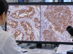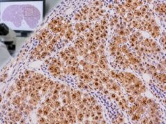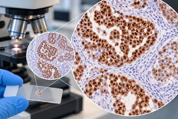
SALL4 immunohistochemistry (IHC) is a tissue stain used mainly to support germ cell differentiation in a tumor. SALL4 is a nuclear transcription factor associated with embryonic and germ cell development, so a strong nuclear stain can be a valuable clue when a pathologist is evaluating a possible seminoma or dysgerminoma, embryonal carcinoma, yolk sac tumor, or a mixed germ cell tumor. It is especially useful in small biopsies and metastatic tumors where the site of origin or tumor subtype is not obvious from routine microscopy alone. A positive SALL4 result does not, by itself, prove that a tumor is a germ cell tumor. Several non-germ cell cancers can express SALL4, and mature teratoma or trophoblastic components may show little or no staining. For that reason, the result is interpreted with the tumor’s microscopic appearance, clinical setting, serum markers, and a focused panel of complementary IHC stains.
- SALL4-positive staining is usually nuclear and supports germ cell differentiation when the morphology and other markers fit.
- SALL4 is broad rather than subtype-specific: it can label seminoma/dysgerminoma, embryonal carcinoma, and yolk sac tumor.
- A positive SALL4 stain is not cancer-specific or germ-cell-specific; selected gastric, ovarian, urothelial, hepatic, renal, and other tumors may also be positive.
- There is no normal “range” or blood level for SALL4 IHC. The pathologist assesses where staining occurs, how strong it is, and how much tumor is stained.
- Interpretation usually requires a panel, commonly including OCT4, PLAP, CD117, CD30, glypican-3, AFP, or other markers chosen for the differential diagnosis.
Table of Contents
- What the SALL4 IHC test detects
- What positive SALL4 staining means
- SALL4 patterns in major germ cell tumors
- When SALL4 is positive outside germ cell tumors
- How pathologists use SALL4 in an IHC panel
- What a negative or equivocal SALL4 result can mean
- What usually happens after SALL4 testing
What the SALL4 IHC test detects
SALL4 IHC detects SALL4 protein in cells preserved in a tissue sample. The stain is performed on a biopsy, surgical specimen, or sometimes a cell-block preparation from cytology. Pathologists look for nuclear staining in tumor cells. This is different from a serum tumor-marker test: SALL4 IHC does not produce a concentration, reference interval, or single numeric cutoff that applies to every laboratory.
SALL4 is particularly useful because many malignant germ cell tumors retain a developmental program that includes this transcription factor. In the right morphologic setting, diffuse nuclear positivity can strongly support germ cell lineage. The marker is often considered alongside an OCT4 IHC test for seminoma and embryonal carcinoma and other stains that narrow the exact germ cell subtype.
The stain is an ancillary diagnostic tool. The diagnosis still begins with hematoxylin-and-eosin microscopy, the patient’s age and tumor location, imaging findings, and—when relevant—serum AFP, beta-hCG, and LDH.
What positive SALL4 staining means
A meaningful positive result is typically described as nuclear staining in the neoplastic cells. Strong, diffuse labeling is common in several primitive germ cell tumors, including seminoma/dysgerminoma, embryonal carcinoma, and yolk sac tumor. The pattern helps confirm lineage, but it does not distinguish all of these entities from one another.
The next question is therefore not simply “Is SALL4 positive?” but “What else is positive or negative?” Seminoma and dysgerminoma commonly pair SALL4 with OCT4 and may also express PLAP and CD117. Embryonal carcinoma usually expresses OCT4 and often CD30, with SOX2 providing another useful clue. Yolk sac tumor is usually SALL4 positive but OCT4 negative and may express glypican-3 and AFP. An IHC panel that includes PLAP can be helpful, but PLAP is less sensitive than SALL4 in some germ cell tumor settings.
Pathologists also note whether staining is diffuse or focal and whether internal control tissues behave as expected. A focal result in a poorly differentiated tumor deserves more cautious interpretation than a classic diffuse pattern in a morphologically compatible tumor.
SALL4 patterns in major germ cell tumors
SALL4 has different practical value across germ cell tumor components. The broad pattern is more important than any single percentage threshold.
| Tumor or component | Typical SALL4 role | Useful companion markers |
|---|---|---|
| Seminoma / dysgerminoma | Usually strong nuclear positivity | OCT4, PLAP, CD117 |
| Embryonal carcinoma | Usually strong nuclear positivity | OCT4, CD30, SOX2 |
| Yolk sac tumor | Usually strong nuclear positivity | Glypican-3, AFP; OCT4 usually negative |
| Teratoma | Variable; mature components may be negative | Marker selection depends on the tissue component |
| Choriocarcinoma / trophoblastic tumor | May be negative or less consistently positive | beta-hCG and other trophoblastic markers |
Mixed germ cell tumors are a key reason a panel matters. A small aggressive component can alter classification and treatment even when another component dominates the specimen. Pathologists therefore target the stain to morphologically suspicious areas rather than relying on a single global label.
When SALL4 is positive outside germ cell tumors
SALL4 is sensitive for many nonteratomatous germ cell tumors, but it is not absolutely specific. Positivity has been documented in subsets of gastric adenocarcinoma, high-grade urothelial carcinoma, serous carcinoma of the ovary, hepatocellular carcinoma, Wilms tumor and other renal tumors, and occasional additional malignancies. Some non-germ cell cancers can stain extensively enough to mimic a germ cell tumor immunophenotype.
This limitation is most important when the clinical location is unusual—for example, a SALL4-positive mass in the liver, stomach, bladder, or ovary of an older adult. The pathologist asks whether the microscopic architecture and the rest of the IHC panel truly support germ cell differentiation. Lack of concordant germ cell markers can be a warning that SALL4 reflects an alternative tumor phenotype rather than germ cell origin.
The reverse problem also exists. Treatment, sampling, and tumor heterogeneity can reduce marker expression. A negative SALL4 result therefore cannot exclude every germ cell component when morphology or clinical data remain strongly suspicious.
How pathologists use SALL4 in an IHC panel
A focused panel is chosen from the differential diagnosis, not from SALL4 alone. For a testicular or mediastinal mass in a young man, the main question may be seminoma versus embryonal carcinoma versus yolk sac tumor. In an ovarian mass in a young patient, dysgerminoma and yolk sac tumor may be weighed against sex cord-stromal and epithelial neoplasms. In a metastatic poorly differentiated carcinoma, the panel may first need to establish whether germ cell lineage is plausible at all.
Useful pairings include SALL4 with OCT4 and CD30 for embryonal carcinoma, or with glypican-3 and AFP for yolk sac tumor. SOX2 can support embryonal carcinoma in the right setting. Cytokeratins are also informative because embryonal carcinoma and yolk sac tumor may be keratin positive, while seminoma often shows a different pattern.
A pathologist may also add organ-lineage markers to exclude mimics. This is the same principle used in a broader tumor immunohistochemistry panel: multiple concordant findings are more reliable than one stain interpreted in isolation.
What a negative or equivocal SALL4 result can mean
A negative result means that convincing nuclear SALL4 staining was not identified in the tumor cells under the laboratory’s validated conditions. It may support a non-germ cell diagnosis, but the meaning depends on what tumor is being considered. Mature teratoma and some trophoblastic components are not expected to show the same strong pattern as primitive germ cell tumors.
An equivocal result can arise from weak staining, very focal staining, crush artifact, necrosis, scant viable tumor, or uncertainty about whether staining is truly in tumor nuclei. In these situations, the pathologist may repeat the stain, test a better tissue block, or rely more heavily on a complementary marker panel. Internal positive controls are important: absence of expected control staining raises a technical concern.
If a pathology report says only “SALL4 positive,” the diagnosis and comment section usually provide the context that matters most. The result should be read together with the final tumor type, other stains, serum tumor markers, imaging, and the anatomic site.
What usually happens after SALL4 testing
SALL4 testing is usually part of diagnosis rather than a treatment-monitoring assay. Once the tissue type and germ cell components are classified, clinical management depends on the tumor site, stage, serum marker profile, patient age, and whether disease is localized or metastatic. Additional imaging, serum AFP and beta-hCG testing, and multidisciplinary review may follow.
A positive stain does not mean that SALL4 itself is a treatment target, and a negative stain does not automatically mean that no germ cell tumor is present. The practical goal is to give the oncology team the most accurate histologic classification possible.
Patients reviewing a report can ask which cells were SALL4 positive, whether staining was diffuse or focal, which other germ cell markers were tested, and what diagnosis the full panel supports. Those questions are more useful than trying to interpret SALL4 as a stand-alone “positive cancer test.”
References
- Pathogenesis and pathobiology of testicular germ cell tumours: a view from a developmental biological perspective with guidelines for pathological diagnostics. 2024 (Review)
- Higher expression of SALL4-A isoform is correlated with worse outcomes and progression of the disease in subtype of testicular germ cell tumours. 2024 (Study)
- Testicular Germ Cell Tumors: Serological and Immunohistochemical Diagnosis 2021 (Review)
- SALL4 expression in germ cell and non-germ cell tumors: a systematic immunohistochemical study of 3215 cases 2014 (Study)
- SALL4 is a novel diagnostic marker for testicular germ cell tumors 2009 (Study)
Disclaimer
SALL4 IHC is a pathology test interpreted in the context of tissue morphology and other clinical and laboratory findings. A positive or negative stain should not be used by itself to diagnose, exclude, stage, or treat a germ cell tumor. Discuss the complete pathology report with the treating clinician or pathologist if the result is unclear.