
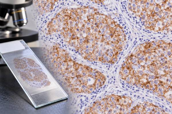
The PLAP IHC test detects placental alkaline phosphatase-related protein in tissue and is used mainly as a supporting marker for germ cell tumors. PLAP has a long history in surgical pathology, particularly in the evaluation of seminoma and germ cell neoplasia in situ. A positive stain can support germ cell differentiation, but it is not specific enough to identify a tumor by itself. Modern germ cell tumor panels often pair PLAP with more sensitive or more lineage-restricted markers such as SALL4, OCT4, SOX17, CD117, CD30, glypican-3, and AFP. The exact value of PLAP depends on the microscopic pattern, the patient’s age and tumor location, and which tumor cells stain. Strong membranous and cytoplasmic staining in a tumor that looks like seminoma is supportive, while focal staining in an unrelated carcinoma may be nonspecific. The final diagnosis therefore comes from the entire immunohistochemical and morphologic picture.
- PLAP positivity supports germ cell tumor differentiation but is not completely specific for germ cell tumors.
- Seminoma is commonly PLAP positive, often with membranous and cytoplasmic staining.
- Embryonal carcinoma can also express PLAP, so PLAP alone does not separate seminoma from embryonal carcinoma.
- SALL4 and OCT4 are often more informative companion markers in modern germ cell tumor workups.
- PLAP IHC requires no special patient preparation because it is performed on fixed tissue from a biopsy or operation.
Table of Contents
- What the PLAP IHC Test Detects
- What Positive PLAP Staining Means
- PLAP in Seminoma and Other Germ Cell Tumors
- How PLAP IHC Is Performed and Reported
- How PLAP Fits Into Marker Panels
- Limitations and False-Positive Patterns
- What Happens After a PLAP Result
What the PLAP IHC Test Detects
PLAP stands for placental alkaline phosphatase. The name reflects the enzyme’s strong physiologic association with placenta, but PLAP or closely related alkaline phosphatase activity can also be expressed in germ cells and a range of tumors. Immunohistochemistry uses an antibody to visualize this protein in a tissue section.
In diagnostic pathology, PLAP is best known as a germ cell tumor marker. It is commonly expressed in seminoma and can be present in germ cell neoplasia in situ, the precursor lesion of most postpubertal-type testicular germ cell tumors. Embryonal carcinoma can also be positive. This overlap is useful for confirming germ cell differentiation but means that PLAP cannot determine the exact subtype on its own.
The test is fundamentally different from a blood alkaline phosphatase measurement. A routine serum alkaline phosphatase level reflects enzyme activity from tissues such as liver and bone and does not substitute for PLAP immunostaining. PLAP IHC is a microscopic tissue test designed to show where the protein is located within a lesion.
Modern practice has reduced reliance on PLAP as a stand-alone “pan-germ-cell” marker because SALL4 IHC is more broadly sensitive for many germ cell tumors and nuclear markers are often easier to interpret. Still, PLAP remains useful when the morphology and panel are concordant, particularly in seminoma-pattern lesions.
The distinction between a supportive marker and a definitive marker matters. PLAP can be expressed in some non-germ-cell tumors, including subsets of gynecologic, gastrointestinal, and other carcinomas. Therefore, a pathologist who sees PLAP positivity in an unusual site asks whether the full immunophenotype and morphology truly support germ cell origin.
What Positive PLAP Staining Means
A positive PLAP result means the cells being assessed show immunoreactivity for PLAP under the laboratory’s validated staining conditions. In seminoma, staining is often diffuse and can involve the cell membrane and cytoplasm. This pattern can be striking, but the pathologist still checks whether the positive cells are actually the tumor cells and whether appropriate controls worked.
The strength and distribution of staining matter. Diffuse strong staining in a classic seminoma is more persuasive than a tiny focus of weak reactivity in a carcinoma with completely different morphology. Reports may describe staining as diffuse, patchy, focal, strong, moderate, weak, membranous, or cytoplasmic. There is no universal treatment threshold comparable with a hormone receptor percentage score in breast cancer.
A positive PLAP stain does not mean that a tumor is definitely seminoma. Embryonal carcinoma can be positive, and PLAP expression has been documented in non-germ-cell tumors. The result therefore answers a narrower question: does the tissue show a protein-expression pattern that can support germ cell differentiation in the right context?
A negative PLAP stain also does not exclude a germ cell tumor. Yolk sac tumors may be PLAP negative or only variably positive, and treated or poorly preserved tumors can lose staining. SALL4, glypican-3, AFP, OCT4, and other markers may be more useful depending on the suspected subtype.
For readers looking at a pathology report, the most important phrase is usually the final integrated diagnosis, not the isolated line “PLAP positive.” If the report says PLAP is positive but OCT4, CD117, CD30, or other stains are listed too, those results are meant to be interpreted together.
PLAP in Seminoma and Other Germ Cell Tumors
Seminoma is the classic setting in which PLAP has been used. Most seminomas show PLAP reactivity, often accompanied by OCT4, SALL4, SOX17, and CD117 expression. This combination fits the biology of a tumor that retains features of early germ-cell differentiation.
Embryonal carcinoma can also express PLAP, but its companion profile is usually different. Embryonal carcinoma is typically OCT4 and SALL4 positive, commonly CD30 positive, and often SOX2 positive. Seminoma more often shows CD117 and SOX17. These differences help separate two tumors that can both be PLAP positive.
Yolk sac tumor is a useful contrast. It is generally better highlighted by SALL4, glypican-3, and AFP than by PLAP. Choriocarcinoma is diagnosed with trophoblastic morphology and markers such as beta-hCG and other trophoblastic proteins. Mature teratoma contains differentiated somatic tissues and does not show a uniform PLAP-positive germ cell phenotype.
This marker logic becomes especially important in mixed germ cell tumors. A single testicular tumor can contain seminoma, embryonal carcinoma, yolk sac tumor, choriocarcinoma, and teratoma in different proportions. The pathologist may apply stains to suspicious areas to make sure a clinically important component is not overlooked. Finding an embryonal carcinoma or yolk sac component changes a lesion from pure seminoma to a nonseminomatous germ cell tumor category.
PLAP can also help identify germ cell neoplasia in situ in seminiferous tubules adjacent to an invasive tumor, although OCT4 and other markers are now widely used for this purpose. The precursor lesion is clinically important because it supports the developmental pathway of most postpubertal-type testicular germ cell tumors.
At extragonadal sites, such as the mediastinum or retroperitoneum, PLAP positivity can support a germ cell tumor but should trigger a full differential diagnosis. The pathologist must distinguish a true primary extragonadal germ cell tumor from metastatic testicular disease and from non-germ-cell tumors that can occasionally express PLAP.
How PLAP IHC Is Performed and Reported
PLAP immunohistochemistry is usually performed on formalin-fixed, paraffin-embedded tissue. The sample may come from an orchiectomy, lymph node, core biopsy, mediastinal mass, retroperitoneal mass, or another suspected tumor site. Thin sections are placed on slides, exposed to a PLAP antibody, and processed with a detection system that creates visible staining where the antigen is present.
The patient does not need to fast, stop medication, or change activity for the stain itself. The practical factors are tissue quality and laboratory technique. Adequate fixation helps preserve proteins. Necrotic tissue, crush artifact, heavy cautery, or prior treatment may reduce interpretability. Laboratories therefore use controls and validated protocols rather than assuming every weak stain reflects tumor biology.
A report may simply list PLAP as positive or negative, or it may describe intensity and distribution. The most useful interpretation usually appears in a diagnostic comment, such as a statement that the immunophenotype supports seminoma or supports germ cell tumor differentiation. PLAP is not generally assigned a standardized numeric score with direct treatment cutoffs.
Pathologists also compare staining with hematoxylin-and-eosin morphology. A stain can only help if the observer knows which cells are supposed to be positive. In a lymph node, for example, a few PLAP-reactive tumor cells may be diagnostically meaningful, whereas nonspecific background or staining in non-neoplastic structures should not be overcalled.
When tissue is scarce, an efficient panel is important. Starting with a targeted set of high-yield markers helps preserve tissue for additional IHC or molecular testing. A general IHC panel for tumor origin may be used first when the lineage is uncertain, followed by specific germ cell stains once the differential narrows.
How PLAP Fits Into Marker Panels
PLAP is strongest when it contributes to a pattern, not when it acts alone. Germ cell tumor panels are chosen according to the clinical setting and morphology, but several marker combinations are common.
For a suspected seminoma, useful findings include OCT4 positivity, SALL4 positivity, SOX17 positivity, CD117 positivity, and PLAP positivity, with CD30 usually negative. For embryonal carcinoma, OCT4, SALL4, CD30, and SOX2 are more characteristic. For yolk sac tumor, SALL4, glypican-3, and AFP are key markers, while OCT4 is generally negative. These profiles are not mathematical formulas; they are diagnostic patterns interpreted by a pathologist.
Serum tests add information that IHC cannot provide. AFP is particularly important. A patient with a testicular tumor and clearly elevated AFP should be managed as having a nonseminomatous component even if the sampled tissue resembles seminoma, because pure seminoma does not produce AFP. Beta-hCG can be elevated in some seminomas and in choriocarcinoma-containing tumors. LDH is less specific but can reflect tumor burden.
The hCG tumor marker test and AFP therefore complement tissue markers rather than compete with them. Tissue stains classify what the cells are; serum markers can help with staging, risk grouping, and follow-up.
PLAP can also be useful when a poorly differentiated metastatic tumor is being considered as germ cell origin. However, because some carcinomas are PLAP positive, broad epithelial markers, organ-specific markers, and germ cell-specific nuclear stains may be needed. A convincing germ cell diagnosis usually rests on concordant morphology plus several markers.
Limitations and False-Positive Patterns
The most important limitation is specificity. PLAP is associated with germ cell tumors but is not exclusive to them. Large tissue studies have shown moderate or strong PLAP expression in a range of non-germ-cell tumors, including subsets of gynecologic and gastrointestinal carcinomas. This means PLAP positivity in an unexpected cancer should not automatically redirect the diagnosis to germ cell tumor.
Sensitivity is also imperfect across germ cell tumor subtypes. PLAP performs well in seminoma and often in embryonal carcinoma, but it is not the best marker for every germ cell neoplasm. SALL4 is more broadly sensitive, while subtype markers provide more discriminating information.
Technical issues can cause false reassurance or false concern. Overstaining can create background that looks positive. Poor fixation can weaken true staining. Treated metastases may lose marker expression. Different antibody clones and laboratory platforms can also yield somewhat different results, so interpretation should be anchored in validated local controls.
Another pitfall is sampling. Mixed tumors can contain PLAP-positive and PLAP-negative components. A needle biopsy that captures only one region cannot prove that no other component exists elsewhere. This is one reason the final diagnosis may be revised or expanded after resection.
PLAP should also not be confused with serum placental alkaline phosphatase testing or routine serum alkaline phosphatase. Tissue IHC is a localization test. The presence of staining does not directly indicate how much enzyme is circulating in blood, nor does it provide a stage or tumor burden measurement.
The practical rule is simple: the more unexpected the PLAP result, the more important it is to verify it with morphology and a complementary panel.
What Happens After a PLAP Result
After PLAP staining, the pathologist integrates the result with all other findings and issues a final diagnosis or recommends additional studies. If the profile supports seminoma, the report may also document tumor size, local extension, lymphovascular invasion, and germ cell neoplasia in situ when applicable. If nonseminomatous components are present, they are identified because they affect classification and clinical management.
Clinicians then combine the pathology with imaging and serum markers. The AFP blood test, beta-hCG, and LDH are commonly part of the testicular germ cell tumor evaluation. A mismatch between tissue and serum findings may lead to pathology review, repeat marker testing, or closer sampling of the specimen.
If a PLAP-positive tumor occurs outside the testis, clinicians may evaluate the testes and other likely primary sites rather than assume the mass is a primary extragonadal germ cell tumor. In mediastinal or retroperitoneal disease, age, imaging, serum markers, and the complete IHC panel are particularly important.
Patients reviewing the report can ask what tumor type the full panel supports, whether OCT4 or SALL4 was positive, whether a nonseminomatous component was found, and whether the serum markers fit the tissue findings. The key question is not “How positive was PLAP?” but “What diagnosis does the entire pattern support?”
PLAP remains a useful marker because it can reinforce a germ cell diagnosis quickly and visually. Its greatest value comes from being used with modern nuclear and subtype-specific markers, not from being treated as a stand-alone proof of seminoma.
Laboratory quality is especially important when PLAP staining is weak or unexpected. The pathologist confirms that the assay controls perform appropriately and that staining is present in the cell compartment expected for the antibody. Germ cell tumors may show membranous and cytoplasmic labeling, but intensity can vary within a lesion. A tiny biopsy, extensive necrosis, crush artifact, or treatment-related change can make a true positive population look much less impressive than it would in a well-preserved resection. In those settings, a second germ cell marker can be more informative than trying to force a borderline PLAP result into a positive-or-negative category.
Sampling also matters because mixed germ cell tumors contain biologically different components. Seminoma, embryonal carcinoma, yolk sac tumor, choriocarcinoma, and teratoma do not share one uniform PLAP pattern. A core biopsy that samples only one component may therefore produce an immunophenotype that differs from the final orchiectomy or from a metastatic deposit. Pathologists reconcile these differences by identifying each component morphologically and applying subtype markers where needed.
After chemotherapy, PLAP can become even less useful as a stand-alone marker. Viable residual tumor may be sparse, antigen expression may change, and mature teratoma or somatic-type transformation may dominate the specimen. The post-treatment diagnosis is based on viable morphology and a panel chosen for the residual tissue rather than on whether the original tumor was PLAP positive.
For patients, this means that a change in PLAP staining between specimens is not automatically evidence that the diagnosis was wrong. Different sampled components, treatment effect, and technical factors can all change the visible pattern. The final report should explain which viable tumor component is present now and which markers support that interpretation.
References
- Pattern of placental alkaline phosphatase (PLAP) expression in human tumors: a tissue microarray study on 12,381 tumors 2021
- Pathogenesis and pathobiology of testicular germ cell tumours: a view from a developmental biological perspective with guidelines for pathological diagnostics 2024 (Review)
- Molecular pathology of testicular germ cell tumours: an update for practicing pathologists 2025 (Review)
- An introduction to the WHO 5th edition 2022 classification of testicular tumours 2022 (Review)
- International Society of Urological Pathology (ISUP) Consensus Conference on Precursor Lesions: Working Group 4 Precursor Lesions of Testis Results of a Premeeting Survey 2026
Disclaimer
PLAP IHC is an ancillary pathology test and should be interpreted with tumor morphology, other immunostains, serum tumor markers, and clinical findings. A PLAP-positive result alone does not prove seminoma or any other specific cancer. Discuss the complete pathology diagnosis with the treating clinician or pathologist.





