
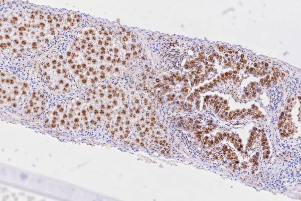
The OCT4 IHC test is a tissue stain that helps pathologists identify certain germ cell tumors, especially seminoma and embryonal carcinoma. OCT4, also called OCT3/4 or POU5F1, is a nuclear transcription factor involved in maintaining pluripotency—the ability of an early cell to develop into different cell types. In routine pathology, strong nuclear OCT4 staining can be a powerful clue that a tumor belongs to the seminoma/germinoma or embryonal carcinoma family. It is also useful for detecting germ cell neoplasia in situ in the testis. A positive result is not interpreted alone, however. The pathologist combines the staining pattern with the tumor’s microscopic appearance, patient age, tumor location, serum markers, and a panel of other immunohistochemical stains. This is especially important because different germ cell tumor components can occur together and may require different treatment or staging considerations.
- OCT4 positivity usually means tumor-cell nuclei are staining, not the cytoplasm.
- Strong, diffuse OCT4 staining supports seminoma or embryonal carcinoma in the right clinical and microscopic setting.
- OCT4 is generally negative in yolk sac tumor, choriocarcinoma, and mature teratoma, so a negative result can help narrow the differential diagnosis.
- OCT4 does not by itself distinguish seminoma from embryonal carcinoma; companion markers such as CD117, SOX17, SOX2, and CD30 are often needed.
- No fasting or special patient preparation is required because OCT4 IHC is performed on a biopsy or surgical tissue specimen.
Table of Contents
- What the OCT4 IHC Test Detects
- What Positive OCT4 Staining Means
- OCT4 in Seminoma and Embryonal Carcinoma
- How OCT4 IHC Is Performed and Reported
- How OCT4 Fits Into Germ Cell Tumor Marker Panels
- Limitations and Diagnostic Pitfalls
- What Happens After an OCT4 Result
What the OCT4 IHC Test Detects
OCT4 immunohistochemistry detects OCT4 protein in cells within a tissue sample. OCT4 is encoded by the POU5F1 gene and acts as a transcription factor that helps maintain the undifferentiated state of embryonic and germ cells. The diagnostically useful signal is nuclear staining. When a pathologist sees appropriately localized nuclear staining in a tumor with compatible morphology, OCT4 can provide strong evidence of a specific germ cell differentiation pattern.
The test is most useful in postpubertal-type germ cell tumors associated with germ cell neoplasia in situ. In the testis, OCT4 commonly labels seminoma, embryonal carcinoma, and germ cell neoplasia in situ. Similar principles apply to related germinomas and embryonal carcinomas at extragonadal sites, such as the mediastinum or central nervous system, although the entire clinical context matters.
OCT4 is not a general “cancer positive” stain. Many cancers are OCT4 negative. Its value comes from the restricted pattern of expression expected in routine surgical pathology. That is why an OCT4 result can be especially helpful when a metastatic tumor is poorly differentiated and the pathologist is deciding whether germ cell origin belongs in the differential diagnosis.
The marker also differs from broad germ cell markers. For example, SALL4 stains a wider range of germ cell tumors, including many yolk sac tumors, whereas OCT4 is concentrated in seminoma/germinoma and embryonal carcinoma. This difference makes the two stains complementary rather than interchangeable.
A practical point is that OCT4 IHC assesses tissue expression; it is not the same as a blood tumor marker. Serum alpha-fetoprotein and beta-hCG answer different questions about tumor components and disease activity. Pathologists and oncologists often integrate the tissue phenotype with serum markers because mixed germ cell tumors can contain more than one histologic component.
What Positive OCT4 Staining Means
A positive OCT4 IHC result means the antibody has produced convincing nuclear staining in the cells being evaluated. In a tumor with the expected appearance and location, strong diffuse nuclear staining supports a diagnosis within the OCT4-positive germ cell tumor spectrum. The two most important malignant tumor types are seminoma and embryonal carcinoma.
“Positive” is not simply a yes-or-no concept. Pathologists assess where the stain is located, which cells are positive, and how much of the tumor is stained. True diagnostic OCT4 expression is nuclear. Cytoplasmic haze, edge artifact, staining in crushed tissue, or nonspecific background should not be mistaken for true tumor-cell positivity. Internal or external controls also need to show that the stain worked as expected.
In classic seminoma, OCT4 is usually strong and widespread in tumor-cell nuclei. Embryonal carcinoma is also typically strongly nuclear positive. Because both can stain, OCT4 is best viewed as a lineage and differentiation marker rather than a single-marker separator between the two. The tumor architecture and cytologic features remain essential.
A negative OCT4 result makes classic seminoma or embryonal carcinoma less likely when the stain is technically adequate, but it does not exclude a germ cell tumor as a category. Yolk sac tumor, choriocarcinoma, and most mature teratoma components are generally OCT4 negative. A broad marker such as SALL4 or a lineage-specific panel may still support germ cell origin.
The result also should not be read as a direct measure of aggressiveness. A tumor with intense OCT4 staining is not necessarily “more positive” biologically or more dangerous than one with weaker staining. In clinical pathology, OCT4 is primarily a diagnostic marker. Stage, anatomic spread, serum tumor markers, histologic components, and treatment response are far more important for prognosis.
OCT4 in Seminoma and Embryonal Carcinoma
OCT4 is particularly useful because seminoma and embryonal carcinoma can overlap morphologically, especially in small biopsies, crushed specimens, or metastatic sites. Both are OCT4 positive, so the pathologist uses the stain as an entry point and then relies on additional features to separate them.
Seminoma usually consists of relatively uniform large cells with clear or pale cytoplasm, distinct cell borders, and prominent nucleoli. Fibrous septa with lymphocytes are common. Embryonal carcinoma often shows greater pleomorphism, more crowded growth, gland-like or papillary structures, brisk mitotic activity, and areas of necrosis. In limited tissue, those distinctions may be incomplete.
Companion stains sharpen the distinction. Seminoma commonly expresses CD117 and often shows a SOX17-positive, SOX2-negative pattern. Embryonal carcinoma usually expresses CD30 and SOX2 and is generally CD117 negative or only variably positive. SOX2 IHC can therefore be helpful when an OCT4-positive tumor needs further classification. PLAP may stain both tumor types and is less specific for separating them.
The distinction matters because a postpubertal testicular tumor containing embryonal carcinoma is classified within the nonseminomatous germ cell tumor group, even if seminoma is also present. Mixed germ cell tumors are common, and a small nonseminomatous component can alter classification and management. This is one reason pathologists sample resection specimens extensively and use immunostains strategically rather than assuming a tumor is uniform.
OCT4 also highlights germ cell neoplasia in situ, the precursor lesion for most postpubertal-type testicular germ cell tumors. Detecting this lesion in adjacent seminiferous tubules can support the relationship between an invasive tumor and the testis. However, interpretation requires care because age, developmental context, and the appearance of the involved tubules matter.
For metastatic disease, the same principle applies: an OCT4-positive poorly differentiated tumor may prompt a focused germ cell panel and correlation with serum AFP, beta-hCG, and LDH. The tissue result is one part of a diagnostic chain, not a substitute for complete classification.
How OCT4 IHC Is Performed and Reported
OCT4 IHC is usually performed on formalin-fixed, paraffin-embedded tissue from a core biopsy, excision, orchiectomy, lymph-node biopsy, or resection. The laboratory cuts a thin tissue section, applies an antibody directed against OCT4, and uses a detection system that creates a colored reaction where the target protein is present. A pathologist then interprets the stain under a microscope.
No special preparation is needed from the patient for the stain itself. The important variables occur before and inside the laboratory: prompt fixation, appropriate formalin exposure, adequate tissue, validated antibody conditions, and reliable controls. Poor fixation or very small samples can reduce staining quality. Decalcification of bone specimens can also alter antigenicity, so the laboratory may interpret results more cautiously in heavily treated tissue.
Reports vary by institution. A pathology report may state “OCT3/4 positive, nuclear,” “diffuse strong nuclear positivity,” or simply list OCT4 among the immunophenotypic findings. Unlike hormone receptor testing in breast cancer, there is no universal clinical percentage threshold that turns OCT4 into a graded treatment score. The pattern is interpreted diagnostically.
The report should be read together with the final diagnosis rather than as a standalone laboratory value. A line that says “OCT4 positive” does not automatically mean seminoma, because embryonal carcinoma is also positive. Likewise, an OCT4-negative tumor can still be a germ cell neoplasm of another subtype.
When tissue is limited, pathologists often choose stains that answer the highest-value questions while preserving material for molecular studies if needed. A broad tumor IHC panel may first establish lineage, followed by a smaller germ cell panel tailored to the morphology. This staged approach reduces unnecessary staining and helps avoid consuming a tiny biopsy.
If the specimen has undergone chemotherapy, the pathologist also considers treatment effect. Viable post-treatment tumor may show altered morphology or reduced antigen expression, and teratoma or somatic-type malignancy may become more prominent. The result should therefore be interpreted in the context of prior therapy.
How OCT4 Fits Into Germ Cell Tumor Marker Panels
OCT4 works best as part of a panel because germ cell tumors are biologically diverse and can be mixed. The panel depends on the suspected tumor, location, patient age, morphology, and serum markers. No single fixed panel is appropriate for every case.
A common diagnostic strategy starts with broad germ cell lineage markers and then adds subtype markers. SALL4 is highly sensitive across many nonteratomatous germ cell tumors. OCT4 narrows attention toward seminoma/germinoma or embryonal carcinoma. CD117 and SOX17 favor seminoma, while CD30 and SOX2 favor embryonal carcinoma. Glypican-3 and AFP support yolk sac differentiation. Beta-hCG and other trophoblastic markers may help identify choriocarcinoma or syncytiotrophoblastic cells.
This explains why a patient can have a germ cell tumor even when one marker is negative. For example, a yolk sac tumor is typically OCT4 negative but may be SALL4 and glypican-3 positive. In contrast, a tumor that is OCT4 positive and CD30 positive with compatible morphology strongly supports embryonal carcinoma. The PLAP IHC test may add supportive evidence but is generally less specific than newer nuclear markers.
Serum markers add another layer. An elevated AFP in a patient with a testicular mass indicates a nonseminomatous component, because pure seminoma does not produce clinically significant AFP. Beta-hCG can be elevated in some seminomas as well as nonseminomatous tumors, so it is less specific for histologic subtype. These blood tests do not replace tissue diagnosis, but discordance between serum and tissue findings can prompt re-review or additional sampling.
The panel also helps distinguish germ cell tumors from mimics. Metastatic carcinoma, lymphoma, melanoma, and sarcoma can all appear poorly differentiated. Pancytokeratin may be positive in embryonal carcinoma, which can make it look epithelial, while CD30 is not exclusive to germ cell tumors. A combination of morphology and multiple markers prevents overreliance on one stain.
In short, OCT4 is most useful when it answers a focused question within a coordinated panel: “Does this tumor show the pluripotent germ cell phenotype expected in seminoma or embryonal carcinoma?”
Limitations and Diagnostic Pitfalls
The main limitation of OCT4 is that positive staining does not distinguish seminoma from embryonal carcinoma. The stain is highly informative but not self-sufficient. A correct diagnosis still depends on morphology, companion markers, and the clinical setting.
Another pitfall is confusing OCT4 with similarly named proteins or transcripts. Diagnostic antibodies are intended to detect the clinically relevant OCT4 protein pattern, but assay performance depends on antibody clone, platform, antigen retrieval, and validation. The laboratory’s own controls and experience matter more than comparing staining intensity across different institutions.
Technical artifacts can also mislead. Poor fixation may cause unexpectedly weak staining. Crushed biopsies can make nuclear localization hard to evaluate. Necrotic tissue may be noninformative. If the expected controls fail, a negative result should not be trusted until the technical problem is resolved.
Biologic heterogeneity matters as well. Mixed germ cell tumors can contain OCT4-positive and OCT4-negative components side by side. A small biopsy may sample only one component. For example, a metastasis may contain embryonal carcinoma while the primary also contains yolk sac tumor or teratoma. Conversely, post-treatment specimens may be dominated by mature teratoma even when the pretreatment tumor had an OCT4-positive component.
OCT4 expression should also be interpreted cautiously outside the classic germ cell tumor setting. Rare non-germ-cell tumors may create diagnostic confusion through aberrant staining or antibody-related issues. A pathologist should never disregard strong contradictory morphology simply because one stain is positive.
The safest rule is panel-based interpretation. If OCT4, SALL4, morphology, clinical location, age, and serum markers point in the same direction, diagnostic confidence rises substantially. If they conflict, the answer may require repeat staining, additional markers, molecular testing, or a larger specimen.
What Happens After an OCT4 Result
The next step depends on the final pathology diagnosis rather than on OCT4 alone. If the stain supports seminoma, the pathology team confirms the overall immunophenotype and evaluates the specimen for other germ cell components, tumor size, local extension, lymphovascular invasion, and precursor lesions when relevant. If embryonal carcinoma is identified, the report documents that component because it places a testicular tumor in the nonseminomatous category.
Clinicians then integrate pathology with imaging and serum tumor markers. AFP, beta-hCG, and LDH are commonly used in testicular germ cell tumor staging and follow-up. The AFP blood test is particularly important because an elevated AFP level is incompatible with a truly pure seminoma and suggests a nonseminomatous element even if that element was not sampled histologically.
Patients should not try to interpret an OCT4 line in isolation from the diagnostic comment. Questions worth asking include: Which cells were OCT4 positive? Was the staining strong and nuclear? What diagnosis did the full marker panel support? Were mixed components found? Do the serum markers agree with the tissue diagnosis? Is more tissue or molecular testing needed?
If results are internally inconsistent, pathology review can be valuable, especially for rare extragonadal tumors or small biopsies. Germ cell tumor classification has major treatment implications and often benefits from expert genitourinary or pediatric pathology review when morphology and immunostains are unusual.
An OCT4-positive result is therefore best understood as a highly useful diagnostic clue. It can strongly support seminoma or embryonal carcinoma, but the final meaning comes from the complete pathology interpretation and the patient’s clinical workup.
A useful final distinction is between diagnostic identity and disease monitoring. OCT4 is usually assessed on a tissue specimen to classify the cells that are present at that moment. It is not ordinarily followed as a serial blood marker after treatment. Follow-up for germ cell tumors instead relies on the final histology, imaging, clinical examination, and serum markers when the tumor produces them. That difference explains why an OCT4 result may remain important in the pathology record even though the clinician never orders an “OCT4 level” later.
Pathology may also be reviewed again if a later specimen looks different from the original tumor. A patient can have a mixed germ cell tumor in which separate components show different marker profiles. Comparing the new tissue with the original slides helps determine whether the apparent change reflects sampling, treatment effect, or a genuinely different component. This is especially relevant when the new specimen contains only teratoma, somatic-type malignancy, or another OCT4-negative element after an earlier OCT4-positive tumor.
References
- Pathogenesis and pathobiology of testicular germ cell tumours: a view from a developmental biological perspective with guidelines for pathological diagnostics 2024 (Review)
- Molecular pathology of testicular germ cell tumours: an update for practicing pathologists 2025 (Review)
- An introduction to the WHO 5th edition 2022 classification of testicular tumours 2022 (Review)
- International Society of Urological Pathology (ISUP) Consensus Conference on Precursor Lesions: Working Group 4 Precursor Lesions of Testis Results of a Premeeting Survey 2026
- OCT4 staining in testicular tumors: a sensitive and specific marker for seminoma and embryonal carcinoma 2004
Disclaimer
OCT4 IHC is a pathology test interpreted in the context of tissue appearance, other stains, and clinical findings. A positive or negative OCT4 result alone cannot establish the complete diagnosis, stage, or treatment plan. Discuss the final pathology report with the treating clinician or pathologist if the significance of the result is unclear.





