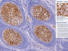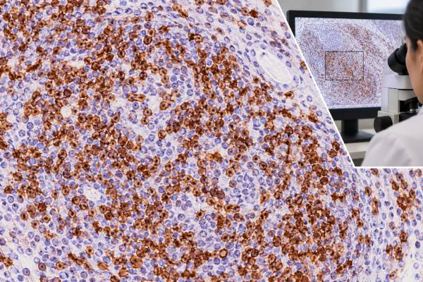
CD3 immunohistochemistry (IHC) is one of the most widely used tissue stains for identifying T-cell lineage. In a normal lymph node, CD3 highlights T lymphocytes in characteristic areas. In a tumor biopsy, pathologists use the stain to determine whether an abnormal lymphoid population is T-cell derived, to map reactive T cells around another cancer, and to help classify T-cell and NK/T-cell lymphomas. A positive CD3 stain does not by itself mean lymphoma because normal and reactive T cells are naturally CD3 positive.
The pattern also has important exceptions. Some T-cell lymphomas lose CD3 or show weaker expression, while anaplastic large cell lymphoma is often CD3 negative despite belonging to the T-cell lymphoma family. Extranodal NK/T-cell lymphoma may show cytoplasmic CD3-epsilon by tissue IHC even though NK cells lack surface CD3. For that reason, CD3 is interpreted with morphology, other T-cell markers, B-cell markers, cytotoxic markers, EBV studies, and clonality or molecular testing when needed.
- CD3 IHC is a broad T-lineage marker that normally stains many mature T lymphocytes.
- Positive staining is not proof of T-cell lymphoma because normal reactive T cells are CD3 positive in almost every lymphoid tissue sample.
- Loss or reduced CD3 can support an abnormal T-cell phenotype, but some genuine T-cell lymphomas are CD3 negative.
- Tissue CD3 positivity does not always mean surface T-cell receptor expression; NK/T-cell lymphoma can show cytoplasmic CD3-epsilon.
- There is no numeric normal range or universal positive cutoff; interpretation depends on which cells stain and how the pattern fits the full panel.
Table of Contents
- What CD3 IHC Detects
- Normal CD3 Staining Pattern
- CD3 in T-Cell Lymphomas
- Important CD3 Exceptions
- CD3 Versus B-Cell Markers
- How to Interpret a CD3 IHC Result
- What Happens After CD3 Testing
What CD3 IHC Detects
CD3 is a group of proteins associated with the T-cell receptor complex. Mature T cells use this complex to recognize antigens and transmit activation signals. In routine paraffin-section IHC, commonly used CD3 antibodies detect components such as CD3 epsilon and create predominantly membranous and sometimes cytoplasmic staining in T cells.
The test is performed on tissue, not as a serum tumor marker. It can be applied to lymph nodes, skin, gastrointestinal biopsies, bone marrow, tonsil, spleen, extranodal masses, and many other specimens.
CD3 helps answer several basic diagnostic questions:
- Is a lymphoid infiltrate predominantly T cells or B cells?
- Are the atypical cells themselves CD3 positive?
- Has an abnormal T-cell population lost a marker that normal T cells should express?
- Are there many reactive T cells surrounding a B-cell lymphoma, Hodgkin lymphoma, carcinoma, or other tumor?
- Does the phenotype support a mature T-cell neoplasm, precursor T-lymphoblastic neoplasm, or NK/T-cell process?
CD3 is therefore often one of the first stains in a tumor immunohistochemistry panel for suspected lymphoma. It provides lineage information quickly, but it rarely provides the final disease name.
An important concept is aberrancy. Normal T cells usually express a coordinated set of antigens, including CD2, CD3, CD5, and CD7, with either CD4 or CD8 in most mature populations. A lymphoma may lose one or more of these markers. The pattern of retained and lost antigens can be more informative than simple CD3 positivity.
Normal CD3 Staining Pattern
In a normal or reactive lymph node, CD3 highlights paracortical T-cell zones between and around B-cell follicles. Small T cells also appear within germinal centers and elsewhere in the node. This organized distribution is expected and often provides a useful internal control.
CD3 positivity is also common in inflammatory infiltrates outside lymph nodes. Skin, stomach, colon, lung, and many other organs may contain scattered or numerous reactive T cells. As a result, a tissue can be “full of CD3-positive cells” without containing a T-cell lymphoma.
Pathologists focus on whether the atypical population is positive. This can be straightforward when a tumor consists of sheets of large cells, but it is more difficult when malignant cells are sparse or mixed with many reactive lymphocytes.
For example, classic Hodgkin lymphoma contains relatively few Hodgkin/Reed-Sternberg tumor cells in a rich inflammatory background. Numerous surrounding T cells may be strongly CD3 positive while the actual tumor cells are CD3 negative. If the stain is interpreted without identifying the cell population, the result can be misleading.
Normal staining also helps judge technical quality. If expected small T cells stain but the suspicious large cells do not, tumor-cell negativity may be real. If no lymphocytes stain at all, the laboratory must consider fixation or technical failure before calling the tumor negative.
CD3 in T-Cell Lymphomas
Most mature T-cell lymphomas show at least some evidence of T-cell lineage, and CD3 is a central marker in that assessment. However, mature T-cell neoplasms are heterogeneous, and antigen loss is common.
Peripheral T-cell lymphoma, not otherwise specified, often expresses CD3 but may show weaker intensity or partial loss compared with background normal T cells. CD2, CD5, and CD7 are assessed at the same time. Loss of CD5 or CD7 is frequent, but no single loss pattern is unique to lymphoma.
T-follicular helper lymphomas, including angioimmunoblastic-type disease, are typically T-cell neoplasms and usually retain CD3. Classification depends on expression of T-follicular helper markers such as PD-1, ICOS, BCL6, CD10, or CXCL13 in the appropriate morphologic and clinical setting. A BCL6 IHC result can therefore have a very different meaning in T-follicular helper cells than it does in a B-cell follicular lymphoma.
Intestinal and hepatosplenic T-cell lymphomas also commonly express CD3, but their combinations of CD4, CD8, CD56, T-cell receptor markers, cytotoxic proteins, and clinical site help distinguish them. Cutaneous T-cell lymphomas may show epidermotropic CD3-positive lymphocytes, but reactive dermatitis can do the same. Distribution, atypia, antigen loss, and T-cell receptor clonality become important.
Precursor T-lymphoblastic lymphoma or leukemia is another setting in which CD3 is important. Cytoplasmic CD3 is considered strong evidence of T-lineage in precursor lymphoid neoplasms. These tumors are usually assessed with markers of immaturity such as TdT and CD1a as well as other T-cell antigens.
The key point is that CD3 answers “does this population show T-cell-associated differentiation?” more reliably than “which exact T-cell lymphoma is this?” The latter requires a broader integrated workup.
T-cell lymphoma diagnosis is often harder than simply finding many CD3-positive cells because reactive immune responses are also rich in T cells. Pathologists look for evidence that the suspicious population is abnormal: loss of other pan-T-cell antigens such as CD2, CD5, or CD7; an unusual CD4/CD8 pattern; cytologic atypia; destructive growth; aberrant expression of markers such as CD30; or a clonal T-cell receptor rearrangement. The exact combination depends on the suspected entity.
For lymphomas of T-follicular helper origin, CD3 confirms T-cell identity while markers such as PD-1, ICOS, BCL6, and sometimes CD10 establish the specialized helper phenotype. For cytotoxic T-cell lymphomas, granzyme B, TIA1, and perforin may be important. Thus, CD3 is usually the start of lineage assessment rather than the endpoint of classification.
Important CD3 Exceptions
Several exceptions prevent CD3 from being used as a simple yes-or-no definition of T-cell lymphoma.
Anaplastic large cell lymphoma
Anaplastic large cell lymphoma (ALCL) is a mature T-cell lymphoma characterized by strong CD30 expression, yet the tumor cells frequently lose CD3. Some cases lose several T-cell antigens and can appear “null” by routine IHC. A CD30 IHC test and, for systemic ALCL, an ALK IHC test are much more central to the classification.
Thus, CD3 negativity does not exclude ALCL when the morphology and CD30 phenotype are characteristic. T-cell receptor gene rearrangement or other molecular findings may help establish lineage in difficult cases.
Extranodal NK/T-cell lymphoma
Extranodal NK/T-cell lymphoma illustrates the difference between tissue IHC and surface immunophenotyping. Most cases are of NK-cell derivation. NK cells lack surface CD3 and a surface T-cell receptor, but they can contain cytoplasmic CD3 epsilon. Paraffin-section IHC may therefore be positive for CD3 even though flow cytometry would not show normal surface CD3.
The typical workup also includes CD56, cytotoxic proteins such as granzyme B or TIA-1, and in situ hybridization for Epstein-Barr virus–encoded RNA. EBV demonstration in tumor cells is critical to the diagnosis in the proper setting.
Reactive T cells in non-T-cell tumors
B-cell lymphomas, Hodgkin lymphoma, carcinomas, and melanomas can contain abundant CD3-positive reactive T cells. The stain may outline a rich immune microenvironment while the malignant cells themselves remain negative. This is one of the most common reasons that cell-by-cell correlation with morphology is essential.
Anaplastic large cell lymphoma is a notable exception because the tumor is of T-cell lineage in many cases yet can lose surface and tissue expression of several conventional T-cell markers, including CD3. The hallmark cells are strongly CD30 positive, and ALK status separates major biologic groups. A CD3-negative, CD30-strong large-cell tumor can therefore still be a T-cell lymphoma once B-cell lineage and other mimics are excluded.
Natural killer (NK) cells create a different nuance. They do not express surface CD3 as normal T cells do, but they contain cytoplasmic CD3 epsilon chains that can be detected by some tissue antibodies. Extranodal NK/T-cell lymphoma may therefore appear CD3 positive by IHC while lacking surface CD3 on flow cytometry. CD56, cytotoxic markers, and EBV testing help resolve that pattern.
CD3 Versus B-Cell Markers
A basic lymphoma panel often pairs CD3 with a B-cell marker such as CD20. The comparison provides a fast view of lineage and tissue architecture.
A CD20 IHC test labels most mature B cells and many B-cell lymphomas. In a reactive node, CD20 highlights follicles while CD3 highlights paracortical zones. In a B-cell lymphoma, tumor cells may be CD20 positive while background T cells are CD3 positive. In a T-cell lymphoma, the reverse pattern may be seen.
| Pattern | What it may suggest | Important caution |
|---|---|---|
| CD3-positive atypical cells, CD20-negative atypical cells | Supports T-cell lineage | Needs other T-cell markers and disease-specific studies |
| CD20-positive tumor cells with scattered CD3-positive small cells | Supports B-cell lymphoma with reactive T cells | CD20 can be lost in some treated or differentiated B-cell tumors |
| Strong CD3 in background, large tumor cells CD3 negative | Could be Hodgkin lymphoma, B-cell lymphoma, ALCL, or another tumor | Do not classify the tumor from background cells |
| Both CD3 and CD20 largely absent in tumor | Requires broader lineage workup | Antigen loss or non-lymphoid tumor is possible |
Other markers may be needed when CD20 or CD3 is lost. PAX5 and CD79a can support B-cell lineage, while CD2, CD5, CD7, T-cell receptor markers, and molecular clonality can support T-cell differentiation.
CD3 staining can also reveal antigen loss, which is often more informative than simple positivity. A suspicious T-cell population may retain CD3 but lose CD5 or CD7, or it may show patchy/weak CD3 compared with nearby normal T cells. Such aberrancy supports a neoplastic process when it occurs in the morphologically abnormal cells, although no single pattern is perfectly specific.
Skin and gastrointestinal biopsies provide common examples of why distribution matters. Numerous CD3-positive lymphocytes can be a normal or inflammatory finding at epithelial surfaces. A diagnosis of cutaneous or intestinal T-cell lymphoma requires the correct cytology, growth pattern, additional phenotype, and clinical setting rather than an increased number of CD3-positive cells alone.
How to Interpret a CD3 IHC Result
The most useful first question is: Which cells are CD3 positive? A pathology report may specify “neoplastic lymphocytes are positive for CD3” or “background small T cells are highlighted by CD3.” Those statements have very different meanings.
A positive tumor-cell result supports T-cell-associated differentiation, but the pathologist still looks for abnormal patterns. Uniform loss of CD5 or CD7, unusual CD4/CD8 expression, or a phenotype that is discordant with normal T-cell maturation can strengthen suspicion for lymphoma.
A negative tumor-cell result is also contextual. It argues against many T-cell lymphomas but does not rule out ALCL and a few other T-cell neoplasms with antigen loss. It can be particularly informative if numerous normal background T cells stain strongly, proving that the assay worked.
CD3 IHC is different from flow cytometry. IHC preserves tissue architecture and shows where cells are located, while flow cytometry measures antigens on individual suspended cells and can distinguish surface from cytoplasmic expression with appropriate methods. The two techniques often complement one another.
Likewise, T-cell receptor gene rearrangement testing answers a different question: clonality. A clonal rearrangement can support a T-cell neoplasm, but reactive clonal expansions can occur and some tumors may yield technically limited results. Neither CD3 IHC nor clonality should be interpreted in isolation.
The distribution of CD3-positive cells can be diagnostically informative even when those cells are reactive. Small T cells commonly surround or rosette around tumor cells in certain B-cell lymphomas, and a T-cell-rich background can obscure a sparse malignant B-cell population. Comparing CD3 and CD20 on adjacent sections often reveals whether the large atypical cells belong to the T-cell compartment or sit within it as a separate population.
When a T-cell neoplasm is suspected, clonality studies can support the diagnosis but are not interpreted in isolation. Reactive immune responses can occasionally produce restricted clones, and some true T-cell lymphomas yield negative or technically limited clonality assays. The final diagnosis still depends on concordance among morphology, immunophenotype, clinical site, EBV status when relevant, and molecular findings.
Clinical site narrows the interpretation further. A CD3-positive neoplasm in skin, intestine, liver/spleen, or lymph node enters different diagnostic pathways because current T-cell lymphoma entities are partly defined by anatomic distribution and clinical behavior. The same immunophenotype can therefore carry different weight in different organs.
What Happens After CD3 Testing
If CD3 identifies an atypical T-cell population, the next studies are chosen according to morphology and clinical site. They may include CD2, CD5, CD7, CD4, CD8, CD30, ALK, cytotoxic markers, T-follicular helper markers, CD56, EBV testing, Ki-67, and T-cell receptor studies.
A Ki-67 IHC test may help show proliferative activity, but a high index does not establish lineage. Similarly, loss of a pan–T-cell marker may raise suspicion without naming the specific disease.
If the tissue is small or heavily necrotic, additional biopsy material may be necessary. T-cell lymphomas often require integration of architecture, immunophenotype, flow cytometry, molecular findings, and clinical distribution. Modern WHO and International Consensus Classification systems increasingly emphasize this integrated approach.
For patients reading a pathology report, useful questions are: Did the tumor cells themselves express CD3? Were any T-cell markers lost? Did the pattern support a specific T-cell lymphoma or only T-cell lineage? Were EBV, ALK, CD30, or clonality tests needed? The final diagnosis is more important than whether the CD3 stain was simply labeled positive or negative.
No special preparation is required for CD3 IHC itself because the stain is performed after a tissue sample has been collected. If lymphoma is suspected before biopsy, preserving fresh tissue for flow cytometry can be useful, so communication between the procedural and pathology teams may improve the completeness of the workup.
References
- The fifth edition of the WHO classification of mature T cell, NK cell and stroma-derived neoplasms 2025 (Review)
- Updates in the Classification of T-cell Lymphomas and Lymphoproliferative Disorders 2023 (Review)
- Basic immunohistochemistry for lymphoma diagnosis. 2022 (Review)
- Extranodal T- and NK-cell lymphomas 2023 (Review)
- Classification and challenges in the histopathological diagnosis of peripheral T-cell lymphomas, emphasis on the WHO-HAEM5 updates 2023 (Review)
Disclaimer
CD3 IHC is a tissue marker used to assess T-cell-associated differentiation and cannot diagnose T-cell lymphoma by itself. Reactive T cells are normally CD3 positive, while some true T-cell lymphomas may lose CD3. The full result should be interpreted with morphology, other immunostains, flow or molecular testing when needed, and clinical findings.