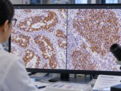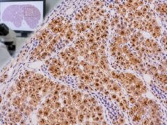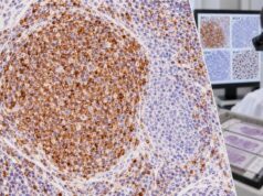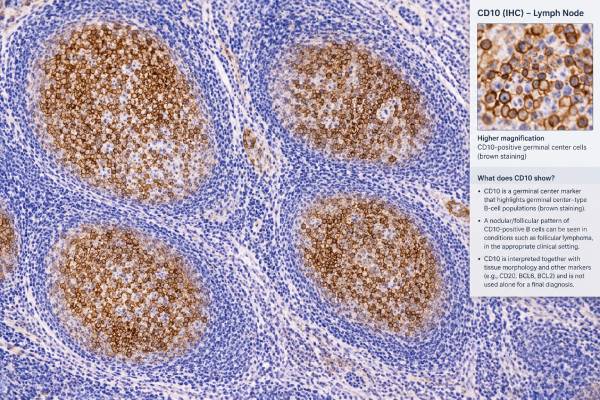
CD10 immunohistochemistry (IHC) is a tissue stain that helps pathologists identify cells with germinal center B-cell differentiation. It is commonly used when evaluating follicular lymphoma, diffuse large B-cell lymphoma (DLBCL), Burkitt lymphoma, and other lymphoid disorders. CD10 is not a cancer-specific protein: normal germinal center B cells and several non-lymphoid tissues can also express it. For that reason, a positive stain is a clue about cell differentiation rather than a diagnosis by itself.
In a lymph node with abnormal follicles, CD10 can support follicular lymphoma when the staining pattern matches the suspicious B-cell population and the rest of the panel fits. The pathologist also considers architecture, BCL6, BCL2, CD20, Ki-67, and sometimes flow cytometry or genetic studies. A negative CD10 stain does not rule out follicular lymphoma, especially in higher-grade, unusual, or biologically distinct cases. The report is therefore best read as an integrated interpretation of several findings rather than a simple positive-versus-negative test.
- CD10 positivity usually supports germinal center differentiation, but it is not specific for follicular lymphoma or even for lymphoma.
- Follicular lymphoma commonly expresses CD10 and BCL6, often together with abnormal BCL2 expression in neoplastic follicles.
- Reactive germinal centers are also CD10 positive, so tissue architecture and companion stains are essential.
- CD10 can help classify DLBCL as germinal center B-cell-like in IHC algorithms, but IHC is only a surrogate for molecular cell-of-origin testing.
- A negative CD10 result does not exclude follicular lymphoma, and additional markers or molecular studies may be needed.
Table of Contents
- What the CD10 IHC Test Detects
- How CD10 Staining Is Read
- CD10 in Follicular Lymphoma
- Reactive Follicles and Common Mimics
- CD10 in DLBCL and Other Lymphomas
- What Positive and Negative CD10 Results Mean
- What Happens After CD10 Testing
What the CD10 IHC Test Detects
CD10 is a cell-surface enzyme also called neutral endopeptidase or membrane metalloendopeptidase. In hematopathology, its most useful role is as a marker of germinal center differentiation. Germinal centers are temporary structures within lymphoid follicles where activated B cells mature, undergo immunoglobulin gene changes, and are selected for improved antibody responses.
Normal germinal center B cells commonly express CD10. Many lymphomas derived from or resembling these cells retain the protein, including follicular lymphoma, Burkitt lymphoma, and a substantial subset of DLBCL. CD10 is also often present on precursor B lymphoblasts, which is why older literature sometimes calls it the common acute lymphoblastic leukemia antigen, or CALLA.
IHC shows where the protein is located in a tissue section while preserving tissue architecture. That is different from flow cytometry, which measures markers on individual cells in suspension. A lymph node may undergo both tests because each supplies different information: IHC shows whether CD10-positive cells are forming abnormal follicles or sheets, while flow cytometry can identify an abnormal B-cell population and assess light-chain restriction.
CD10 is rarely interpreted alone. A lymphoma workup may start with a broad tumor IHC panel and then add lineage and maturation markers based on the morphology. In suspected B-cell lymphoma, CD20 or PAX5 can establish B-cell identity, while CD10, BCL6, MUM1, BCL2, cyclin D1, and other markers help narrow the subtype.
An important limitation is that CD10 is not confined to lymphocytes. It may be expressed in renal tumors, endometrial stromal tumors, some carcinomas, certain sarcomas, and other tissues. A positive stain in an unknown tumor therefore cannot be translated automatically into “germinal center lymphoma.” The pathologist must first confirm which cells are staining and what type of tumor is present.
How CD10 Staining Is Read
CD10 IHC is usually performed on formalin-fixed, paraffin-embedded biopsy tissue. The laboratory applies an antibody against CD10, develops a visible reaction, and evaluates the tumor cells alongside internal and external controls. True staining is usually membranous, sometimes with cytoplasmic accentuation depending on the specimen and assay.
The report may describe CD10 as positive or negative and may add the percentage, intensity, or distribution of positive tumor cells. Unlike a blood chemistry test, there is no universal “normal range.” Interpretation depends on the diagnostic setting and on the scoring method used by the laboratory.
The location of the positive cells matters as much as the percentage. In a normal or reactive lymph node, CD10 highlights germinal centers while the surrounding mantle-zone and interfollicular B cells are mostly negative. In follicular lymphoma, the abnormal follicular B-cell population often shows widespread CD10 expression, creating a pattern that matches the neoplastic follicles. In DLBCL, pathologists generally score the proportion of large tumor cells that stain rather than the overall percentage of all cells in the biopsy.
Preanalytic factors can affect the result. Prolonged or inadequate fixation, decalcification, scant tissue, crush artifact, necrosis, and low tumor content can weaken or distort staining. A negative result is less reassuring when the specimen is technically compromised. This is one reason pathology reports sometimes recommend repeat staining, another marker, or additional testing rather than treating one negative stain as definitive.
When CD10 is being used in the Hans IHC algorithm for DLBCL cell-of-origin classification, a threshold of 30% positive tumor cells is commonly used. That cutoff belongs to that specific algorithm; it should not be applied automatically to follicular lymphoma or to every laboratory protocol.
CD10 in Follicular Lymphoma
CD10 is one of the core stains used to recognize follicular lymphoma because most conventional cases show a germinal center phenotype. The typical neoplastic follicles contain B cells positive for CD20, CD10, and BCL6. BCL2 is also commonly expressed in the abnormal follicles, in contrast with many reactive germinal centers, where BCL2 is normally low or absent.
That combination is more informative than any single marker. BCL6 staining provides another marker of germinal center differentiation, while BCL2 IHC helps show the abnormal survival-protein pattern typical of many conventional follicular lymphomas. The morphology must still show a compatible follicular or partly follicular lymphoid proliferation.
The classic genetic event in conventional follicular lymphoma is a BCL2 rearrangement, most often involving IGH and BCL2. It leads to inappropriate BCL2 expression in germinal center B cells that would otherwise be subject to apoptosis. However, modern classifications recognize biologic diversity. Roughly 10%–15% of conventional-appearing follicular lymphomas may lack the classic BCL2 rearrangement, and some have different genetic or phenotypic features.
CD10 expression is not perfectly uniform. It can be weaker in interfollicular tumor cells than within follicles. Some grade 3 follicular lymphomas, diffuse areas, pediatric-type follicular lymphomas, cutaneous follicle center lymphomas, and other follicular-pattern B-cell neoplasms may have reduced or absent CD10. A negative stain therefore changes the differential diagnosis but does not end it.
Pathologists may use additional germinal center markers such as HGAL or LMO2 when CD10 and BCL6 are equivocal or when morphology strongly suggests a germinal center-derived tumor. FISH for BCL2 or BCL6 rearrangements can also help in selected cases. The exact workup depends on age, site, grade, architecture, and the competing diagnoses.
The distinction between follicular lymphoma and reactive follicular hyperplasia is especially important. Both can contain many CD10-positive cells. Features favoring lymphoma include crowded or back-to-back follicles, loss of normal follicular polarization, reduced tingible-body macrophages, a monotonous cell population, abnormal BCL2 expression, a clonal B-cell population, and supportive genetics. No one feature is sufficient in every case.
Reactive Follicles and Common Mimics
A positive CD10 stain can be completely normal in a reactive lymph node. Germinal centers formed during infection, vaccination, autoimmune activity, or other immune stimulation usually contain many CD10-positive B cells. The key is whether those cells are arranged in normal reactive follicles or form an abnormal clonal proliferation.
Reactive follicles usually preserve a recognizable architecture: they have variable sizes and shapes, polarized dark and light zones, numerous tingible-body macrophages, and intact mantle zones. BCL2 is typically negative or weak in the germinal center B cells, although background T cells and mantle-zone cells remain positive. Follicular lymphoma tends to replace normal architecture with more uniform neoplastic follicles and often shows BCL2 expression within the follicular B-cell population.
Several other lymphomas can also be CD10 positive, so the stain cannot identify follicular lymphoma by itself. The differential may include Burkitt lymphoma, DLBCL, B-lymphoblastic leukemia/lymphoma, and lymphomas of T-follicular helper origin in which some neoplastic T cells express CD10. Rarely, other mature B-cell lymphomas show aberrant CD10 expression.
A useful practical safeguard is to confirm lineage. If the suspicious CD10-positive cells are large and atypical, CD20 or PAX5 IHC can support B-cell differentiation. If a T-cell lymphoma is in the differential, CD3 and other T-cell markers are necessary. The staining pattern must be assigned to the actual neoplastic cells, not simply to the background population.
Small biopsies can be especially challenging because architecture may be only partly represented. A core biopsy containing a few CD10-positive follicles may not show whether the entire node is replaced or whether the follicles are reactive. In those situations, flow cytometry, clonality testing, FISH, or a larger biopsy may be needed if the available material cannot support a confident classification.
CD10 in DLBCL and Other Lymphomas
In DLBCL, CD10 serves mainly as a cell-of-origin surrogate marker rather than as proof of one specific disease. DLBCL, not otherwise specified, can be divided biologically into germinal center B-cell-like (GCB) and activated B-cell-like categories by gene-expression methods. IHC algorithms were developed to approximate those molecular groups when gene-expression testing is unavailable.
The widely used Hans algorithm evaluates CD10, BCL6, and MUM1/IRF4. If at least 30% of tumor cells express CD10, the case is assigned to the GCB group in that algorithm. CD10-negative cases are further sorted using BCL6 and MUM1. The method is practical, but it is not identical to molecular profiling, and discordance can occur. A pathology report may therefore specify that the result is an IHC-based GCB/non-GCB classification.
CD10 also appears in other important settings:
| Setting | Typical CD10 role | What else is needed |
|---|---|---|
| Follicular lymphoma | Supports germinal center phenotype | Architecture, BCL6, BCL2, B-cell markers, sometimes FISH |
| DLBCL | Helps assign IHC cell-of-origin group | BCL6, MUM1 and broader lymphoma classification |
| Burkitt lymphoma | Often strong and diffuse | BCL6, BCL2 pattern, very high Ki-67, MYC testing, morphology |
| B-lymphoblastic leukemia/lymphoma | Common precursor B-cell marker | TdT, CD34, PAX5/CD79a and genetic classification |
| Reactive germinal centers | Normal positive pattern | Preserved architecture and non-neoplastic companion findings |
Burkitt lymphoma is usually CD10 and BCL6 positive, with an extremely high Ki-67 proliferation index, often approaching 100%. Yet those findings are not enough to make the diagnosis. Burkitt lymphoma requires characteristic morphology and a compatible MYC rearrangement context, while high-grade B-cell lymphomas can mimic the phenotype.
In precursor B-lymphoblastic leukemia/lymphoma, CD10 is common but maturity markers may differ from those in mature B-cell lymphoma. TdT and often CD34 identify immaturity, and flow cytometry or genetic studies usually provides more precise classification. Thus, the same positive CD10 stain can represent very different biology depending on the rest of the phenotype.
Another nuance is that treatment can alter the tissue pattern over time. A recurrent or transformed follicular lymphoma may not reproduce the exact CD10 intensity seen in the original biopsy, and a transformed large-cell component can differ from the low-grade follicular areas. Comparing current tissue with prior slides can help determine whether an apparent phenotypic shift represents tumor evolution, sampling, or a different process.
The same principle applies to small extranodal samples. A limited biopsy may show CD10-positive B cells without enough architecture to decide whether they form a true neoplasm. When the distinction carries major treatment consequences, pathologists may recommend a larger specimen rather than forcing a definitive diagnosis from an incomplete pattern.
What Positive and Negative CD10 Results Mean
A positive CD10 result means the tested cells contain detectable CD10 protein. In a B-cell lymphoma, that often points toward germinal center differentiation, but the final meaning depends on cell size, architecture, companion markers, and genetics. In a reactive lymph node, the same positive result may simply highlight normal germinal centers.
A negative CD10 result means the assay did not detect convincing protein expression in the relevant cells. It does not mean the tissue is normal and does not exclude lymphoma. Many B-cell lymphomas are expected to be CD10 negative, including most marginal zone lymphomas, lymphoplasmacytic lymphomas, and many activated B-cell-like DLBCLs. Even follicular lymphoma can lose or lack CD10 in selected settings.
An equivocal or limited result can occur when tumor is scant, the staining is weak, tissue preservation is poor, or positive reactive cells make it difficult to identify the tumor population. The pathologist may repeat the stain, compare serial sections, or use a different germinal center marker.
Common interpretation mistakes include assuming that any CD10-positive lymphoid cell is malignant, treating CD10 negativity as proof against follicular lymphoma, and applying a DLBCL percentage cutoff to other diseases. Another mistake is reading the stain without confirming lineage: CD10 expression outside B cells can have a completely different implication.
CD10 itself is not a stand-alone prognosis test. Prognosis in follicular lymphoma depends on clinical stage, tumor burden, symptoms, grade, transformation, molecular features, and treatment response. In DLBCL, cell-of-origin information contributes biologic context, but treatment and prognosis are not determined by CD10 staining alone.
Because CD10 is also expressed in several non-lymphoid tissues, an unexpected positive stain in a poorly differentiated tumor should prompt lineage confirmation before a lymphoma diagnosis is considered. Broad cytokeratins, PAX8, stromal markers, or other organ-directed stains may be more appropriate depending on the site and morphology.
What Happens After CD10 Testing
The next step after CD10 IHC depends on the diagnostic question. For suspected follicular lymphoma, the pathologist usually integrates CD10 with morphology, CD20, BCL6, BCL2, Ki-67, and the follicular dendritic cell network. Flow cytometry may demonstrate a clonal B-cell population, while FISH can test for BCL2 or other rearrangements when the phenotype is atypical.
For DLBCL, CD10 is only one component of a larger classification workup. BCL6 and MUM1 may be scored for an IHC cell-of-origin algorithm, while MYC and BCL2 protein expression and MYC/BCL2 rearrangement testing are considered when indicated by current classification and laboratory practice. Clinical staging then establishes disease extent.
If a report says “CD10 positive,” useful questions include: Which cells are positive? Is the pattern follicular or diffuse? What do BCL6 and BCL2 show? Are the abnormal cells definitely B cells? Was clonality or FISH performed? The final diagnostic line and interpretive comment usually carry more clinical meaning than the isolated marker result.
Patients generally do not need special preparation for CD10 IHC because the test is performed on tissue already obtained by biopsy or surgery. The main practical issue is whether the specimen contains enough well-preserved tumor for a complete panel. When tissue is limited, pathologists often prioritize stains and molecular tests to preserve material for the highest-value questions.
The most important takeaway is that CD10 is a pattern and differentiation marker. It becomes diagnostically powerful when it agrees with architecture and other markers, but it should never be treated as a one-marker verdict on whether lymphoma is present or which lymphoma a patient has.
References
- Diagnosis and Classification of Follicular Lymphoma and Related Entities. 2025 (Review)
- Follicular lymphoma: 2023 update on diagnosis and management. 2023 (Review)
- Heterogeneity or change in cell of origin in diffuse large B-cell lymphomas determined using hans algorithm. 2024
- Value of GCET1, HGAL (GCET2), and LMO2 in the Determination of Germinal Center Phenotype in Diffuse Large B-cell Lymphoma 2023
- The International Consensus Classification of Mature Lymphoid Neoplasms: a report from the Clinical Advisory Committee 2022 (Consensus Classification)
Disclaimer
CD10 IHC is a pathology tool and cannot diagnose or exclude lymphoma by itself. Its meaning depends on the biopsy appearance, the cells that stain, companion immunostains, and sometimes flow cytometry or molecular testing. Discuss the integrated pathology report with the treating clinician or pathologist for interpretation in the individual clinical setting.