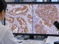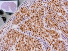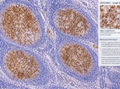
BCL2 immunohistochemistry (IHC) is a tissue stain that detects the anti-apoptotic BCL2 protein in lymphoid cells. Pathologists use it most often when classifying B-cell lymphomas, especially follicular lymphoma and diffuse large B-cell lymphoma (DLBCL). The meaning of a positive result depends on where the positive cells are located, how many tumor cells stain, how intense the staining is, and what other markers and genetic tests show.
BCL2 is normally expressed in many mature lymphocytes, so positivity by itself is not evidence of cancer. Its diagnostic value comes from patterns. Normal reactive germinal centers are usually BCL2 negative, while conventional follicular lymphoma often shows abnormal BCL2 expression within neoplastic follicles. In DLBCL, BCL2 can be measured along with MYC because co-expression may identify a higher-risk “double-expressor” phenotype. That is not the same as a lymphoma with MYC and BCL2 gene rearrangements, which requires cytogenetic or molecular testing.
- BCL2 IHC shows BCL2 protein in tissue, usually as cytoplasmic staining in lymphoid cells.
- BCL2-positive follicles can support follicular lymphoma because normal reactive germinal centers are usually BCL2 negative.
- BCL2 positivity alone does not diagnose lymphoma; many normal lymphocytes naturally express BCL2.
- In DLBCL, BCL2 is often interpreted with MYC, commonly using tumor-cell percentage cutoffs, but thresholds and prognostic impact depend on the setting.
- BCL2 protein expression is not the same as a BCL2 gene rearrangement; FISH or another genetic method is needed to identify rearrangements.
Table of Contents
- What BCL2 IHC Detects
- Normal and Abnormal BCL2 Staining Patterns
- BCL2 in Follicular Lymphoma
- BCL2 in Diffuse Large B-Cell Lymphoma
- BCL2 Protein Versus BCL2 Gene Rearrangement
- How to Read a BCL2 Pathology Result
- What Happens After BCL2 Testing
What BCL2 IHC Detects
BCL2 is a protein that helps cells resist programmed cell death, or apoptosis. That function is useful in normal immune biology because some lymphocytes need to survive after activation. In lymphoma, however, abnormal BCL2 activity can help malignant cells persist when they otherwise would die.
IHC detects the BCL2 protein in a biopsy. The test is commonly performed on formalin-fixed, paraffin-embedded lymph node, bone marrow, skin, or extranodal tissue. A laboratory applies a BCL2 antibody, develops the stain, and the pathologist assesses the tumor cells under the microscope.
The result is not a serum concentration. There is no normal blood range, therapeutic range, or toxic level. Instead, the pathologist evaluates a staining pattern. Important questions include:
- Are the positive cells actually the neoplastic cells?
- Is staining cytoplasmic, as expected for BCL2?
- Is it diffuse, patchy, or limited to a subset of cells?
- What percentage of tumor cells is positive?
- Does the pattern fit the tissue architecture?
- How do BCL2 results compare with BCL6, CD10, MYC, CD20, and other markers?
BCL2 is almost never interpreted alone. It belongs within a tumor IHC panel designed to test a specific differential diagnosis. In a follicular proliferation, the question may be reactive hyperplasia versus follicular lymphoma. In large-cell lymphoma, the question may be cell of origin, double expression, or whether molecular testing for a high-grade genetic subtype is needed.
Normal and Abnormal BCL2 Staining Patterns
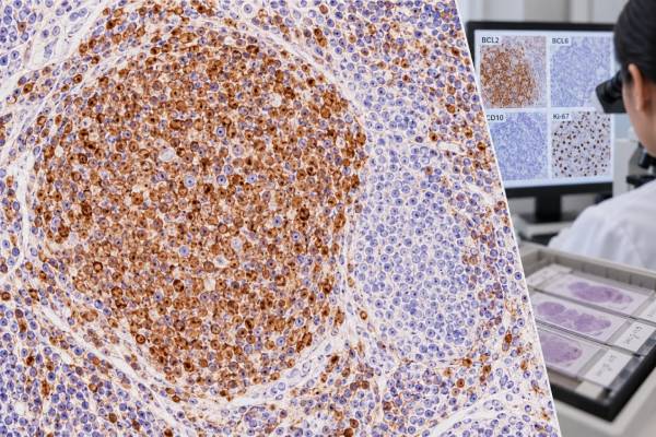
Understanding normal lymph node architecture is essential for interpreting BCL2. In a reactive follicle, germinal-center B cells normally downregulate BCL2 so cells that fail selection can undergo apoptosis. As a result, the germinal center is typically BCL2 negative or markedly weaker than the surrounding mantle-zone and interfollicular lymphocytes, which remain BCL2 positive.
That contrast creates one of the classic diagnostic uses of the stain. If follicles that look suspicious for lymphoma show strong BCL2 expression throughout the germinal-center B-cell population, the pattern is abnormal and can support conventional follicular lymphoma.
The distinction is not absolute. Reactive conditions can alter staining, and not every follicular lymphoma expresses BCL2. Interpretation becomes especially difficult in small biopsies, partially involved nodes, pediatric and young-adult settings, or unusual follicular lymphoma variants. The pathologist therefore compares BCL2 with architecture and other germinal-center markers rather than making a diagnosis from one stain.
In diffuse lymphomas, there is no helpful follicular map. The pathologist instead estimates how many malignant cells show convincing cytoplasmic BCL2 staining and how strong it is. Small reactive T cells and non-neoplastic B cells in the background may also be positive, so cell recognition matters.
A useful internal control is often present on the same slide. If normal small lymphocytes stain but the suspected tumor cells do not, that supports a true negative result. If nothing on the slide stains, technical failure must be considered.
BCL2 in Follicular Lymphoma
Conventional follicular lymphoma is a germinal-center B-cell neoplasm. Most classic cases carry an IGH::BCL2 rearrangement, historically called t(14;18), which places BCL2 under immunoglobulin regulatory control and drives abnormal protein expression. IHC often shows BCL2-positive neoplastic follicles that stand out from the BCL2-negative germinal centers expected in reactive lymphoid tissue.
A CD10 IHC test and BCL6 IHC help establish germinal-center differentiation. CD20 usually confirms B-cell lineage. The diagnosis then depends on the combination of abnormal follicular architecture, cytology, immunophenotype, and sometimes FISH or molecular findings.
BCL2 staining is particularly useful when the differential is reactive follicular hyperplasia. In a reactive node, germinal centers usually contain a mixture of centrocytes and centroblasts and preserve polarization and tingible-body macrophages. BCL2 negativity within those germinal centers fits normal biology. In follicular lymphoma, follicles are often crowded, monotonous, back-to-back, and less polarized, while the neoplastic follicle cells frequently express BCL2.
However, a BCL2-negative follicle is not automatically benign. Several situations can produce genuine follicular lymphoma without the usual BCL2 pattern. These include a subset of conventional follicular lymphomas lacking BCL2 rearrangement and distinct entities or variants recognized in modern classifications, such as pediatric-type follicular lymphoma. Some t(14;18)-negative follicular lymphomas have different genetic pathways and clinicopathologic features.
Likewise, weak or technically inconsistent staining should not be overread. Antibody clone, fixation, and tissue quality can affect sensitivity. If the architecture strongly suggests lymphoma but BCL2 is negative, the pathologist can use other markers, clonality studies, and FISH or sequencing when indicated.
BCL2 also cannot grade follicular lymphoma by itself. Grading and current classification rely on morphology and the specific disease entity. A strongly positive BCL2 stain tells the pathologist about phenotype, not how many centroblasts are present or how the patient should be treated.
BCL2 in Diffuse Large B-Cell Lymphoma
In DLBCL, BCL2 is common but has a different purpose. The tumor grows diffusely rather than in follicles, so the pathologist generally assesses the proportion of large lymphoma cells with cytoplasmic BCL2 expression. BCL2 can contribute to prognostic stratification and is often interpreted with MYC protein expression.
Many laboratories use the term double-expressor lymphoma when DLBCL shows high MYC and BCL2 protein expression by IHC. Commonly used research and clinical thresholds are at least 40% MYC-positive tumor cells and at least 50% BCL2-positive tumor cells, although methods and cutoffs vary. A pathology report may therefore provide percentages instead of only “positive” or “negative.”
Double expression has often been associated with more aggressive biology and worse outcomes in patients treated with standard immunochemotherapy, but the effect is not uniform across all molecular subgroups and treatment settings. Age, stage, International Prognostic Index, cell-of-origin biology, and genetic subtype remain important. BCL2 positivity alone should not be presented as a personalized survival prediction.
The cell-of-origin workup may include CD10, BCL6, and MUM1/IRF4. MUM1 IHC, for example, contributes to common IHC algorithms that approximate germinal-center B-cell versus non-germinal-center phenotypes. These algorithms are useful but are not identical to gene-expression profiling.
BCL2 also needs to be considered with Ki-67 proliferation staining. BCL2 protects against cell death, while Ki-67 indicates how many cells are cycling. A tumor can show high values for both because rapid proliferation and resistance to apoptosis are separate biologic features.
Double expressor is not double hit
This distinction is essential. “Double expressor” refers to protein levels measured by IHC. “Double hit” historically referred to rearrangements involving MYC plus BCL2 and/or BCL6 detected by FISH or another genetic method. Modern classifications have refined the terminology and disease boundaries, particularly for tumors with MYC and BCL2 rearrangements.
A DLBCL can be a double expressor without having MYC and BCL2 rearrangements. Conversely, a tumor with rearrangements may not meet a laboratory’s chosen IHC threshold in every case. IHC cannot substitute for the genetic assay when the diagnostic question is whether a rearrangement is present.
BCL2 expression in DLBCL must also be separated from the concept of a double-expressor lymphoma. In routine practice, the term usually refers to DLBCL in which MYC and BCL2 proteins are coexpressed above established IHC thresholds, commonly MYC in at least 40% of tumor cells and BCL2 in at least 50%. This phenotype can be associated with more aggressive clinical behavior in some cohorts, but it is not the same as a MYC/BCL2 “double-hit” genetic lymphoma.
The distinction changes what test comes next. IHC measures protein abundance, whereas FISH identifies rearrangements. A tumor can be a double expressor without either rearrangement, and a rearranged tumor does not always show a perfectly predictable IHC pattern. Current lymphoma classification therefore uses morphology, IHC, and genetics as separate but complementary layers rather than allowing a BCL2 stain to stand in for cytogenetic testing.
BCL2 Protein Versus BCL2 Gene Rearrangement
BCL2 IHC and BCL2 FISH measure different things. IHC asks whether the protein is present in the cells. FISH asks whether the BCL2 gene locus has undergone a detectable rearrangement. Sequencing can provide still other information about mutations, copy-number changes, or broader genomic context.
In conventional follicular lymphoma, protein positivity often correlates with an IGH::BCL2 rearrangement, but the relationship is not perfect. BCL2 protein can be increased through mechanisms other than the classic translocation, and some rearranged tumors may show weak or absent staining because of technical or biologic factors.
In aggressive B-cell lymphoma, genetic testing matters because certain MYC/BCL2-rearranged tumors fall into specific high-grade categories under current classification systems. This has implications for diagnosis, prognosis, and management that are not captured by protein staining alone.
| Test | What it measures | Typical use |
|---|---|---|
| BCL2 IHC | BCL2 protein expression in cells | Follicular pattern assessment, DLBCL protein expression |
| BCL2 FISH | BCL2 locus rearrangement | Confirming t(14;18)-type biology or assessing aggressive lymphoma genetics |
| Broader molecular testing | Multiple genes, mutations, rearrangements, or signatures | Resolving complex classification or defining molecular subtype |
The tests are complementary. If the pathology report says “BCL2 positive by IHC,” it should not be translated into “BCL2 rearranged” unless a genetic test demonstrated that alteration.
BCL2 interpretation is also shaped by the normal cells surrounding a lymphoma. Small mantle-zone lymphocytes and many interfollicular T cells normally express BCL2 and can serve as internal positive controls. In a follicular lesion, the question is whether the germinal center B cells themselves are abnormally positive. In a large-cell lymphoma, the pathologist must distinguish staining in the large tumor cells from staining in numerous small reactive lymphocytes.
Technical quality matters because fixation, antigen retrieval, antibody clone, and scoring method can affect intensity. When a result will be used with a percentage cutoff, laboratories validate how tumor cells are counted and how weak staining is handled. This helps explain why a report may provide an estimated percentage rather than a laboratory “normal range.”
How to Read a BCL2 Pathology Result
Start with the final diagnosis, not the stain list. A BCL2 result makes sense only after identifying which lymphoma or reactive process the pathologist is considering.
For a follicular process, ask whether the neoplastic follicles express BCL2 and whether CD10 and BCL6 support germinal-center B-cell differentiation. A CD20 stain can confirm that the suspicious cells belong to the B-cell compartment. If BCL2 is negative, the report may explain whether the morphology still supports follicular lymphoma or whether additional genetic studies were performed.
For DLBCL, look for a percentage or descriptive estimate of tumor-cell staining. If MYC is also reported, determine whether the report calls the tumor a double expressor. Then check separately for FISH results involving MYC, BCL2, and BCL6. Those are different layers of information.
Common interpretations include:
- BCL2 positive in neoplastic follicles: supports follicular lymphoma in a compatible biopsy.
- BCL2 negative in reactive germinal centers: expected normal pattern.
- Diffuse BCL2 in DLBCL: common; significance depends on percentage, MYC, and clinical context.
- BCL2 negative tumor with positive small lymphocytes: likely a true tumor-negative result if controls are appropriate.
- BCL2 positive without a BCL2 rearrangement: possible and not contradictory because protein expression has multiple causes.
Do not use BCL2 staining to estimate how long an individual patient will live. Prognosis is multifactorial, and modern lymphoma classification increasingly integrates genetics, phenotype, clinical risk, and treatment response.
BCL2 also has different weight in different lymphoma families. In follicular lymphoma, abnormal expression inside neoplastic follicles is a powerful diagnostic clue. In DLBCL, the percentage of BCL2-positive large cells contributes to a prognostic phenotype and may help decide which genetic studies are appropriate. In several other small B-cell lymphomas, BCL2 is commonly positive but much less specific, so the stain adds little unless interpreted with lineage and subtype markers.
This context prevents a common misunderstanding: a “high BCL2” tissue result is not comparable to a high blood test. It describes the proportion and intensity of protein expression in tumor cells and does not provide a circulating concentration, treatment target level, or universal risk category.
Another important distinction is between diagnostic significance and treatment targeting. Although BCL2 is a drug target in some hematologic cancers, routine BCL2 IHC in a lymphoma biopsy is not automatically a companion diagnostic for a BCL2 inhibitor. Drug selection depends on the exact disease, approved indication, molecular context, prior therapy, and current clinical guidelines.
What Happens After BCL2 Testing
After BCL2 IHC, the pathologist completes whatever studies are needed to answer the diagnostic question. In suspected follicular lymphoma, that may include CD20, CD10, BCL6, Ki-67, and BCL2 FISH. In DLBCL or another aggressive B-cell lymphoma, the panel may include MYC, BCL6, MUM1, EBV testing, and FISH for rearrangements.
If the biopsy is very small, damaged, or decalcified, staining may be difficult to interpret. A larger biopsy can be important when architecture is needed or when too few viable tumor cells remain for genetic studies.
The hematologist then interprets the final pathologic subtype along with stage, symptoms, blood results, imaging, age, organ function, and performance status. BCL2 expression may contribute useful prognostic information, but treatment is not selected from BCL2 IHC alone in most routine lymphoma settings.
For a patient reading a pathology report, three questions usually clarify the result: Was BCL2 positive in the actual tumor cells? Was the stain being used for diagnosis, prognosis, or both? Were MYC/BCL2 gene rearrangements tested separately? Keeping protein expression and genetics separate prevents one of the most common misunderstandings in lymphoma reports.
References
- The International Consensus Classification of Mature Lymphoid Neoplasms: a report from the Clinical Advisory Committee 2022 (Consensus Classification)
- Fifth edition WHO classification: mature B-cell neoplasms. 2025 (Review)
- Follicular lymphoma and marginal zone lymphoma: how many diseases? 2022 (Review)
- Immunohistochemistry-based investigation of MYC, BCL2, and Ki-67 protein expression and their clinical impact in diffuse large B-cell lymphoma in upper Northern Thailand 2024
- From Biopsy to Diagnosis: Navigating Aggressive B-Cell Lymphomas in Practice 2025 (Review)
Disclaimer
BCL2 IHC is one component of lymphoma diagnosis and risk assessment. A positive or negative result cannot identify a lymphoma subtype or predict an individual outcome without morphology, other immunostains, genetics, and clinical information. Review the complete pathology report with the treating hematologist, oncologist, or pathologist.