
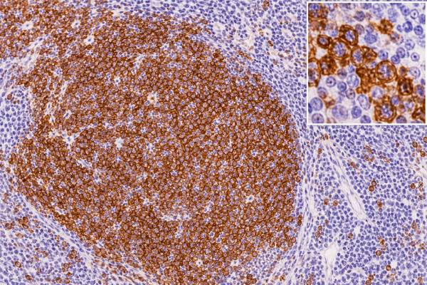
CD20 immunohistochemistry (IHC) is one of the most widely used tissue stains for identifying mature B cells and B-cell lymphomas. A positive result can show that suspicious lymphoid cells have B-cell differentiation, helping separate many B-cell lymphomas from T-cell lymphomas and non-lymphoid tumors. CD20 is also clinically familiar because it is the target of drugs such as rituximab, obinutuzumab, and other anti-CD20 antibodies.
CD20 positivity alone does not identify a specific lymphoma. Normal B cells are positive, most mature B-cell lymphomas express the marker, and a small number of tumors can show unusual or reduced expression. Some B-cell neoplasms—especially those with plasmablastic or plasma-cell differentiation, precursor B-cell disease, or prior anti-CD20 treatment—may be weak or negative. Pathologists therefore interpret CD20 with morphology and other stains such as PAX5, CD79a, CD3, CD10, BCL6, BCL2, cyclin D1, and Ki-67. The final diagnosis depends on the whole immunophenotype, not one marker.
- CD20 positivity usually supports mature B-cell differentiation when the staining is present in the suspicious tumor cells.
- Most mature B-cell lymphomas are CD20 positive, but CD20 does not distinguish one B-cell lymphoma subtype from another.
- CD20 negativity does not exclude B-cell lineage, especially in plasmablastic tumors, precursor B-cell neoplasms, or disease sampled after anti-CD20 therapy.
- Reactive lymph nodes normally contain many CD20-positive B cells, so positivity by itself is not proof of lymphoma.
- CD20 can be both a diagnostic marker and a treatment target, but treatment decisions depend on the specific disease and full clinical context.
Table of Contents
- What the CD20 IHC Test Detects
- How CD20 Staining Is Read
- CD20 in B-Cell Lymphomas
- CD20 With Other Lineage Markers
- Why a B-Cell Lymphoma Can Be CD20 Negative
- CD20 as a Treatment Target
- What Happens After CD20 Testing
What the CD20 IHC Test Detects
CD20 is a membrane protein encoded by the MS4A1 gene. It appears during B-cell maturation, is present on most mature B cells, and is usually lost as B cells complete differentiation into plasma cells. This developmental pattern explains both its diagnostic usefulness and its limitations.
In a tissue biopsy, CD20 IHC highlights the membranes of cells that express the protein. In a normal lymph node, B-cell follicles are strongly positive, while most T cells in interfollicular areas are negative. That normal compartmental pattern gives the pathologist an internal reference. If a biopsy is replaced by a sheet or abnormal nodular population of CD20-positive cells, the finding can support a mature B-cell process.
CD20 is a lineage-associated marker, not a malignancy marker. Reactive B cells are positive, so the stain cannot tell benign from malignant by itself. The pathologist must determine whether the positive cells have abnormal morphology, architecture, distribution, or coexpression of other markers.
It is also not absolutely lineage-specific. Rare non-B-cell neoplasms can show aberrant CD20 staining, and technical artifacts can occur. Conversely, some genuine B-cell tumors lack CD20. For those reasons, B-cell identity is stronger when several findings agree.
CD20 is commonly part of an initial tumor IHC panel when lymphoma is suspected. CD3 provides a complementary T-cell map, while PAX5 and CD79a can support B-cell differentiation. Once lineage is established, subtype-specific markers and molecular tests answer the next questions.
The result has a second role beyond diagnosis: CD20 is an accessible cell-surface target for monoclonal antibody therapy. That connection makes the marker clinically important, but pathology interpretation and treatment eligibility are not identical questions.
How CD20 Staining Is Read
CD20 IHC is usually performed on formalin-fixed, paraffin-embedded tissue from a lymph node, bone marrow core, extranodal mass, or other biopsy. The commonly used L26 antibody recognizes an intracellular portion of the CD20 molecule and produces crisp membranous staining in positive cells.
A pathologist evaluates which cells stain, how many stain, how strongly they stain, and how they are distributed. A report may say diffuse positive, strong positive, variable, focal, weak, or negative. There is no single “normal range” or universal numerical cutoff that applies to every lymphoma.
The pattern can be more useful than a raw percentage. Examples include:
- Strong staining of nearly all atypical small lymphocytes in a diffuse infiltrate.
- Strong staining of abnormal follicles in follicular lymphoma.
- Diffuse membrane staining in large atypical cells of many DLBCLs.
- Positive background B cells but negative large tumor cells in a T-cell lymphoma or classic Hodgkin lymphoma.
- Weak or absent staining in a plasmablastic neoplasm despite evidence of B-cell origin from other markers or genetics.
Technical quality matters. Fixation, decalcification, tissue necrosis, crush artifact, and very small samples can reduce antigen detection. Bone specimens exposed to harsh decalcification may be especially difficult. Internal normal B cells can help show whether the stain worked, but even a technically successful stain must be assigned to the correct cells.
Flow cytometry can measure CD20 on suspended cells and is often used alongside IHC in lymphoid specimens. The two methods are complementary rather than interchangeable. IHC preserves architecture; flow cytometry rapidly assesses multiple markers and clonality. Discordance can occur because of sampling, different antibody epitopes, low antigen density, or therapy-related change.
CD20 in B-Cell Lymphomas
Most mature B-cell lymphomas express CD20, making it one of the most useful first-line markers in hematopathology. However, CD20 tells the pathologist that a population looks B-cell-like; it usually does not tell which B-cell lymphoma is present.
Common CD20-positive mature B-cell neoplasms include follicular lymphoma, mantle cell lymphoma, marginal zone lymphoma, chronic lymphocytic leukemia/small lymphocytic lymphoma, hairy cell leukemia, Burkitt lymphoma, and most diffuse large B-cell lymphomas. Their classification requires additional features.
For example, a follicular lymphoma often combines CD20 with CD10 and BCL6, while many conventional cases also show abnormal follicular BCL2 expression. Mantle cell lymphoma commonly expresses CD20 with cyclin D1 and SOX11. CLL/SLL usually expresses CD20 more weakly than many other mature B-cell lymphomas and coexpresses markers such as CD5 and CD23.
DLBCL usually shows a mature B-cell phenotype that includes CD20, but morphology and a larger panel are required. CD10, BCL6, and MUM1 may be used for IHC-based cell-of-origin assignment. MYC, BCL2, EBV studies, and rearrangement testing may be indicated depending on morphology and current classification rules. Ki-67 IHC provides proliferation information but does not replace subtype-defining tests.
CD20 is particularly helpful when the tissue contains a mixture of normal and abnormal lymphocytes. In a T-cell-rich background, highlighting a sparse population of atypical CD20-positive large cells can reveal a B-cell lymphoma that might otherwise be overlooked. The opposite situation also occurs: abundant reactive CD20-positive small B cells can surround tumor cells of another lineage, so cell-by-cell correlation with the H&E slide is essential.
Current lymphoma classifications increasingly integrate genetics, viral status, site, and clinical setting. CD20 remains foundational because it is fast, robust, and visually maps B cells, but it is only the first layer of classification.
CD20 With Other Lineage Markers
No single B-cell marker is perfect. Pathologists often use CD20 with PAX5, CD79a, and sometimes OCT2 or BOB1 when lineage is uncertain. These proteins are expressed at different stages of B-cell development, so their strengths and weaknesses complement one another.
PAX5 IHC is a nuclear B-cell transcription-factor stain. It is retained in many B-cell neoplasms that may have reduced CD20, although it becomes weak in classic Hodgkin lymphoma and is usually lost with terminal plasma-cell differentiation. CD79a is another useful B-lineage marker and can persist in some CD20-negative B-cell tumors.
CD3 is commonly paired with CD20 in an initial lymphoma panel because it maps T cells. A reactive node often shows an organized distribution: CD20-positive follicles and CD3-positive interfollicular T cells. A lymphoma can disturb or replace that pattern. CD3 staining is therefore valuable not only for identifying T-cell neoplasms but also for showing the background around B-cell tumors.
A simplified comparison illustrates why panels matter:
| Marker | Main diagnostic role | Important limitation |
|---|---|---|
| CD20 | Mature B-cell membrane marker | Can be lost with plasmacytic differentiation or after anti-CD20 therapy |
| PAX5 | Nuclear B-cell lineage marker | Weak in classic Hodgkin lymphoma; usually lost in plasma cells |
| CD79a | B-cell receptor-associated lineage marker | Can persist in plasma-cell differentiation and is not perfectly specific |
| CD3 | T-cell lineage marker | Does not identify B-cell subtype; cytoplasmic expression has special contexts |
Classic Hodgkin lymphoma illustrates the need for this approach. HRS cells are B-cell derived but often lack CD20 and show only weak PAX5, while CD30 is strong and CD15 is often positive. A CD20-negative result in that setting is therefore expected rather than evidence against B-cell origin at the molecular level.
Why a B-Cell Lymphoma Can Be CD20 Negative
A CD20-negative lymphoma can still be a B-cell neoplasm. The most important explanations are normal biology of the maturation stage, plasmablastic differentiation, unusual lymphoma subtype, prior therapy, or technical limitations.
Precursor B-lymphoblastic leukemia/lymphoma often lacks strong CD20 because the cells are immature. Markers such as TdT, PAX5, CD79a, and CD19, together with flow cytometry and genetics, are more informative for lineage and classification in this setting.
At the other end of B-cell maturation, plasma cells normally lose CD20. Neoplasms with plasmablastic or plasmacytic differentiation can therefore lose conventional mature B-cell markers. Plasmablastic lymphoma, primary effusion lymphoma, ALK-positive large B-cell lymphoma, and some other aggressive B-cell neoplasms may be CD20 negative or weak while expressing plasma-cell markers such as CD138 and MUM1/IRF4. Viral studies and molecular findings can be critical.
Treatment can also change antigen expression. Anti-CD20 antibodies apply selective pressure to CD20-positive tumor cells. At relapse, some lymphomas show reduced or absent CD20 because of biologic selection, altered protein expression, or other resistance mechanisms. This is why a pathology team may repeat immunophenotyping on a new biopsy rather than assume the original marker profile is unchanged.
Technical causes must be excluded before interpreting a negative stain biologically. Poor preservation or decalcification may weaken CD20. If internal B-cell controls are also absent, the stain may not be reliable. If normal B cells stain appropriately but the tumor does not, true antigen loss is more convincing.
When CD20 is negative but morphology suggests lymphoma, pathologists deliberately broaden the panel instead of abandoning B-cell lineage. PAX5, CD79a, OCT2, BOB1, immunoglobulin studies, EBV/HHV8 testing, ALK, and molecular clonality can help depending on the differential.
There are also situations in which CD20 expression is heterogeneous rather than uniformly present or absent. A lymphoma may contain strongly positive regions and weak or negative regions, particularly after therapy or with plasmacytic differentiation. Small samples can miss that variation. When treatment selection or classification depends on the phenotype, a pathologist may comment on heterogeneity or compare several blocks.
CD20 should also be distinguished from CD19. Both are B-cell-associated surface proteins, but they appear at somewhat different stages of B-cell development and are targeted by different therapies. A tumor can lose CD20 while retaining CD19 or other B-cell markers. That is why modern relapse workups often reassess multiple antigens rather than assuming that one lineage marker represents the entire B-cell program.
CD20 as a Treatment Target
CD20 is not only a diagnostic marker; it is also a therapeutically useful surface antigen. Rituximab, the best-known anti-CD20 monoclonal antibody, transformed treatment of many B-cell lymphomas by adding targeted immune-mediated cell killing to chemotherapy or other systemic regimens. Newer anti-CD20 antibodies are used in selected B-cell malignancies as well.
A pathology report showing CD20 expression can support the biologic rationale for anti-CD20 therapy, but the IHC result is not a stand-alone treatment prescription. Treatment depends on the exact lymphoma, stage, symptoms, prior therapy, age, organ function, and current clinical guidelines. Some diseases receive anti-CD20 therapy routinely; others do not, even when CD20 is present.
The amount of staining also does not translate directly into a drug dose. Laboratories generally do not use a universal IHC percentage threshold for rituximab eligibility across all B-cell lymphomas. Clinicians consider the established treatment evidence for the diagnosed entity and may use flow cytometry or repeat tissue assessment if antigen expression is uncertain.
Loss of CD20 after treatment has practical implications. If lymphoma relapses after rituximab or another anti-CD20 agent, a new biopsy may reveal CD20-negative disease. In that situation, repeating the marker panel can confirm that the recurrent tumor is related to the original lymphoma while identifying a phenotype that may affect subsequent treatment choices.
CD20-targeted therapy can also deplete normal B cells. That is part of the drug’s mechanism and explains some effects on immune function. Those treatment considerations are managed clinically and should not be confused with what the IHC stain itself measures in the biopsy.
The amount of CD20 expression can also differ by lymphoma subtype before any treatment. CLL/SLL often shows dimmer CD20 than follicular lymphoma or DLBCL, while plasmacytic areas may fade as cells approach terminal differentiation. A pathologist interprets that relative intensity as part of the phenotype but does not convert it into a universal numeric severity scale.
When flow cytometry and IHC disagree, both specimens and methods are reviewed. Flow may sample a different portion of the lesion or lose fragile large cells, while IHC may be affected by fixation. The final lineage assignment should reflect the best-supported interpretation across all available material.
What Happens After CD20 Testing
The next diagnostic step depends on what the biopsy looks like. If abnormal cells are strongly CD20 positive, the pathologist usually adds markers that distinguish among mature B-cell lymphoma subtypes. Small-cell processes may require CD5, CD10, CD23, cyclin D1, SOX11, BCL2, and other stains. Large-cell processes may require CD10, BCL6, MUM1, MYC, BCL2, CD30, EBV studies, and molecular testing.
Flow cytometry can determine whether the B-cell population is clonal and can define surface markers that are difficult to assess by IHC. FISH or sequencing may identify rearrangements or mutations required for classification. These tests answer questions that CD20 cannot answer on its own.
If CD20 is unexpectedly negative, the pathologist checks tissue quality and uses alternate B-cell markers. Prior therapy is important history: knowing that the patient previously received an anti-CD20 drug can immediately change how a negative stain is interpreted. A new biopsy may also be compared with the original tumor’s morphology and genetics.
For patients reviewing a report, useful questions include: Are the tumor cells CD20 positive, or only the background B cells? Which other B-cell markers are positive? What specific lymphoma diagnosis does the full panel support? Was flow cytometry or molecular testing performed? Is the result relevant to an anti-CD20 treatment option in this particular disease?
No special patient preparation is required for CD20 IHC itself because the stain is performed on tissue after the biopsy. The important procedural preparation relates to obtaining the biopsy, not to the immunostain. When lymphoma is suspected, preserving fresh tissue for flow cytometry may be helpful, so coordination before biopsy can sometimes improve diagnostic yield.
A CD20 result is most useful when read as part of a layered diagnosis: first identify the abnormal lymphoid population, then establish lineage, then classify the lymphoma, and finally connect that classification with staging and treatment. CD20 is a powerful marker within that sequence, but it is not the entire answer.
References
- Basic immunohistochemistry for lymphoma diagnosis 2022 (Review)
- Fifth Edition of the World Health Classification of Tumors of the Hematopoietic and Lymphoid Tissues: B-cell Neoplasms 2024 (Review)
- CD20-Negative Large B-Cell Lymphomas: The Diagnostic Challenge of Tumors with Downregulation of Mature B-Cell Marker Expression. 2025 (Review)
- Diffuse large B cell lymphoma (DLBCL): epidemiology, pathophysiology, risk stratification, advancement in diagnostic approaches and prospects: narrative review 2025 (Review)
- Diagnostic utility of the B-cell lineage markers CD20, CD79a, PAX5, and CD19 in paraffin-embedded tissues from lymphoid neoplasms 2009
Disclaimer
CD20 IHC is a pathology test used to assess B-cell differentiation and cannot diagnose a lymphoma subtype or determine treatment by itself. Results must be interpreted with tissue morphology, other immunostains, clinical history, and molecular or flow-cytometric studies when needed. Discuss the integrated pathology diagnosis and treatment implications with the treating clinician or hematopathologist.





