Calretinin immunohistochemistry (IHC) is a tissue stain used most often to support mesothelial differentiation, especially when a pleural, peritoneal, or other serosal tumor could be mesothelioma. A convincing mesothelial pattern usually includes both nuclear and cytoplasmic staining in the tumor cells. Calretinin is sensitive for many epithelioid mesotheliomas, but it is not completely specific and can be positive in several other tumors.
The most important limitation is that calretinin shows lineage, not malignancy. Reactive benign mesothelial cells can be strongly positive, so the stain cannot separate reactive mesothelial proliferation from mesothelioma by itself. Pathologists therefore combine it with other mesothelial markers such as WT1 or D2-40, carcinoma markers such as claudin-4, and malignancy-associated tests such as BAP1, MTAP, or CDKN2A FISH when needed. The final diagnosis depends on tissue invasion, morphology, clinical and imaging findings, and the complete marker panel.
- Calretinin IHC is mainly a mesothelial-lineage marker used in the diagnosis of pleural and peritoneal tumors.
- True supportive staining usually involves both the nucleus and cytoplasm of the cells being evaluated.
- Positive calretinin does not prove mesothelioma because benign mesothelial cells and several nonmesothelial tumors can also stain.
- Calretinin cannot reliably distinguish benign from malignant mesothelial proliferation; BAP1, MTAP, CDKN2A, and invasion are more useful for that question.
- There is no numeric normal range or universal percent-positive cutoff; interpretation depends on staining pattern and the differential diagnosis.
Table of Contents
- What Calretinin IHC Shows
- How Positive Calretinin Staining Looks
- Calretinin in Mesothelioma
- Calretinin Versus Carcinoma Markers
- Benign Versus Malignant Mesothelial Proliferation
- Other Tumors That Can Be Calretinin Positive
- How to Interpret a Calretinin Result
What Calretinin IHC Shows
Calretinin is a calcium-binding protein encoded by CALB2. It is normally expressed in mesothelial cells and selected cells in the nervous system and other tissues. Its relatively strong expression in mesothelial cells made it one of the classic positive markers used to distinguish mesothelioma from metastatic carcinoma involving a serosal surface.
The test is performed on tissue, not blood. A laboratory applies an antibody to a formalin-fixed, paraffin-embedded biopsy or cell-block section. Positive cells develop visible staining that the pathologist evaluates under the microscope.
Calretinin is most useful when the question is what lineage does this tumor show? Common situations include:
- A pleural biopsy that could represent mesothelioma or metastatic lung adenocarcinoma.
- A peritoneal tumor that could be mesothelioma or metastatic adenocarcinoma.
- An effusion cell block containing atypical epithelioid cells.
- A poorly differentiated serosal tumor with uncertain origin.
Because no mesothelial marker is perfectly specific, calretinin is usually part of a tumor immunohistochemistry panel. The panel deliberately includes both positive mesothelial markers and markers that favor the most likely competing carcinoma.
The test does not answer whether the patient has been exposed to asbestos, how advanced a tumor is, or whether a mesothelial proliferation is invasive. Those questions require history, imaging, morphology, and additional testing.
How Positive Calretinin Staining Looks
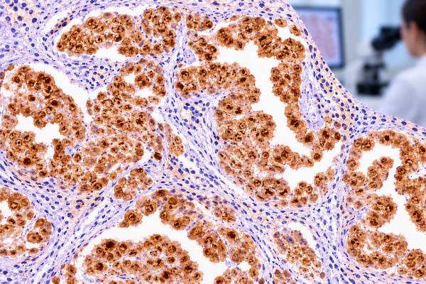
A classic positive mesothelial pattern shows nuclear and cytoplasmic staining in the same cells. Nuclear labeling is particularly important. Cytoplasmic staining alone can be less specific and may occur in tumors that are not mesothelial.
In epithelioid mesothelioma, staining may be strong and diffuse, although expression can vary. Sarcomatoid mesothelioma is more difficult because many traditional mesothelial markers, including calretinin, are less sensitive in spindle-cell tumors. A negative result in a sarcomatoid pleural neoplasm therefore does not exclude mesothelioma.
Pathologists assess several features:
- Whether the tumor cells, rather than only background mesothelial cells, stain.
- Whether staining is nuclear plus cytoplasmic.
- How diffuse or focal the staining is.
- Whether internal controls are appropriate.
- Whether companion mesothelial and carcinoma markers agree.
On a pleural biopsy, benign surface mesothelium may stain strongly while an underlying metastatic carcinoma remains negative. This can be diagnostically useful, but it also creates a pitfall if the surface cells are mistaken for the tumor population.
There is no universal threshold such as “more than 10% equals mesothelioma.” The value of the result depends on the distribution and cellular pattern and on how it fits with the rest of the panel.
Calretinin in Mesothelioma
Calretinin is especially sensitive in epithelioid mesothelioma, the most common histologic type. Recent systematic evidence continues to support its usefulness as a positive mesothelial marker, but modern guidelines emphasize that it should not be used alone.
A typical panel for an epithelioid pleural tumor may include two markers that support mesothelial differentiation and two that support carcinoma, selected according to the leading differential. Calretinin is often paired with WT1 IHC or D2-40 IHC. Cytokeratin 5/6 or newer markers such as HEG1 may also be used in appropriate laboratories.
The morphology still leads the diagnosis. Epithelioid mesothelioma may form tubules, papillae, trabeculae, solid nests, or other patterns. Mesothelioma can also be biphasic, containing epithelioid and sarcomatoid components, or predominantly sarcomatoid. The sensitivity of individual stains changes across those patterns.
For a pleural tumor, calretinin positivity becomes more persuasive when several findings line up:
- The lesion is centered on the pleural surface rather than forming a dominant lung parenchymal mass.
- Tumor morphology fits mesothelial differentiation.
- Calretinin shows nuclear and cytoplasmic staining in tumor cells.
- Another mesothelial marker is positive.
- Strong carcinoma markers such as claudin-4 are negative.
Even this combination establishes mesothelial lineage more directly than malignancy. Invasion into fat, chest wall, lung, or other structures can establish mesothelioma morphologically. When invasion is not demonstrable, malignancy-specific markers become especially important.
A modern mesothelioma panel is deliberately balanced rather than built from mesothelial markers alone. For an epithelioid pleural tumor, the pathologist generally wants at least two markers that support mesothelial differentiation and two that support carcinoma, chosen for the most likely competing primary. Claudin-4 is particularly useful because strong membranous expression favors carcinoma and is usually absent in mesothelioma. MOC31 or BerEP4 may also support epithelial carcinoma in the appropriate setting.
Organ-specific markers then refine the comparison. TTF-1 and Napsin A support lung adenocarcinoma, GATA3 may support breast or urothelial origin in the right context, and PAX8 can support renal, thyroid, or Müllerian primaries. The goal is not to prove mesothelioma with one positive stain but to build a coherent phenotype that fits the pleural tumor and excludes the most plausible metastases.
Calretinin Versus Carcinoma Markers
The classic diagnostic challenge is distinguishing epithelioid mesothelioma from metastatic adenocarcinoma, especially lung adenocarcinoma. Both can involve the pleura, form gland-like structures, and express broad-spectrum cytokeratins.
Modern practice uses opposing marker sets. Calretinin, WT1, D2-40, CK5/6, and HEG1 can support mesothelial lineage. Claudin-4 is a strong epithelial marker that is typically positive in carcinomas and negative in mesothelioma. MOC-31 and BerEP4 may also support carcinoma but can be less absolute than claudin-4.
If lung adenocarcinoma is the main alternative, TTF-1 IHC and Napsin A IHC can be highly useful because convincing positivity favors pulmonary adenocarcinoma rather than mesothelioma.
A simplified comparison is shown below.
| Marker | Epithelioid mesothelioma | Lung adenocarcinoma |
|---|---|---|
| Calretinin | Often positive, usually nuclear and cytoplasmic | Usually negative, but occasional positivity occurs |
| WT1 | Often positive | Usually negative |
| D2-40 | Often positive | Usually negative |
| Claudin-4 | Negative | Usually positive |
| TTF-1 / Napsin A | Negative | Often positive in lung adenocarcinoma |
These are tendencies rather than guarantees. High-grade serous carcinoma, squamous carcinoma, renal tumors, breast tumors, and other metastases require different marker combinations. That is why the panel should be tailored to the patient’s imaging and morphology.
Cytology specimens illustrate this limitation clearly. Mesothelial cells in a pleural or peritoneal effusion commonly express calretinin whether they are reactive or malignant. Positive staining can confirm that an atypical cell population is mesothelial, but cytologic atypia alone may still be insufficient to prove mesothelioma. Cell-block BAP1 or MTAP loss can increase specificity for malignancy, while a negative result on those loss markers does not exclude it.
Tissue invasion remains a central morphologic criterion. Mesothelial cells infiltrating fat, skeletal muscle, lung parenchyma, or other structures in an appropriate pattern support invasive mesothelioma. Superficial biopsies may not contain the interface needed to evaluate invasion, so the pathology team may recommend a deeper or larger specimen when the distinction will change management.
Benign Versus Malignant Mesothelial Proliferation
Calretinin does not reliably distinguish reactive mesothelial cells from mesothelioma. Both can be strongly positive because both are mesothelial. This is the single most important interpretive limitation of the test.
Reactive mesothelial proliferation can become highly cellular and cytologically atypical in infection, inflammation, pneumothorax, infarction, surgery, or other pleural injury. Conversely, early mesothelioma can look deceptively bland. A lineage marker cannot resolve that biologic difference.
When tissue shows unequivocal stromal invasion by mesothelial cells, morphology can establish malignancy. In superficial biopsies or cytology samples, invasion may not be assessable. In those cases, pathologists often use malignancy-associated tests:
- BAP1 IHC: loss of nuclear BAP1 expression in a mesothelial proliferation is highly specific for malignancy, although many mesotheliomas retain BAP1.
- MTAP IHC: loss of cytoplasmic MTAP can serve as a surrogate for CDKN2A-region deletion in appropriate settings.
- CDKN2A FISH: homozygous deletion is highly specific for malignant mesothelial proliferation.
- NF2/Merlin-related testing: can add sensitivity in selected laboratories.
These tests have the opposite performance problem from calretinin: loss of BAP1 or MTAP can be highly specific for malignancy but is not sensitive enough to identify every mesothelioma. Therefore, retained expression does not prove a lesion is benign.
The histologic subtype also changes how much confidence a negative calretinin result should carry. Epithelioid mesothelioma has the highest sensitivity for classic mesothelial markers. Sarcomatoid mesothelioma often loses calretinin, WT1, and D2-40, while retaining broad-spectrum cytokeratin in many cases. Biphasic mesothelioma can show a strong epithelioid component and a much less reactive spindle-cell component within the same tumor. Sampling only one area can therefore create an incomplete immunophenotypic picture.
This heterogeneity is why current guidelines emphasize selecting stains according to morphology. A spindle-cell pleural tumor may need keratins, GATA3, BAP1/MTAP-related studies, and markers that exclude sarcoma or melanoma rather than the same panel used for an epithelioid effusion-based differential.
Other Tumors That Can Be Calretinin Positive
Calretinin is not exclusive to mesothelioma. Positive staining has been described in sex cord-stromal tumors of the ovary and testis, adrenal cortical neoplasms, some neural and nerve-sheath tumors, selected breast and lung carcinomas, and other uncommon neoplasms.
This matters most when the tumor is outside the pleura or when clinical imaging suggests another primary. A calretinin-positive pelvic mass, for example, should not automatically be labeled peritoneal mesothelioma because ovarian sex cord-stromal tumors can express the marker. Likewise, a pleural tumor with only focal cytoplasmic staining but strong organ-specific carcinoma markers should not be forced into a mesothelial diagnosis.
Sarcomatoid lesions create another challenge. Calretinin and D2-40 may be positive in some sarcomas, and sarcomatoid mesothelioma may lose classic mesothelial markers. Current mesothelioma guidelines therefore emphasize demonstrating broad cytokeratin expression and carefully excluding other spindle-cell malignancies rather than relying on calretinin alone.
Calretinin also has useful diagnostic roles beyond the pleura, but those roles are context-specific. Sex cord-stromal tumors may show nuclear and cytoplasmic positivity, and adrenal cortical neoplasms can express the marker. In a pelvic or retroperitoneal mass, those possibilities can be more important than mesothelioma. A pathologist therefore chooses companion stains based on the organ, patient history, and microscopic pattern rather than applying a fixed “mesothelioma panel” to every calretinin-positive tumor.
The safest rule is to treat calretinin as evidence of a differentiation program, not as an organ barcode. The more unusual the anatomic site or morphology, the more important it becomes to confirm the interpretation with independent markers.
When the differential is mesothelioma versus lung adenocarcinoma, the most useful evidence comes from opposing marker sets. Calretinin, WT1, and D2-40 can support mesothelial differentiation, while TTF-1 IHC and Napsin A IHC support pulmonary adenocarcinoma. Claudin-4 provides a broad carcinoma marker that is often especially helpful when organ-specific stains are negative.
The interpretation becomes more difficult when treatment, necrosis, or poor preservation reduces staining. A biopsy with only a few viable tumor cells may not support a large panel, and each additional stain consumes tissue that could be needed for molecular testing. Pathologists therefore prioritize the smallest set of markers that can answer the main lineage question, then add malignancy-specific or organ-specific tests only when necessary.
A repeat biopsy is sometimes the correct next step rather than an ever-expanding panel. This is most likely when the original sample is superficial, lacks the tumor-normal interface needed to show invasion, or contains too few viable cells for reliable BAP1, MTAP, FISH, and other studies. Adequate tissue can resolve both the diagnosis and the histologic subtype more confidently.
The final diagnosis may also incorporate molecular findings that calretinin cannot provide. Recurrent alterations involving BAP1, CDKN2A, and NF2 pathways can support mesothelioma biology and, in selected settings, help distinguish malignant from reactive mesothelial proliferations. These tests complement lineage IHC rather than replacing it, because molecular abnormalities answer a different question from whether the cells show mesothelial differentiation.
How to Interpret a Calretinin Result
A pathology report that says “calretinin positive” should be read in context. The most useful details are which cells stained, whether the pattern was nuclear and cytoplasmic, and which other markers were performed.
Common result patterns include:
- Diffuse nuclear and cytoplasmic staining in an epithelioid pleural tumor: supports mesothelial differentiation.
- Calretinin positive with WT1/D2-40 positive and claudin-4 negative: a coherent mesothelial phenotype that may strongly support mesothelioma when morphology and clinical findings fit.
- Calretinin positive in surface mesothelial cells but negative in deeper tumor: suggests the deeper tumor may be a nonmesothelial metastasis.
- Calretinin negative in a sarcomatoid pleural tumor: does not exclude sarcomatoid mesothelioma because sensitivity is lower in this subtype.
- Calretinin positive in a reactive effusion: expected and not proof of cancer.
After lineage is established, the pathologist asks the separate question of malignancy. If invasion is absent or uncertain, BAP1, MTAP, and CDKN2A testing may be recommended. The final report may also specify histologic subtype because epithelioid, biphasic, and sarcomatoid mesotheliomas differ in clinical behavior and treatment planning.
For patients, the key question is not simply “Was calretinin positive?” but “What diagnosis did the full panel support?” A positive lineage marker can be very helpful, yet it is only one step in the diagnostic pathway.
For patients, the wording “calretinin positive” is therefore best treated as a clue about cell type. The final pathology line should state whether the overall findings support mesothelioma, another mesothelial lesion, metastatic carcinoma, or an unresolved differential. When the diagnosis remains uncertain, asking which additional stain, molecular test, or tissue sample would resolve the uncertainty is more useful than focusing on the calretinin result alone.
Staging and treatment planning occur after the tissue diagnosis is established. Calretinin intensity does not determine mesothelioma stage, operability, or a treatment dose. Imaging, anatomic extent, histologic subtype, overall health, and other clinical factors guide those decisions, which is another reason the marker should not be treated as a numerical severity test.
References
- Guidelines for Pathologic Diagnosis of Mesothelioma: 2023 Update of the Consensus Statement From the International Mesothelioma Interest Group 2024 (Guideline)
- Diagnostic performance of immunohistochemistry markers for malignant pleural mesothelioma diagnosis and subtypes. A systematic review and meta-analysis 2024 (Systematic Review)
- Diagnostic Challenges in the Pathological Approach to Pleural Mesothelioma 2025 (Review)
- Diffuse Pleural Mesothelioma: Advances in Molecular Pathogenesis, Diagnosis, and Treatment. 2024 (Review)
- Clinical and molecular validation of BAP1, MTAP, P53, and Merlin immunohistochemistry in diagnosis of pleural mesothelioma 2022
Disclaimer
Calretinin IHC is a tissue marker of mesothelial differentiation and cannot diagnose mesothelioma by itself or reliably separate benign from malignant mesothelial cells. The result must be interpreted with morphology, other immunostains, clinical and imaging findings, and malignancy-specific testing when needed. Discuss the complete pathology diagnosis with the treating clinician or pathologist.