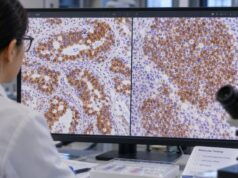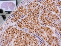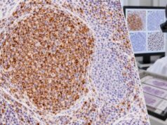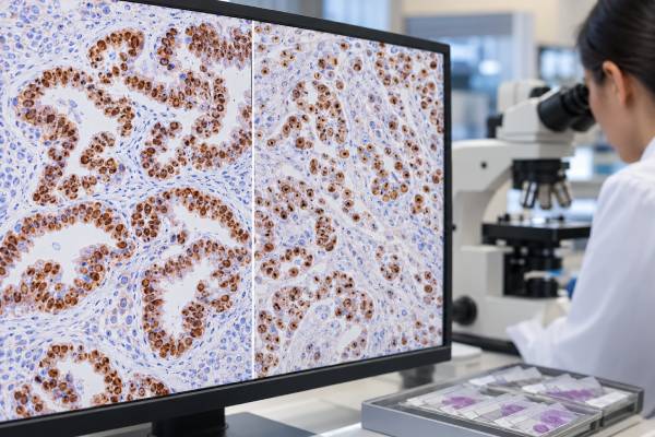
WT1 immunohistochemistry (IHC) is a nuclear tissue stain with two major diagnostic uses: supporting serous Müllerian carcinoma, especially tubo-ovarian high-grade serous carcinoma, and supporting mesothelial differentiation in malignant mesothelioma. WT1 is the Wilms tumor 1 transcription factor, but in routine adult tumor pathology the stain is far more commonly used in ovarian, fallopian tube, peritoneal, and pleural differentials than to diagnose Wilms tumor. The key pattern is nuclear staining; cytoplasmic staining is generally not interpreted as equivalent evidence. WT1 is useful but not specific enough to stand alone. Some uterine serous carcinomas express WT1, not every ovarian serous carcinoma is positive, and mesothelioma diagnosis requires a panel because reactive mesothelial cells can also express WT1. Pathologists combine the result with morphology and markers such as PAX8, p53, ER, calretinin, D2-40, cytokeratin 5/6, BAP1, claudin-4, and other carcinoma markers.
- Meaningful WT1 positivity is nuclear staining in the tumor cells.
- Tubo-ovarian high-grade serous carcinoma is commonly WT1 positive, often together with PAX8 and abnormal p53 staining.
- Epithelioid mesothelioma is often WT1 positive, but mesothelioma requires both mesothelial and carcinoma-exclusion markers.
- WT1 is not specific for ovarian cancer or mesothelioma, and reactive mesothelial cells can also stain.
- Cytoplasmic WT1 staining should not be interpreted like true nuclear positivity.
Table of Contents
- What the WT1 IHC test detects
- WT1 in ovarian and tubal serous carcinoma
- WT1 in malignant mesothelioma
- How WT1-positive ovarian carcinoma is distinguished from mesothelioma
- Other WT1-positive and negative patterns
- How pathologists build a WT1-based panel
- What the WT1 result means for care
What the WT1 IHC test detects
WT1 IHC detects the Wilms tumor 1 protein in fixed tissue. In most diagnostic applications discussed here, pathologists look for nuclear staining. The location of the signal matters because nonspecific or cytoplasmic staining does not carry the same tissue-lineage significance.
WT1 is used when morphology suggests a serous Müllerian carcinoma or a mesothelial neoplasm. It can also appear in other tumor contexts, so the stain is not ordered as a universal cancer marker. There is no blood reference range or patient preparation because the assay is performed on tissue.
The pathologist compares WT1 with the location, architecture, and other immunostains. A PAX8 IHC stain is often central in Müllerian differentials, while mesothelioma uses a very different companion panel.
WT1 in ovarian and tubal serous carcinoma
High-grade serous carcinoma of the ovary, fallopian tube, and peritoneum commonly expresses WT1. The typical diagnostic profile includes PAX8 positivity, WT1 positivity, and an abnormal p53 pattern reflecting the near-universal TP53 alteration characteristic of high-grade serous carcinoma.
An abnormal p53 IHC pattern may show diffuse overexpression, complete absence, or—less commonly—a cytoplasmic pattern. WT1 answers a different question: it supports serous Müllerian differentiation.
WT1 is particularly helpful in metastatic disease. A pleural or peritoneal carcinoma in a woman may raise breast, lung, gastrointestinal, mesothelial, and Müllerian possibilities. PAX8 plus WT1, interpreted with ER, p53, GATA3, TTF-1, and other markers, can strongly support tubo-ovarian origin when the overall pattern fits.
WT1 in malignant mesothelioma
Many epithelioid mesotheliomas show nuclear WT1 staining, making WT1 one of the established positive mesothelial markers. It is usually paired with calretinin, D2-40, or CK5/6 rather than used alone. An IHC stain for calretinin and D2-40 are common companions.
The opposing side of the panel uses epithelial carcinoma markers such as claudin-4, MOC31, or Ber-EP4, with organ-specific markers when a particular metastatic carcinoma is suspected. The aim is to show a coherent mesothelial phenotype while excluding the leading mimics.
WT1 positivity does not prove malignancy because reactive mesothelial cells can express WT1. In difficult pleural proliferations, BAP1 loss, MTAP loss, or CDKN2A homozygous deletion by FISH may provide evidence supporting malignant mesothelioma, depending on the specimen and laboratory approach.
How WT1-positive ovarian carcinoma is distinguished from mesothelioma
WT1 can be positive in both serous Müllerian carcinoma and mesothelioma, so the clinical site and companion markers are decisive. PAX8 strongly favors Müllerian origin over mesothelioma in most routine settings. Calretinin and D2-40 support mesothelial differentiation but can have exceptions.
In a peritoneal tumor, morphology may show papillary architecture in either disease. A panel including PAX8, ER, WT1, calretinin, D2-40, claudin-4, and BAP1/MTAP-related testing can clarify the distinction. Claudin-4 is a useful epithelial marker that generally favors carcinoma over mesothelioma.
The pathologist also considers prior gynecologic cancer, imaging of the ovaries and tubes, diffuse pleural or peritoneal growth, and cytology. No single WT1-positive cell population should override these broader findings.
Other WT1-positive and negative patterns
WT1 expression is not exclusive to ovarian serous carcinoma or mesothelioma. Some uterine serous carcinomas are positive, although historically WT1 was used as a discriminator because ovarian serous carcinoma is positive more often. Other neoplasms and normal tissues can also express WT1 depending on antibody clone and pattern.
A negative WT1 stain does not completely exclude high-grade serous carcinoma or mesothelioma. Tumor heterogeneity, subtype, fixation, and treatment can affect expression. Sarcomatoid mesotheliomas in particular may lose classic mesothelial markers, making diagnosis more difficult.
The location of staining remains crucial. In mesothelioma differentials, nuclear WT1 is the meaningful pattern. Cytoplasmic-only reactivity should not be used as evidence of mesothelial or Müllerian lineage.
How pathologists build a WT1-based panel
For suspected tubo-ovarian high-grade serous carcinoma, a typical panel may include PAX8, WT1, p53, ER, and p16, with additional stains dictated by the differential. For suspected mesothelioma, at least two mesothelial markers and two carcinoma markers are often used so both sides of the differential are tested.
The exact panel changes with morphology. If lung adenocarcinoma is the main alternative, TTF-1 and Napsin A may be added. If breast carcinoma is considered, GATA3 and breast-lineage markers may be more useful. If the tumor is gastrointestinal, CDX2 and SATB2 may enter the panel.
This is why WT1 should be interpreted as part of a tailored tumor-origin IHC panel rather than a stand-alone marker.
What the WT1 result means for care
WT1 IHC mainly helps classify the tumor correctly. A diagnosis of tubo-ovarian high-grade serous carcinoma leads to gynecologic cancer staging and molecular testing pathways, including BRCA1/2 and homologous recombination assessment where appropriate. A diagnosis of mesothelioma leads to a different staging system and treatment strategy.
The intensity of WT1 staining itself does not determine stage and is not a routine stand-alone predictive biomarker for standard therapy. Prognostic research exists, but individual management depends on the complete diagnosis, stage, resectability, molecular findings, and patient factors.
If a pathology report says “WT1 positive,” ask whether the staining was nuclear, which cells were positive, and which companion markers establish whether the tumor is Müllerian, mesothelial, or another lineage. That integrated pattern is the clinically useful result.
References
- Study of High Levels of Wilms’ Tumor 1 (WT1) Expression in Ovarian Cancers. 2026 (Study)
- Diagnostic performance of immunohistochemistry markers for malignant pleural mesothelioma diagnosis and subtypes. A systematic review and meta-analysis 2024 (Systematic Review)
- P53 and TLR4 expression are prognostic markers informing progression free survival of advanced stage high grade serous ovarian cancer 2024 (Study)
- Evaluation of the incidence and clinical significance of WT-1 expression in uterine serous carcinoma 2022 (Study)
- GATA3 is a useful immunohistochemical marker for distinguishing sarcomatoid malignant mesothelioma from lung sarcomatoid carcinoma and organizing pleuritis 2021 (Study)
- The immunohistochemical diagnosis of mesothelioma: a comparative study of epithelioid mesothelioma and lung adenocarcinoma. 2003 (Study)
Disclaimer
WT1 IHC is an ancillary tissue test and cannot diagnose ovarian cancer or mesothelioma by itself. Nuclear staining must be interpreted with morphology, the anatomic site, and a complementary panel. Reactive mesothelial cells and other tumors may also express WT1.